. 2009 Jan;181(1):65-79.
doi: 10.1534/genetics.108.095513. Epub 2008 Nov 10.

Pds1p is required for meiotic recombination and prophase I progression in Saccharomyces cerevisiae

Affiliations

Pds1p is required for meiotic recombination and prophase I progression in Saccharomyces cerevisiae

Katrina F Cooper et al. Genetics. 2009 Jan.

Abstract

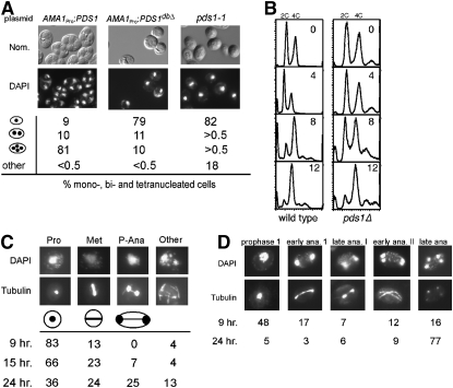
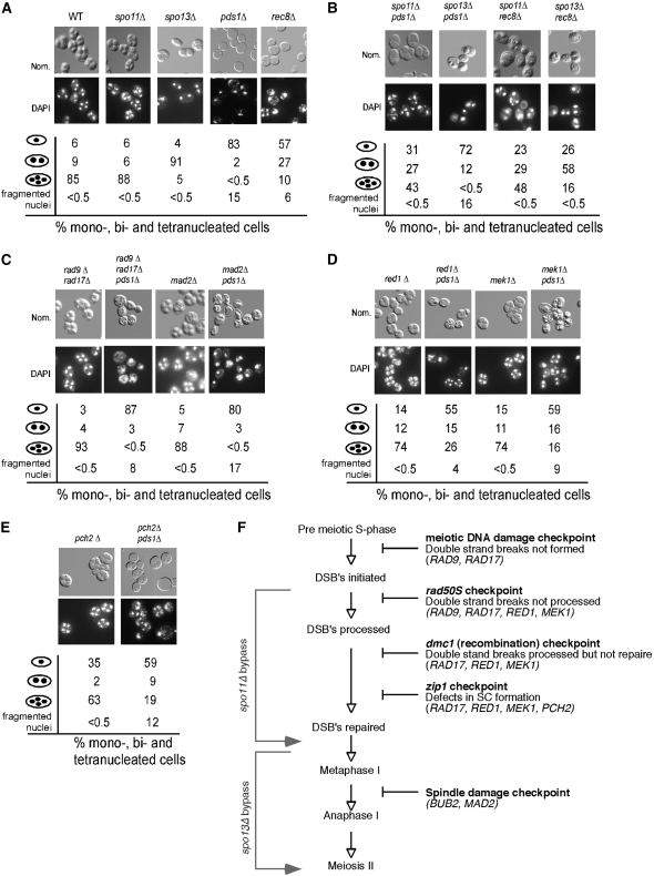
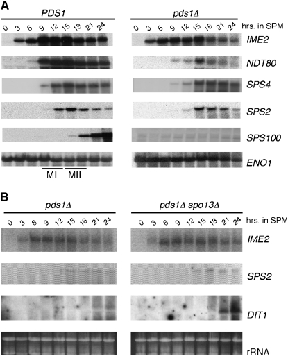
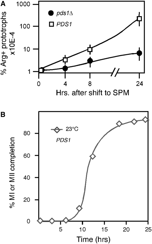
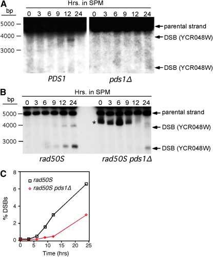
Sister-chromatid separation at the metaphase-anaphase transition is regulated by a proteolytic cascade. Destruction of the securin Pds1p liberates the Esp1p separase, which ultimately targets the mitotic cohesin Mcd1p/Scc1p for destruction. Pds1p stabilization by the spindle or DNA damage checkpoints prevents sister-chromatid separation while mutants lacking PDS1 (pds1Delta) are temperature sensitive for growth due to elevated chromosome loss. This report examined the role of the budding yeast Pds1p in meiotic progression using genetic, cytological, and biochemical assays. Similar to its mitotic function, Pds1p destruction is required for metaphase I-anaphase I transition. However, even at the permissive temperature for growth, pds1Delta mutants arrest with prophase I spindle and nuclear characteristics. This arrest was partially suppressed by preventing recombination initiation or by inactivating a subset of recombination checkpoint components. Further studies revealed that Pds1p is required for recombination in both double-strand-break formation and synaptonemal complex assembly. Although deleting PDS1 did not affect the degradation of the meiotic cohesin Rec8p, Mcd1p was precociously destroyed as cells entered the meiotic program. This role is meiosis specific as Mcd1p destruction is not altered in vegetative pds1Delta cultures. These results define a previously undescribed role for Pds1p in cohesin maintenance, recombination, and meiotic progression.


Figures

F<sc>igure</sc> 1.—
Figure 1.—
Pds1p regulates meiotic progression. (A) Pds1p destruction is required for meiotic progression. Wild-type culture (RSY335) harboring either wild-type or nondegradable Pds1p mutant (PDS1dbΔ) plasmids (pMSC13 and pMSC14, respectively) under the control of the meiosis-specific AMA1 promoter and pds1-1 cells (KCY348) were induced to enter meiosis at 23°. After 24 hr, nuclear divisions and spore formation were analyzed by DAPI staining (bottom) or Nomarski imaging (top), respectively. Percentages of the culture containing one, two or three-plus-four, or fragmented nuclei are indicated. Fragmented nuclei were scored as described in materials and methods. (B) Pds1p is not required for premeiotic S phase. Wild-type (RSY335) and pds1Δ (RSY787) diploid strains were induced to enter meiosis at 23° and samples were taken at the times indicated (in hours) and then analyzed by FACS. Peaks of 2C (unreplicated) and 4C (replicated) are indicated. (C and D) Terminal meiotic arrest phenotypes of pds1Δ mutants. Wild-type (RSY335, D) and pds1Δ (RSY787, C) diploid strains were induced to enter meiosis at 23° and samples were taken at the times indicated. Nuclear and spindle morphologies were determined by DAPI staining (top) and indirect immunofluorescence of tubulin (bottom). Cells were scored as described in materials and methods. For all morphology quantitations presented, the standard deviations were ≤6% for all values. Magnification is ×1000. (D) All strains used (except KCY348, which is derived from the A364A background) are isogenic to RSY335, our standard SK1/303 wild type.
F<sc>igure</sc> 2.—
Figure 2.—
Recombination initiation is required for the pds1Δ-induced meiotic arrest. (A) The meiotic progression and spore-wall assembly of wild-type (RSY335), spo11Δ (KCY198), spo13Δ (RSY767), pds1Δ (RSY787), and rec8Δ (KCY385) strains were assayed after 24 hr at 23° by DAPI analysis (bottom) and Nomarski imaging (top), respectively. Population percentages of mono-, bi-, tri-, and tetra-nucleated cells as well as irregular nuclei (scored as fragmented nuclei) in each culture are indicated. (B) Same as in A except that spo11Δ pds1Δ (KCY207), spo13Δ pds1Δ (RSY795), spo11Δ rec8Δ (KCY398), and spo13Δ rec8Δ (KCY399) strains were assayed for nuclear division and spore formation. (C) Same as in A except that rad9Δ rad17Δ (KCY450), rad9Δ rad17Δ pds1Δ (KCY453), mad2Δ (RSY740), and mad2Δ pds1Δ (RSY864) strains were assayed for nuclear division and spore formation. (D) Same as in A except that red1Δ (RSY1355), red1Δ pds1Δ (RSY1358), mek1Δ (RSY1356), and mek1Δ pds1Δ (RSY1359) strains were examined. (E) Same as in A except that pch2Δ (RSY1536) and pch2Δ pds1Δ (RSY1537) strains were examined. For all morphology quantitations presented, the standard deviations were ≤9% for all values. Magnification is ×1000. All the strains used are isogenic to RSY335, the SK1/W303 parent. (F) Diagram depicting the meiotic checkpoints tested with the bypass function of the genes assayed indicated.
F<sc>igure</sc> 3.—
Figure 3.—
The pds1Δ prophase arrest does not inhibit middle meiotic gene expression. (A) Wild-type (RSY335) and pds1Δ (RSY787) SK1/W303 cells were induced to enter meiosis at 23° and samples were taken at the times indicated (in hours). Total RNA was prepared and the transcript expression profiles of IME2, NDT80, SPS2, SPS4, and SPS100 were analyzed by Northern analysis. The approximate times of meiosis I (MI) and meiosis II (MII) as determined by DAPI analysis are indicated in the wild-type strain. ENO1 serves as a loading control. (B) Northern analysis of IME2, SPS2, and DIT1 mRNA expression in pds1Δ (RSY787) and pds1Δ spo13Δ (RSY795) SK1/W303 strains. (Bottom) Ethidium-bromide-stained rRNA is shown as a loading control.
F<sc>igure</sc> 4.—
Figure 4.—
Pds1p is required for meiotic recombination. (A) Three individual cultures of wild-type (KCY257) and pds1Δ (KCY274) SK1 strains containing heteroalleles at the arg4 locus were induced to enter meiosis at 23°. Samples were taken at the time points indicated following meiotic induction, serial diluted, and plated onto SD medium lacking arginine or complete SD medium. The resulting colonies were counted and the percentage of Arg+ prototrophs recovered was calculated (see supplemental Table S1). Plating dilutions for all experiments were adjusted on the basis of known recombination frequencies in wild-type strains at these loci. (B) Appearance of bi- and tetra-nucleated cells in wild-type cells in the SKI background at 23°.
F<sc>igure</sc> 5.—
Figure 5.—
Pds1p is required for normal double-strand-break formation. (A) Physical detection of DSB at the YCR048W recombination hotspot in wild-type (KCY428) and pds1Δ (KCY430) SK1 derivatives. The strains were induced to enter meiosis at 23° and samples were taken at the time points indicated (in hours). DNA extracts were digested with AseI and probed with sequences specific to YCR048W as described (Borde et al. 2004). Arrows indicate predicted recombination–restriction enzyme double-strand-break fragments. The asterisk denotes a nonspecific cross-hybridizing band. (B) Same as in A except that rad50S (KCY427) and rad50S pds1Δ (KCY429) SKI strains were examined. (C) Graphic representation of quantified DSB percentages with respect to parental signal in B.
F<sc>igure</sc> 6.—
Figure 6.—
Pds1p is required for normal SC formation. (A) Live-cell overlay images (GFP and Nomaski) of wild type (RSY335) and pds1Δ (RSY1433) strains expressing GFP-Zip1p 6 hr after transfer to SPM. The finger-like structure present in the wild-type cells represents individual chromosomes with formed SCs. (B) Fixed pds1Δ cells (RSY1433) harboring the GFP-ZIP1 expression plasmid 7.5 hr after transfer to SPM stained with DAPI (blue) examined under the fluorescent microscope. An expanded region indicated by the box is provided on the right (×600 magnification). (C) The cells described in B were subjected to indirect immunofluorescence staining for the nucleolar protein Nop1p. The images shown represent deconvoluted Z-stacks (0.6 μm slices) of Nomarski, GFP-Zip1p (green), Nop1p (red), and nuclei (blue). Magnification is ×1000.
F<sc>igure</sc> 7.—
Figure 7.—
Pds1p does not associate with chromatin during recombination. REC8-HA-tagged SK1/W303 strain (KCY392) harboring the Pds1p-FLAG expression plasmid (pKC7000) was grown to mid-log phase at 30° in synthetic acetate medium and then transferred to SPM and incubated at the same temperature. Samples were taken at the time points indicated (in hours) and whole-cell lysates (W) fractionated through a sucrose cushion to generate a chromatin bound pellet (P) and unbound supernatant (S) were analyzed by Western blotting and probed for the presence of Pds1p-FLAG (arrows, top) or Rec8p-HA (arrow, bottom). Molecular weight markers (in kilodaltons) are given on the left.
F<sc>igure</sc> 8.—
Figure 8.—
Mcd1p is cleaved prematurely in pds1Δ strains. (A) ubr1Δ REC8-3HA (KCY447) and ubr1Δ pds1Δ REC8-3HA (KCY448) SK1/W303 strains were induced to enter meiosis at 23° and samples were taken for Western analysis at the time points indicated (in hours). Full-length Rec8p-3HA and cleavage products are indicated by arrows. Asterisks indicate nonspecific cross-reactive bands observed with the HA antibody. The blot was stripped and reprobed for the presence of Tub1p for a loading control. (B) Return-to-growth recombination experiments as described in Figure 3 were performed on SK1/W303 pds1Δ culture (KCY274) at 23° harboring either Rec8p-HA expressed from a high-copy plasmid (pKC7001) or vector control (YEplac181). (C) Increasing REC8 gene dosage increases Rec8p production. Western blot analysis of Rec8p in a pds1Δ strain harboring either a single-copy (REC8 int) or a multi-copy (REC8 OE) HA-epitope-tagged REC8 allele following transfer to SPM medium as indicated (in hours). Tub1p serves as a loading control. (D) Increased gene dosage of ESP1 does not alter Rec8p degradation. Wild-type (RSY392) and pds1Δ (RSY448) SK1/W303 diploid strains containing the high-copy ESP1 plasmid (pKC7003) were induced to enter meiosis at 23° and samples were taken at the times indicated (in hours). Rec8p-3HA levels were monitored by Western blot analysis of total protein extracts. Tub1p serves as a loading control. (E) Mcd1p is prematurely destroyed in pds1Δ cells. Wild-type (RSY392) and pds1Δ (RSY448) SK1/W303 diploid strains were induced to enter meiosis at 23° and samples were taken for Western analysis at the time points indicated (in hours). Full-length Mcd1p was visualized by Western blot probing with antibodies directed against Mcd1p. The blot was stripped and reprobed for Tub1p for a loading control.

Similar articles

Cited by

References

    1. Agarwal, R., and O. Cohen-Fix, 2002. Phosphorylation of the mitotic regulator Pds1/securin by Cdc28 is required for efficient nuclear localization of Esp1/separase. Genes Dev. 16 1371–1382. - PMC - PubMed
    1. Alani, E., R. Padmore and N. Kleckner, 1990. Analysis of wild-type and rad50 mutants of yeast suggests an intimate relationship between meiotic chromosome synapsis and recombination. Cell 61 419–436. - PubMed
    1. Alexandru, G., W. Zachariae, A. Schleiffer and K. Nasmyth, 1999. Sister chromatid separation and chromosome re-duplication are regulated by different mechanisms in response to spindle damage. EMBO J. 18 2707–2721. - PMC - PubMed
    1. Arora, C., K. Kee, S. Maleki and S. Keeney, 2004. Antiviral protein Ski8 is a direct partner of Spo11 in meiotic DNA break formation, independent of its cytoplasmic role in RNA metabolism. Mol. Cell 13 549–559. - PubMed
    1. Baskerville, C., M. Segal and S. I. Reed, 2008. The protease activity of yeast separase (esp1) is required for anaphase spindle elongation independently of its role in cleavage of cohesin. Genetics 178 2361–2372. - PMC - PubMed

Publication types

MeSH terms