. 2009 Jan 2;284(1):685-695.
doi: 10.1074/jbc.M806124200. Epub 2008 Nov 10.

How does arrestin assemble MAPKs into a signaling complex?

Affiliations

How does arrestin assemble MAPKs into a signaling complex?

Xiufeng Song et al. J Biol Chem. .

Abstract

Arrestins bind active phosphorylated G protein-coupled receptors, precluding G protein activation and channeling signaling to alternative pathways. Arrestins also function as mitogen-activated protein kinase (MAPK) scaffolds, bringing together three components of MAPK signaling modules. Here we have demonstrated that all four vertebrate arrestins interact with JNK3, MKK4, and ASK1, but only arrestin3 facilitates JNK3 activation. Thus, the functional specificity of arrestins is not determined by differential binding of the kinases. Using receptor binding-impaired mutant, we have shown that free arrestin3 readily promotes JNK3 phosphorylation. We identified key arrestin-binding elements in JNK3 and ASK1 and investigated the molecular interactions of arrestin2 and arrestin3 and their individual domains with the components of the two MAPK cascades, ASK1-MKK4-JNK3 and c-Raf-1-MEK1-ERK2. We found that both arrestin domains interact with all six kinases. These findings shed new light on the mechanism of arrestin-mediated MAPK activation and the spatial arrangement of the three kinases on arrestin molecule.


Figures

FIGURE 1.
FIGURE 1.
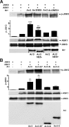
Arrestin3 is the only isoform that enhances ASK1-mediated JNK3 activation. COS-7 cells were transfected with HA-JNK3 alone, or with HA-ASK1, or together with FLAG-tagged arrestin3 (Arr3), arrestin2 (Arr2), arrestin1 (Rod), or arrestin4 (Cone). Cell lysates were immunoblotted for phospho-JNK (p-JNK3), HA, or FLAG. The intensity of the phospho-JNK band in three independent experiments was quantified and statistically analyzed. Means ± S.D. are shown. ****, p < 0.0001, as compared with the basal level in cells co-expressing ASK1 and JNK3 without arrestin.
FIGURE 2.
FIGURE 2.
ASK1, MKK4, and JNK3 bind arrestin3. A, COS-7 cells were transfected with FLAG-arrestin3 (FLAG-Arr3) with GFP-JNK3, HA-ASK1, HA-MKK4, or with indicated combinations of these kinases. Cell lysates were immunoprecipitated with rabbit anti-FLAG antibody, and then immunoblotted with mouse anti-HA, anti-GFP and anti-FLAG antibodies. B, COS-7 cells were transfected with HA-MKK4 with indicated FLAG-tagged arrestins. Cell lysates were immunoprecipitated and immunoblotted as in panel A. C, HEK-293A cells were transfected with HA-ASK-NLS alone or with indicated FLAG-tagged arrestins. Arrestins were visualized with M2 anti-FLAG, HA-ASK1 with rat anti-HA high affinity antibody, followed by Alexa 593 anti-mouse (Red) and Alexa 488 anti-rat (Green) secondary antibodies, respectively. The representative images show arrestins (red), HA-ASK1-NLS (green), and both channels merged. At least 20 cells expressing indicated proteins were scored for the subcellular distribution of HA-ASK1-NLS. Means ± S.D. (n = 3) of the fraction of cells with more ASK1 in the cytoplasm than in the nucleus are shown. ****, p < 0.0001.
FIGURE 3.
FIGURE 3.
Free arrestin scaffolds ASK1-MKK4-JNK3 module. A, COS-7 cells were transfected with HA-JNK3 alone, or with HA-ASK1, or together with arrestin3 (Arr3), arrestin3-NES- (Arr3-NES-), arrestin2 (Arr2), or arrestin2-NES+ (Arr2-NES+). Cell lysates were analyzed as in Fig. 1 (untagged arrestin3 was detected with F4C1 mouse monoclonal antibody). Means ± S.D. (n = 3) of the intensity of phospho-JNK3 band are shown. Both arrestin3 (****, p < 0.0001) and arrestin3-NES- (**, p < 0.01) enhance JNK3 activation, as compared with cells expressing ASK1+JNK3 without arrestin; JNK3 activation enhanced by arrestin3-NES- was significantly different from arrestin3 (###, p = 0.0004). B, COS-7 cells were transfected with HA-JNK3 + HA-ASK1, or together with FLAG-tagged arrestin3, arrestin3-D7, arrestin3-N-domain, or arrestin3-C-domain. The lysates were analyzed as in Fig. 1. Means ± S.D. (n = 3) of the intensity of phospho-JNK3 band are shown. Statistical analysis shows that arrestin3 (**, p = 0.0053) and arrestin3-D7 (**, p = 0.0018) enhance JNK3 activation, whereas arrestin3-N (p = 0.5769) and arrestin3-C (p = 0.3629) do not.
FIGURE 4.
FIGURE 4.
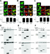
ASK1, MKK4, and JNK3 interact with both domains of arrestin. A-C, HEK-293A cells were transfected with HA-ASK-NLS or GFP-JNK3 individually or with FLAG-tagged full-length arrestin3 (FLAG-Arr3), its N- (FLAG-Arr3-N) or C-domain (FLAG-Arr3-C), arrestin2-NES+ (Arr2-NES), arrestin2 N-domain-NES (Arr2-N-NES), or arrestin2 C-domain-NES (Arr2-C-NES). Immunocytochemistry was done as described in Fig. 2C. GFP-JNK3 was visualized by its intrinsic fluorescence. Representative images are shown. The localization of GFP-JNK and HA-ASK1-NLS was quantified, as described in the legend to in Fig. 2C. Means ± S.D. (n = 3) of the fraction of cells with more JNK3 or ASK1 in the cytoplasm than in the nucleus also shown. (****, p < 0.0001). D-F, COS-7 cells were transfected with HA-ASK1, HA-MKK4, or GFP-JNK3 alone, or together with FLAG-tagged arrestin3 and its domains. Immunoprecipitation and Western blot analysis were performed as described in the legend to Fig. 2A.
FIGURE 5.
FIGURE 5.
N- and C termini of JNK3α2 regulate its binding to arrestin3. A, Truncated JNK3 constructs have deletions in the N- (40-464) or C terminus (1-402), or in both termini (40-402). B, COS-7 cells were transfected GFP-tagged JNK3α2 constructs shown in A with FLAG-arrestin3 (FLAG-Arr3). Immunoprecipitation and Western blot analysis were performed as described in the legend to Fig. 2A. C and D, HEK-293A cells were transfected with GFP-JNK3-(40-402), GFP-JNK3-(40-464), or GFP-JNK3-(1-402) alone, or with FLAG-arrestin3 (Arr3), arrestin2 with engineered NES (Arr2NES), and N- (Arr3N) or C-domain (Arr3C) of arrestin3, respectively. Proteins were visualized, as described in the legend to Fig. 4. The localization of GFP-JNK3 constructs was quantified, as described in the legend to in Fig. 2C. Means ± S.D. (n = 3) of the fraction of cells with more GFP-JNK3 in the cytoplasm than in the nucleus are shown. ****, p < 0.0001. Representative images are shown in D.
FIGURE 6.
FIGURE 6.
Kinase and C-domains are the key arrestin-binding elements in ASK1. A, Constructs containing different domains of ASK1: N domain (ASK1-N), N and Kinase domains (ASK1-(N+K)), C domain (ASK1-C), C and kinase domains (ASK1-(C+K)). B and C, COS-7 cells were transfected with HA-tagged ASK1 constructs shown in panel A, together with FLAG-arrestin3 (FLAG-Arr3) (panel B), or HA-ASK1-(C+K) with full-length arrestin3 (Arr3) or its N- (Arr3-N) and C-domain (Arr3-C) (panel C), respectively. Immunoprecipitation and Western blot analysis were performed as described in the legend to Fig. 2A.
FIGURE 7.
FIGURE 7.
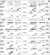
ERK-2, MEK-1 and c-Raf-1 interact with both domains of arrestin2 and arrestin3. COS-7 cells were transfected with plasmids encoding FLAG-tagged arrestin2 (A2), arrestin2 N-domain (A2N), arrestin2 C-domain (A2C), arrestin3 (A3), arrestin3 N-domain (A3N), or arrestin3 C-domain (A3C). HA-tagged ERK2, MEK1, and c-Raf1 were co-expressed with indicated FLAG-tagged arrestins. Arrestins were immunoprecipitated with M2 anti-FLAG antibody, and precipitates were probed for ERK2-HA (A), MEK1-HA (B) or c-Raf-1-HA (C). Alternatively, individually expressed kinases were immunoprecipitated with anti-HA antibody, and the precipitates were probed for arrestins using anti-FLAG antibody (A-C). The relative expression of each protein was confirmed by immunoblotting cell lysates (shown in the two lower blots in each panel). Asterisks, nonspecific band (heavy chain of the antibody).
FIGURE 8.
FIGURE 8.
Arrestin3-specific residues on the non-receptor-binding side of the molecule. The residues that are specific for arrestin3 (absent in the other three mammalian subtypes) were identified based on sequence alignment in (1). The positions of residues that represent nonconservative and conservative substitutions (as compared with arrestin2) are highlighted in red and green, respectively. Because the structure of arrestin3 was not solved, high-resolution structure of arrestin2 (PDB ID 1G4M) (27) was used to generate this picture. Three views are shown: upper panel, receptor-binding side (view down the concave sides of both domains); middle panel, side view; and lower panel, non-receptor-binding “back” of the arrestin molecule where signaling molecules interacting with the arrestin-receptor complex bind.
FIGURE 9.
FIGURE 9.
Model of the MAPK signaling module organized by arrestin. Front (A) and side (B) views of the three-dimensional model of arrestin complex with the three MAP kinases are shown. Arrestin (green) is shown as an elongated two-domain molecule. All the kinases are shown with thick cylinders representing kinase domain, and thin ones representing N- and C-terminal elements. MAPKKK (pink) is represented as ASK1 dimer in the JNK3 pathway with its C and kinase domain interacting with arrestin, and MAPK (purple) with its N terminus interacting with arrestin. MAPKK (yellow) also interacts with both domains of arrestin.

References

    1. Gurevich, E. V., and Gurevich, V. V. (2006) Genome Biol. 7 236. - PMC - PubMed
    1. Shenoy, S. K., and Lefkowitz, R. J. (2003) Biochem. J. 375 503-515 - PMC - PubMed
    1. Gurevich, V. V., and Gurevich, E. V. (2004) Trends Pharmacol. Sci. 25 59-112 - PubMed
    1. Carman, C. V., and Benovic, J. L. (1998) Curr. Opin. Neurobiol. 8 335-344 - PubMed
    1. DeFea, K. A. (2007) Annu. Rev. Physiol. 69 535-560 - PubMed

Publication types

MeSH terms