

Activation of Toll-like receptors 2, 3, and 4 on human melanoma cells induces inflammatory factors
- PMID: 19001446
- PMCID: PMC3480738
- DOI: 10.1158/1535-7163.MCT-08-0582
Activation of Toll-like receptors 2, 3, and 4 on human melanoma cells induces inflammatory factors
Abstract
Toll-like receptors (TLR) have been shown to be expressed on various types of cancers; however, their functional activity is not known. We examined TLR profiles of human melanoma cells and showed that TLR2, TLR3, and TLR4 were found to be highly expressed. By PCR array analysis, specific stimulation of TLR2, TLR3, and TLR4 on melanoma cells showed significant activation of the adaptor protein MyD88, as well as downstream signal transduction factors nuclear factor-kappaB and inflammatory response-related factors. Specific ligand activation of TLR2, TLR3, and TLR4 was shown to induce cell migration. Peripheral blood lymphocytes and melanoma purified RNA was shown to activate TLR3 on melanoma cells. These studies show expression and functional activity of specific TLRs on melanoma cells and as potential therapeutic targets to control tumor progression.
Conflict of interest statement
The authors applied for patent ownership. No other potential conflicts of interest were disclosed.
Figures

 , TLR4. D, six melanoma cells were stained with FITC-LPS and visualized by fluorescence light microscopy. PBL from healthy individuals was used as a positive control, and PANC1 was used as a negative control. C, representative melanoma cell lines fluorescence-activated cell sorting analysis of TLR2, TLR3, TLR4, and early endosome antigen 1. Early endosome antigen 1 is endosome protein and a positive control of TLR3. PANC1 was a negative control. Left curve shaded area is the isotype control.
 , TLR4. D, six melanoma cells were stained with FITC-LPS and visualized by fluorescence light microscopy. PBL from healthy individuals was used as a positive control, and PANC1 was used as a negative control. C, representative melanoma cell lines fluorescence-activated cell sorting analysis of TLR2, TLR3, TLR4, and early endosome antigen 1. Early endosome antigen 1 is endosome protein and a positive control of TLR3. PANC1 was a negative control. Left curve shaded area is the isotype control.
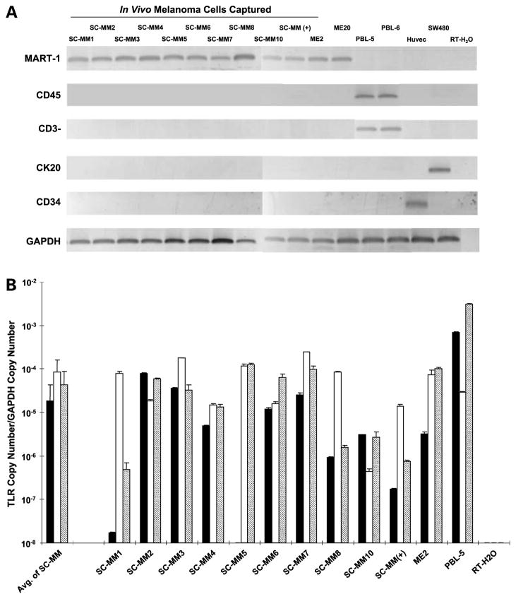 , TLR4.
 , PIC; ▤, zymosan.
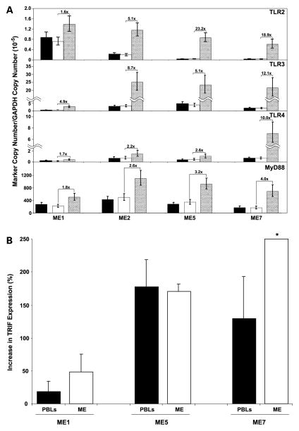 , melanoma cells cultured with supernatant from PHA stimulated PBLs. Small bars, 95% confidence intervals. The Y axis is the copy number of each gene over the copy number of the housekeeping gene GAPDH. B, expression level (mean) of TRIF mRNA in three melanoma lines (ME1, ME5, and ME7) cultured with supernatant of ■ PBLs (three donors) stimulated with PHA-L and □ ME (two melanoma cell lines ME1 and ME5) analyzed by qRT-PCR. The Y axis is the percentage of increase of TRIF mRNA expression in cells cultured with RNA compared with cells without RNA. Small bars, SD. *, actual mean increase of TRIF expression was 566 ± 497% for ME7 line incubated with melanoma cell RNA.
Similar articles
-
Hyperglycemia induces Toll-like receptor-2 and -4 expression and activity in human microvascular retinal endothelial cells: implications for diabetic retinopathy.J Diabetes Res. 2014;2014:790902. doi: 10.1155/2014/790902. Epub 2014 Dec 31. J Diabetes Res. 2014. Retraction in: J Diabetes Res. 2020 Oct 3;2020:5071954. doi: 10.1155/2020/5071954. PMID: 25610879 Free PMC article. Retracted.
-
MyD88-dependent and -independent signalling via TLR3 and TLR4 are differentially modulated by Δ9-tetrahydrocannabinol and cannabidiol in human macrophages.J Neuroimmunol. 2020 Jun 15;343:577217. doi: 10.1016/j.jneuroim.2020.577217. Epub 2020 Mar 20. J Neuroimmunol. 2020. PMID: 32244040
-
Toll-like receptor stimulation in cardiomyoctes decreases contractility and initiates an NF-kappaB dependent inflammatory response.Cardiovasc Res. 2006 Dec 1;72(3):384-93. doi: 10.1016/j.cardiores.2006.09.011. Epub 2006 Sep 23. Cardiovasc Res. 2006. PMID: 17054926
-
TLR3-Dependent Activation of TLR2 Endogenous Ligands via the MyD88 Signaling Pathway Augments the Innate Immune Response.Cells. 2020 Aug 17;9(8):1910. doi: 10.3390/cells9081910. Cells. 2020. PMID: 32824595 Free PMC article.
-
Variations within Toll-like receptor (TLR) and TLR signaling pathway-related genes and their synergistic effects on the risk of Guillain-Barré syndrome.J Peripher Nerv Syst. 2022 Jun;27(2):131-143. doi: 10.1111/jns.12484. Epub 2022 Feb 23. J Peripher Nerv Syst. 2022. PMID: 35138004
Cited by
-
B7-H3 associated with tumor progression and epigenetic regulatory activity in cutaneous melanoma.J Invest Dermatol. 2013 Aug;133(8):2050-8. doi: 10.1038/jid.2013.114. Epub 2013 Mar 8. J Invest Dermatol. 2013. PMID: 23474948 Free PMC article.
-
Salivary proteomics of canine oral tumors using MALDI-TOF mass spectrometry and LC-tandem mass spectrometry.PLoS One. 2019 Jul 18;14(7):e0219390. doi: 10.1371/journal.pone.0219390. eCollection 2019. PLoS One. 2019. PMID: 31318878 Free PMC article.
-
KDM3A promotes inhibitory cytokines secretion by participating in TLR4 regulation of Foxp3 transcription in lung adenocarcinoma cells.Oncol Lett. 2017 May;13(5):3529-3537. doi: 10.3892/ol.2017.5949. Epub 2017 Mar 29. Oncol Lett. 2017. PMID: 28521455 Free PMC article.
-
Cancer Cells Expressing Toll-like Receptors and the Tumor Microenvironment.Cancer Microenviron. 2009 Sep;2 Suppl 1(Suppl 1):205-14. doi: 10.1007/s12307-009-0022-y. Epub 2009 Aug 15. Cancer Microenviron. 2009. PMID: 19685283 Free PMC article.
-
Small-Molecule Natural Product Physachenolide C Potentiates Immunotherapy Efficacy by Targeting BET Proteins.Cancer Res. 2021 Jun 15;81(12):3374-3386. doi: 10.1158/0008-5472.CAN-20-2634. Epub 2021 Apr 9. Cancer Res. 2021. PMID: 33837043 Free PMC article.
References
-
- Medzhitov R, Preston-Hurlburt P, Janeway CA., Jr A human homologue of the Drosophila Toll protein signals activation of adaptive immunity. Nature. 1997;388:394–7. - PubMed
-
- Takeda K, Kaisho T, Akira S. Toll-like receptors. Annu Rev Immunol. 2003;21:335–76. - PubMed
-
- Molteni M, Marabella D, Orlandi C, Rossetti C. Melanoma cell lines are responsive in vitro to lipopolysaccharide and express TLR-4. Cancer Lett. 2006;235:75–83. - PubMed
Publication types
MeSH terms
Substances
Grants and funding
LinkOut - more resources
Full Text Sources
Other Literature Sources
Medical
Research Materials
