. 2008 Dec 1;121(Pt 23):3971-80.
doi: 10.1242/jcs.038414. Epub 2008 Nov 11.

Two distinct regions of Mto1 are required for normal microtubule nucleation and efficient association with the gamma-tubulin complex in vivo

Affiliations

Two distinct regions of Mto1 are required for normal microtubule nucleation and efficient association with the gamma-tubulin complex in vivo

Itaru Samejima et al. J Cell Sci. .

Abstract

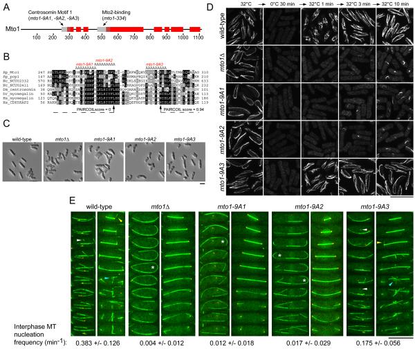
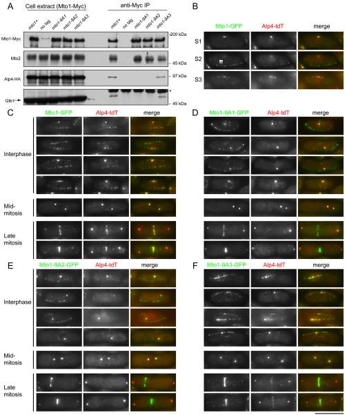
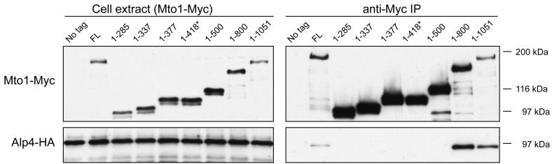
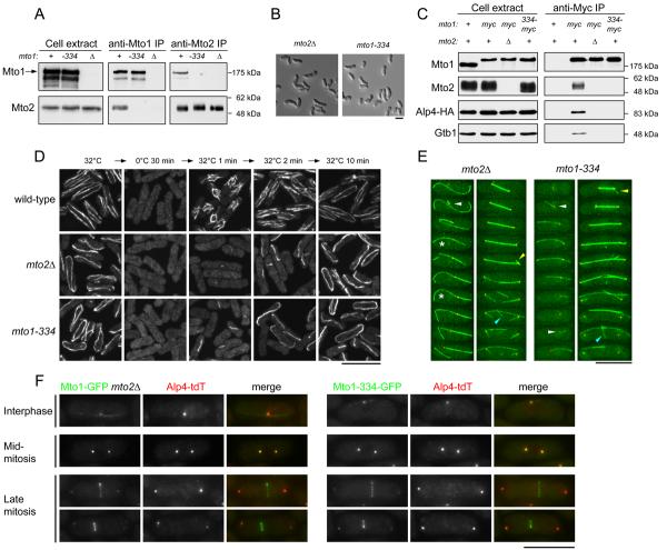
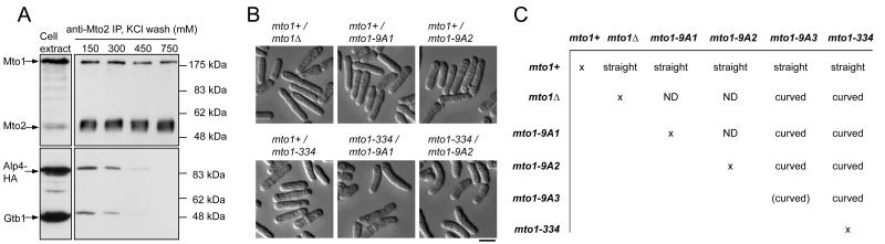
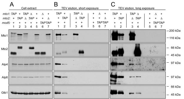
Cytoplasmic microtubule nucleation in the fission yeast Schizosaccharomyces pombe involves the interacting proteins Mto1 and Mto2, which are thought to recruit the gamma-tubulin complex (gamma-TuC) to prospective microtubule organizing centres. Mto1 contains a short amino-terminal region (CM1) that is conserved in higher eukaryotic proteins implicated in microtubule organization, centrosome function and/or brain development. Here we show that mutations in the Mto1 CM1 region generate mutant proteins that are functionally null for cytoplasmic microtubule nucleation and interaction with the gamma-TuC (phenocopying mto1Delta), even though the Mto1-mutant proteins localize normally in cells and can bind Mto2. Interestingly, the CM1 region is not sufficient for efficient interaction with the gamma-TuC. Mutation within a different region of Mto1, outside CM1, abrogates Mto2 binding and also impairs cytoplasmic microtubule nucleation and Mto1 association with the gamma-TuC. However, this mutation allows limited microtubule nucleation in vivo, phenocopying mto2Delta rather than mto1Delta. Further experiments suggest that Mto1 and Mto2 form a complex (Mto1/2 complex) independent of the gamma-TuC and that Mto1 and Mto2 can each associate with the gamma-TuC in the absence of the other, albeit extremely weakly compared to when both Mto1 and Mto2 are present. We propose that Mto2 acts cooperatively with Mto1 to promote association of the Mto1/2 complex with the gamma-TuC.


Figures

Fig. 1
Fig. 1
Two of three mutations in the Centrosomin motif 1 (CM1) region of Mto1 abolish cytoplasmic MT nucleation. (A) Schematic of Mto1 protein, showing CM1 and Mto2-binding region in grey. Predicted coiled-coils (PAIRCOIL score > 0.5) are indicated in red (Berger et al., 1995). Amino acid residue numbers are indicated below. (B) Alignment of Mto1 CM1 region with selected related proteins in fission yeast, Neurospora, Drosophila, zebrafish and human, with the amino acids mutated to alanine in the mto1-9A1, mto1-9A2, and mto1-9A3 mutants indicated above. The mto1-9A3 mutation lies at the beginning of the first predicted coiled-coil of Mto1. (C) Wild-type and mto1 mutant cell morphology (strains KS515, KS1017, KS2007, KS2010, KS2013) after growth to stationary phase and refeeding with fresh medium. (D) Anti-tubulin immunofluorescence time-course of MT regrowth after cold-induced depolymerization in wild-type and mutant strains (strains KS515, KS1017, KS2007, KS2010, KS2013). (E) Stills from movies of strains expressing GFP-tubulin and the SPB component Sad1-dsRed (strains KS2863, KS3609, KS3604, KS3605, KS3607; see supplementary material Movies 1-5). Interphase cells are shown on the left of each pair (40 sec intervals) and mitotic cells on the right (100 sec intervals). White arrowheads indicate examples of interphase MT nucleation. Yellow arrowheads indicate examples of mitotic astral MTs. Blue arrowheads indicate example of PAA MTs. Asterisks indicate bend-breakage events in which a single MT or MT bundle breaks into two. Values underneath images indicate average number of interphase nucleation events per cell per minute (see Materials and Methods). Bars, 10 μm.
Fig. 2
Fig. 2
Mto1-9A1 and Mto1-9A2 mutant proteins do not interact with the γ-tubulin complex (γ-TuC). (A) Anti-Myc immunoprecipitates of Myc-tagged wild-type and mutant Mto1 proteins (strains KS1517, KS1370, KS2290, KS2292, KS2294), probed with antibodies to the indicated proteins. The band migrating near Mto1-Myc on the anti-Myc blot of cell extract, “no tag” lane is an unrelated protein that is not precipitated by the anti-Myc antibody. IgG on the anti-γ-tubulin blot is marked by asterisk. (B) Individual sections (S1, S2, S3) of a single interphase cell expressing Mto1-GFP and Alp4-tdTomato from endogenous promoters (strain KS3957). The SPB is apparent in S1. Note in S2 that some Mto1-GFP satellites do not contain visible Alp4-tdT (middle section, arrowheads). (C-F) Maximum projections of cells expressing Mto1-GFP, Mto1-9A1-GFP, Mto1-9A2-GFP, or Mto1-9A3-GFP, together with Alp4-tdT (strains KS3957, KS3961, KS3962, KS3965) in interphase and mitosis. Bar, 10 μm.
Fig. 3
Fig. 3
The CM1 region of Mto1 is not sufficient for interaction with the γ-TuC. Anti-Myc immunoprecipitates of Myc-tagged carboxy-terminal truncations of Mto1 (strains KS1366, KS1517, KS1910, KS1822, KS1824, KS1825, KS1914, KS1916, KS1933), probed with antibodies to Mto1-Myc and γ-TuC protein Alp4-HA. “FL” indicates full-length Mto1 (1-1115). Abnormally fast migration of Mto1-(1-418)-Myc (marked with asterisk) is due to a reduced number of Myc tags.
Fig. 4
Fig. 4
Mto2 binding to Mto1 is required for efficient interaction of Mto1 with the γ-TuC. (A) anti-Mto1 and anti-Mto2 immunoprecipitates from wild-type, mto1-334 and mto1Δ cells (strains KS516, KS2272, KS1017), probed with anti-Mto1 and anti-Mto2 antibodies. (B) mto2Δ and mto1-334 mutant cell morphology (strains KS977, KS3734) after growth to stationary phase and refeeding with fresh medium. (C) Anti-Myc immunoprecipitates of Mto1, Mto1-Myc or Mto1-334-Myc, in the mto2 backgrounds shown (strains KS1370, KS1517, KS2169, KS3742), probed with antibodies to the indicated proteins. (D) Anti-tubulin immunofluorescence time-course of MT regrowth after cold-induced depolymerization in wild-type, mto2Δ and mto1-334 cells (strains KS516, KS976, KS2272). (E) Stills from movies of mto2Δ and mto1-334 cells expressing GFP-tubulin and Sad1-dsRed (strains KS2785, KS3765; see supplementary material Movies 6 and 7). Interphase cells are shown on the left of each pair (40 sec intervals) and mitotic cells on the right (100 sec intervals). White arrowheads indicate interphase MT nucleation, restricted to the SPB. Yellow arrowheads indicate mitotic astral MTs. Blue arrowheads indicate examples of weak nucleation of MTs from eMTOC. Asterisks indicate bend-breakage events in which a single MT or MT bundle breaks into two. (F) Maximum projections of cells expressing Mto1-GFP in mto2Δ background or Mto1-334-GFP in wild-type mto2+ background, together with Alp4-tdT (KS4074, KS4039). In some but not all mto2Δ and mto1-334 mutants, very faint Alp4-tdT could be observed at the equator, which may represent sub-detection amounts of γ-TuC that are likely responsible for the very small amount of equatorial MT nucleation observed in these mutants (see Fig. 4E). Bars, 10 μm.
Fig. 5
Fig. 5
Mto1 and Mto2 form a stable complex in which Mto1 must contain both an intact CM1 region and an Mto2-binding region for normal function. (A) Anti-Mto2 immunoprecipitates from alp4-HA cells (strain KS1370), washed with the indicated concentrations of KCl and probed with antibodies to the proteins shown. (B) DIC images of representative diploid strains of the indicated genotypes described in (C) after growth to stationary phase and refeeding with fresh medium. Bar, 10 μm. (C) Cell morphology of diploids combining the different mto1 alleles shown in the rows and columns in trans (strains KS3786 × KS3780, KS3785 × KS3775, KS3786 × KS3789, KS3786 × KS3777, KS3795 × KS3784, KS3781 × KS3776, KS3780 × KS3784, KS3774 × KS3777, KS3775 × KS3784, KS3788 × KS3776, KS3789 × KS3784, KS3776 × KS3778, KS3777 × KS3784, reading from left to right). mto1-9A3 did not trans-complement any of the other mto1 mutant alleles, probably because mto1-9A3 is itself a partial loss-of-function allele, as shown in the morphology of mto1-9A3/mto1-9A3 homozygous diploids (see also Figs. 1 and 2; supplementary material Movie 4). Other homozygous diploids were not constructed; ND, not determined.
Fig. 6
Fig. 6
Cooperative binding of the Mto1/2 complex to γ-TuC. Purification of TAP-tagged Mto1 and Mto2 in wild-type and mutant backgrounds (strains KS3524, KS3956, KS4323, KS4326, KS516, KS4267, KS4270), probed with antibodies to the indicated proteins. Differently-migrating forms of Mto1 and Mto2 represent TAP-tagged, cleaved TAP-tagged, and untagged forms of the proteins. Vertical lines indicate blank lanes.

Similar articles

Cited by

References

    1. Anders A, Lourenco PC, Sawin KE. Noncore components of the fission yeast gamma-tubulin complex. Mol. Biol. Cell. 2006;17:5075–93. - PMC - PubMed
    1. Bahler J, Wu JQ, Longtine MS, Shah NG, McKenzie A, 3rd, Steever AB, Wach A, Philippsen P, Pringle JR. Heterologous modules for efficient and versatile PCR-based gene targeting in Schizosaccharomyces pombe. Yeast. 1998;14:943–51. - PubMed
    1. Berger B, Wilson DB, Wolf E, Tonchev T, Milla M, Kim PS. Predicting coiled coils by use of pairwise residue correlations. Proc. Natl. Acad. Sci. U S A. 1995;92:8259–63. - PMC - PubMed
    1. Bond J, Roberts E, Springell K, Lizarraga SB, Scott S, Higgins J, Hampshire DJ, Morrison EE, Leal GF, Silva EO, et al. A centrosomal mechanism involving CDK5RAP2 and CENPJ controls brain size. Nat. Genet. 2005;37:353–5. - PubMed
    1. Brune C, Munchel SE, Fischer N, Podtelejnikov AV, Weis K. Yeast poly(A)-binding protein Pab1 shuttles between the nucleus and the cytoplasm and functions in mRNA export. Rna. 2005;11:517–31. - PMC - PubMed

Publication types

MeSH terms