. 2008 Dec 1;121(Pt 23):3912-21.
doi: 10.1242/jcs.035360. Epub 2008 Nov 11.

The NIMA-family kinase Nek6 phosphorylates the kinesin Eg5 at a novel site necessary for mitotic spindle formation

Affiliations

The NIMA-family kinase Nek6 phosphorylates the kinesin Eg5 at a novel site necessary for mitotic spindle formation

Joseph Rapley et al. J Cell Sci. .

Abstract

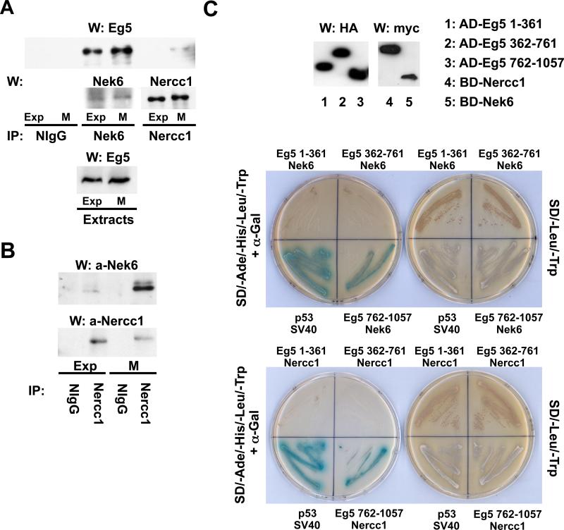
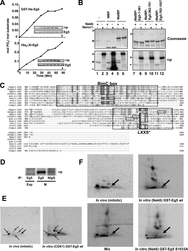
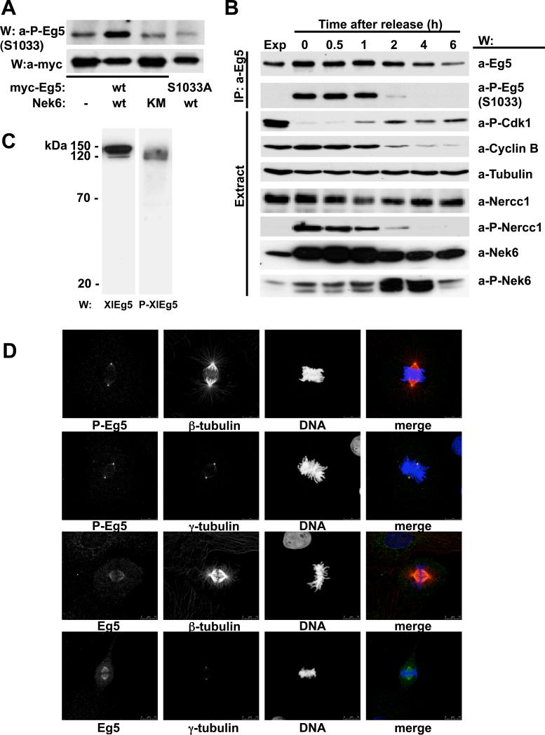
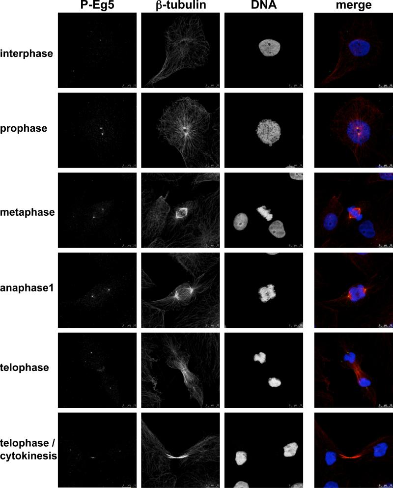
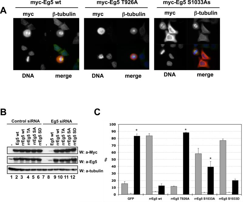
Nek6 and Nercc1 (also known as Nek9) belong to the NIMA family of protein kinases. Nercc1 is activated in mitosis, whereupon it binds, phosphorylates and activates Nek6. Interference with Nek6 or Nercc1 in mammalian cells causes prometaphase-metaphase arrest, and depletion of Nercc1 from Xenopus egg extracts prevents normal spindle assembly. Herein we show that Nek6 is constitutively associated with Eg5 (also known as Kinesin-5 and Kif11), a kinesin that is necessary for spindle bipolarity. Nek6 phosphorylated Eg5 at several sites in vitro and one of these sites, Ser1033, is phosphorylated in vivo during mitosis. Whereas CDK1 phosphorylates nearly all Eg5 at Thr926 during mitosis, Nek6 phosphorylates approximately 3% of Eg5, primarily at the spindle poles. Eg5 depletion caused mitotic arrest, resulting in cells with a monopolar spindle. This arrest could be rescued by wild-type Eg5 but not by Eg5[Thr926Ala]. Despite substantial overexpression, Eg5[Ser1033Ala] rescued 50% of cells compared with wild-type Eg5, whereas an Eg5[Ser1033Asp] mutant was nearly as effective as wild type. Thus, during mitosis Nek6 phosphorylates a subset of Eg5 polypeptides at a conserved site, the phosphorylation of which is crucial for the mitotic function of Eg5.


Figures

Figure 1
Figure 1. Eg5 interacts with Nek6 and Nercc1
(A) Immunoprecipitates were prepared from exponentially growing (Exp) or nocodazole-arrested mitotic (M) U2OS cell extracts using either normal rabbit IgG (NIgG), anti-Nek6 or anti-Nercc1 antibodies and analyzed by immunoblot with the indicated antibodies. Eg5 in the corresponding extracts is shown in the lower panel. (B) Immunoprecipitates were prepared from exponentially growing (Exp) or nocodazole-arrested mitotic (M) U2OS cells using NIgG or anti-Nercc1 antibody, and immunoblotted for Nek6 and Nercc1. (C) The ability of Eg5 head (Eg5 1-361), stalk (Eg5 362-761) and tail (Eg5 762-1057) domains to interact with Nek6 or Nercc1 was assessed using two hybrid by histidine and adenine prototrophy plus expression of α-galactosidase activity (left plate in both pictures). Fusion protein expression was verified by immunoblot (upper panel). AD, BD: Gal4 activation and binding domains. The interaction between p53 and SV40 is a positive control.
Figure 2
Figure 2. Nek6 phosphorylates Eg5 in vitro at a conserved site that is phosphorylated during mitosis
(A) Recombinant GST-HsEg5 or His6-XlEg5 were phosphorylated by FLAG-Nek6. The insets show 32P incorporation with time (upper, left to right with no kinase in rightmost lane) and a coomassie blue stain of the substrate (lower). (B) Myelin basic protein (MBP, positive control), Maltose-binding protein (MalBP), or MalBP-fusion proteins containing the Eg5 segments indicated were incubated with [γ-32P]ATP/Mg2+ and either FLAG-Nek6 (lanes 1, 3, 5, 7, 9, 11) or preactivated FLAG-Nercc1 (2, 4, 6, 8, 10, 12) for 30 min followed by SDS-PAGE. The upper panels show Coomassie blue stains of the gels and the lower panels the corresponding autoradiographs. Open arrowheads indicate Nercc1 and filled arrowheads, Nek6. Asterisks indicate the location of MBP (lanes 3,4) or MalBP-fusion proteins (7-12). The left gel is 12% acrylamide, the right gel 7.5%; Nek6 is run off the latter. (C) HsEg5, human Eg5; MmEg5, mouse Eg5; XLEg5, Xenopus laevis Eg5; KLP61F, Drosophila melanogaster KLP61F; bmk-1, Caenorhabditis elegans bmk-1; BimC, Aspergillus nidulans BimC; KIP1, Cin8p, Saccharomyces cerevisiae KIP1 and Cin8p; Cut7, Schizosaccharomyces pombe Cut7. Identical and conserved residues are shaded; regions conserved around the C-terminal Cdk1 site (BimC box) and Nek6 site (LXXS*) are boxed. (D) Using NlIgG and anti-HsEg5 antibodies, immunoprecipitates were prepared from extracts of 32P-labelled U2OS cells growing exponentially (Exp) or incubated overnight in 0.25mM nocodazole (M), and subjected to SDS-PAGE; a PhosphoImage of the gel is shown. (E) Left, two-dimensional (2-D) tryptic phosphopeptide map of endogenous 32P-Eg5 immunoprecipitated from 32P-labeled mitotic U2OS cells, visualized by PhosphorImager. Right, phosphopeptide map of purified recombinant 32P-GST-Eg5 phosphorylated in vitro by immunoprecipitated mitotic Cdk1. The grey arrows mark phosphopeptides attributed to Cdk1; the black arrow indicates a 32P-peptide evident in digests of mitotic Eg5 that is not present in digests of Cdk1-phosphorylated 32P-Eg5. (F) Top left, 2-D phosphopeptide map of endogenous Eg5 immunoprecipitated from 32P-labeled mitotic U2OS cells; top and bottom right, 2-D maps of recombinant GST-Eg5 wt (top right) and GSTEg5[S1033A] (bottom right) phosphorylated in vitro by FLAG-Nek6; note in the lower right map the absence of the most anodally migrating 32P-peptide seen in the upper right map, which encompasses Ser1033-P. Bottom left, a mixture of comparable amounts of the digests of mitotic 32P-Eg5 and FLAG-Nek6 phosphorylated 32P-Eg5. The black arrow marks the phosphopeptide common to the two digests, which contains serine 1033. In the digests of mitotic 32P-Eg5 (E, left; F, upper left), this spot contains ~3% of total 32P, estimated by PhosphoImager.
Figure 3
Figure 3. Eg5[Ser1033] phosphorylation in vivo
(A) U2OS cells were cotransfected with either myc-Eg5 wt (lanes 1-3) or myc-Eg5[S1033A] (lane 4) and empty plasmid (lane1), FLAG-Nek6 wt (lane 2 and 4) or FLAG-Nek6 [K74M,K75M] (lane 3). 24 hours later, anti-Myc-immunoprecipitates were subjected to immunoblot with anti-Myc (lower panel) and anti-Eg5[Ser1033-P] antibodies (top panel). (B) U2OS cells growing exponentially were either untreated (first lane, Exp) or arrested with nocodazole (0.25mM overnight; lane 2); arrested cells were allowed to exit mitosis in nocodazole-free medium and extracted at the times indicated (lanes 3 to 7). Eg5 immunoprecipitates (top two rows) and cell extracts (bottom seven rows) were analyzed by immunoblot with the antibodies indicated (a-P-Eg5(S1033), anti-Eg5[serine1033P]; a-P-Cdk1, anti-Cdk1[Tyr15P]; a-P-Nercc1, anti-Nercc1[Thr210P]; a-P-Nek6, anti-Nek6[Ser206P]. (C) Extracts from XL177 Xenopus laevis cells were resolved by SDS-PAGE and immunoblotted with either anti-XlEg5 (left) or anti-XlEg5[Ser1046-P] (right). (D) XL177 cells growing exponentially were fixed and stained with antibodies to XlEg5 (lower two rows), XlEg5[Ser1046P] (upper two rows), and either β- or γ-tubulin as indicated. DNA is stained with DAPI. Representative cells in metaphase are shown; bar, 10 μm.
Figure 4
Figure 4. Phosphoserine 1046 immunolocalization in XL177 cells in interphase and different phases of mitosis
As in Figure 3D; bar, 10 μm.
Figure 5
Figure 5. Eg5[Ser1033] phosphorylation is required for normal Eg5 function in mitotic spindle assembly
(A) U2OS cells transiently expressing Myc-Eg5 wt, Thr926Ala or Ser1033Ala were stained for Myc, β-tubulin and DNA. A representative transfected cell in mitosis is shown in each case, except for Myc-Eg5 S1033A, where a mitotic plus two interphase transfected cells are shown. (B). Hela cells were co-transfected with either control oligonucleotides (lanes 1-6) or siRNA oligonucleotides directed against HsEg5 (lanes 7-12), together with empty plasmid (lanes 1,7) or plasmids encoding Myc- Eg5 wt, unmodified (lanes 2 and 8) or Myc-Eg5 variants rendered RNAi-resistant (Eg5 wild type, rrwt; Eg5[Thr926Ala], rrTA; Eg5[Ser1033Ala], rrSA; Eg5[Ser1033Asp], rrSD). Cell lysates were immunoblotted with anti-Myc (upper panel) or anti-Eg5 (middle panel) to determine recombinant and total Eg5 expression, respectively, and with anti-α-tubulin antibodies to evaluate loading (lowest panel). (C) HeLa cells were co-transfected with siRNA oligonucleotides directed against HsEg5 and plasmids encoding Myc-GFP (control) or several RNAi-resistant Myc-Eg5 (rrEg5) variants. Forty hours after transfection, cells were fixed and stained with anti-Myc and anti-β-tubulin antibodies and with DAPI. Myc-positive cells were scored as being in interphase (grey columns), in normal mitosis (white columns), or in an abnormal mitosis (mostly monopolar spindles; black columns). The figure represents the mean of three independent experiments, wherein >100 cells were scored for each point in each of the experiments; error bars indicate S.E.M.; The fraction of abnormal mitoses (black columns) in cells expressing rrEg5 wildtype was compared to each of the other conditions by Student's t test; p values < 0.05 are marked with an asterisk. The comparison of S1033A with 1033D gave a p value of 0.068.

Similar articles

Cited by

References

    1. Belham C, Roig J, Caldwell JA, Aoyama Y, Kemp BE, Comb M, Avruch J. A mitotic cascade of NIMA family kinases. Nercc1/Nek9 activates the Nek6 and Nek7 kinases. J Biol Chem. 2003;278:34897–34909. - PubMed
    1. Belham C, Comb MJ, Avruch J. Identification of the NIMA family kinases NEK6/7 as regulators of the p70 ribosomal S6 kinase. Curr Biol. 2001;11:1155–1167. - PubMed
    1. Bishop JD, Han Z, Schumacher JM. The Caenorhabditis elegans Aurora B kinase AIR-2 phosphorylates and is required for the localization of a BimC kinesin to meiotic and mitotic spindles. Mol Biol Cell. 2005;16:742–756. - PMC - PubMed
    1. Blangy A, Lane HA, d'Hérin P, Harper M, Kress M, Nigg EA. Phosphorylation by p34cdc2 regulates spindle association of human Eg5, a kinesin-related motor essential for bipolar spindle formation in vivo. Cell. 1995;83:1159–1169. - PubMed
    1. Blangy A, Arnaud L, Nigg EA. Phosphorylation by p34cdc2 protein kinase regulates binding of the kinesin-related motor HsEg5 to the dynactin subunit p150. J Biol Chem. 1997;272:19418–19424. - PubMed

Publication types