. 2008 Dec 1;121(Pt 23):3981-90.
doi: 10.1242/jcs.033431. Epub 2008 Nov 11.

The leukemogenic t(8;21) fusion protein AML1-ETO controls rRNA genes and associates with nucleolar-organizing regions at mitotic chromosomes

Affiliations

The leukemogenic t(8;21) fusion protein AML1-ETO controls rRNA genes and associates with nucleolar-organizing regions at mitotic chromosomes

Rachit Bakshi et al. J Cell Sci. .

Abstract

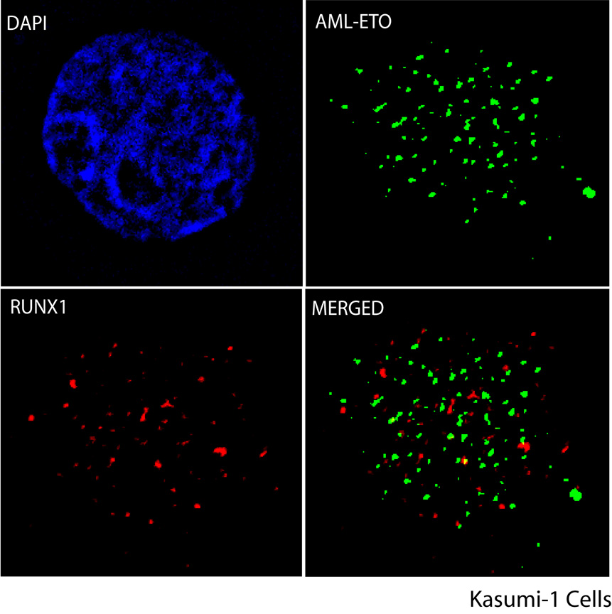
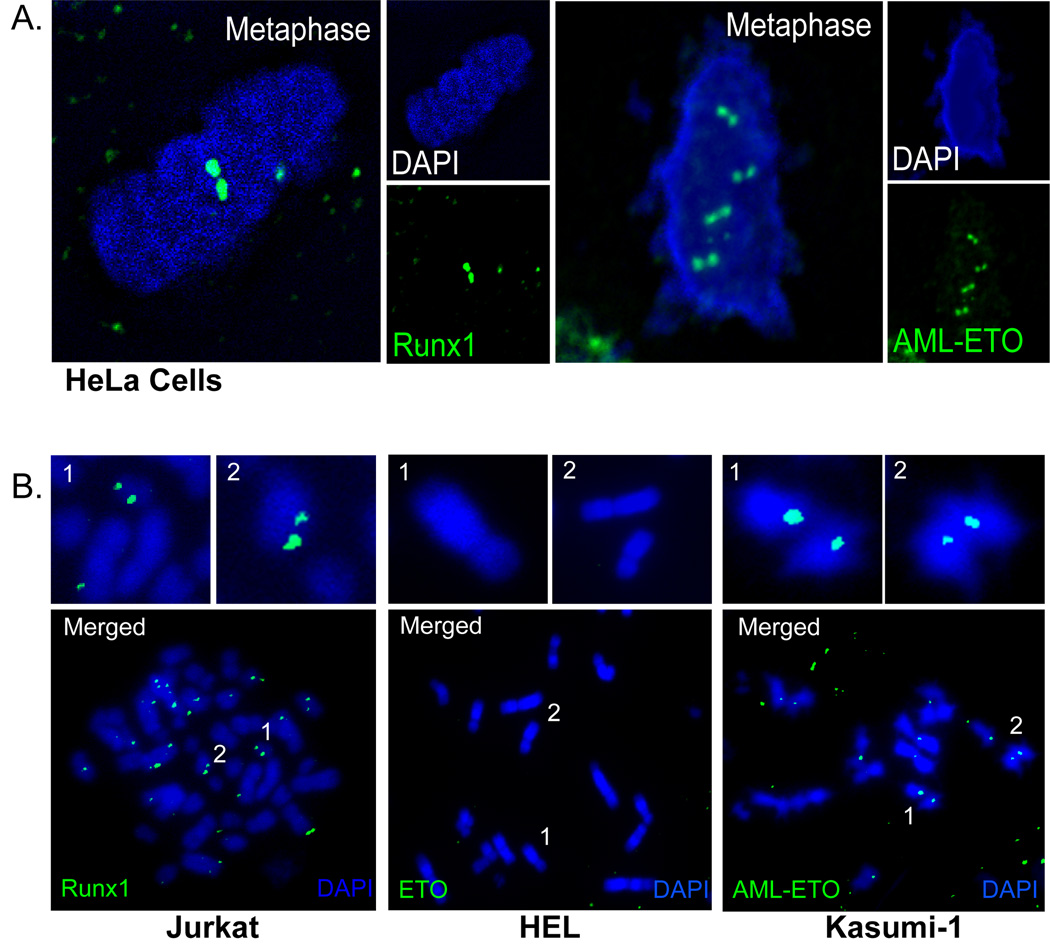
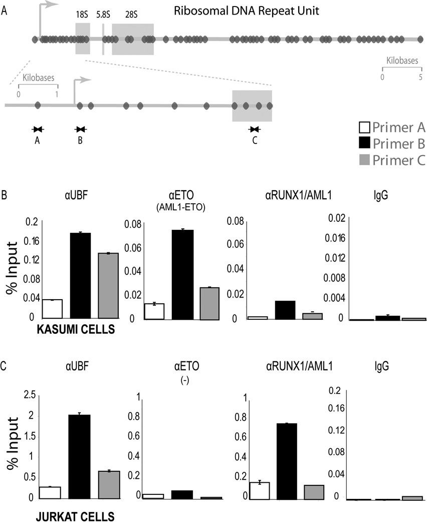
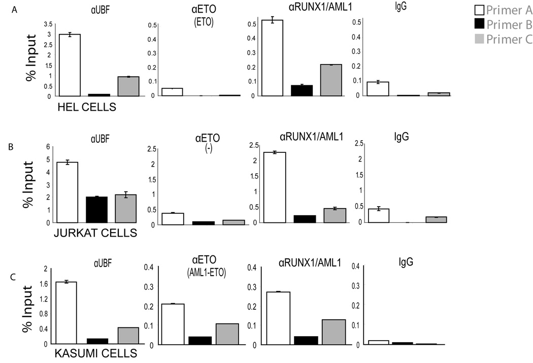
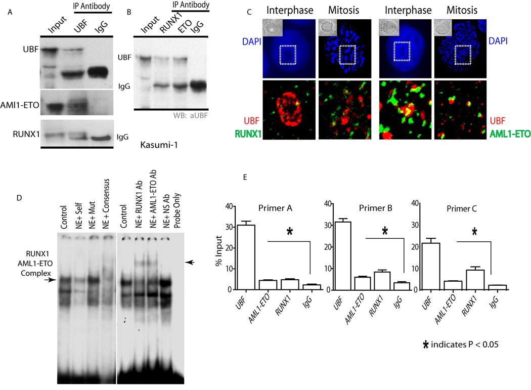
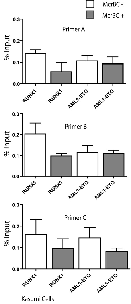
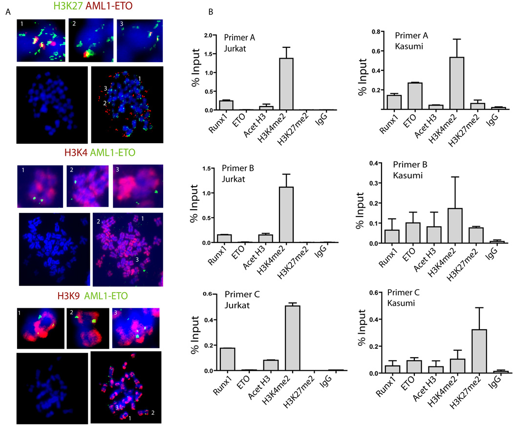
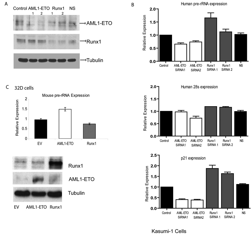
RUNX1/AML1 is required for definitive hematopoiesis and is frequently targeted by chromosomal translocations in acute myeloid leukemia (AML). The t(8;21)-related AML1-ETO fusion protein blocks differentiation of myeloid progenitors. Here, we show by immunofluorescence microscopy that during interphase, endogenous AML1-ETO localizes to nuclear microenvironments distinct from those containing native RUNX1/AML1 protein. At mitosis, we clearly detect binding of AML1-ETO to nucleolar-organizing regions in AML-derived Kasumi-1 cells and binding of RUNX1/AML1 to the same regions in Jurkat cells. Both RUNX1/AML1 and AML1-ETO occupy ribosomal DNA repeats during interphase, as well as interact with the endogenous RNA Pol I transcription factor UBF1. Promoter cytosine methylation analysis indicates that RUNX1/AML1 binds to rDNA repeats that are more highly CpG methylated than those bound by AML1-ETO. Downregulation by RNA interference reveals that RUNX1/AML1 negatively regulates rDNA transcription, whereas AML1-ETO is a positive regulator in Kasumi-1 cells. Taken together, our findings identify a novel role for the leukemia-related AML1-ETO protein in epigenetic control of cell growth through upregulation of ribosomal gene transcription mediated by RNA Pol I, consistent with the hyper-proliferative phenotype of myeloid cells in AML patients.


Figures

Figure 1
Figure 1. RUNX1/AML1 and AML1-ETO are targeted to distinct subnuclear locations
Immunofluorescence microscopy for endogenous RUNX1/AML1 (red) and AML1-ETO (green) with DAPI staining (blue) in Kasumi-1 cells as well as merged images are shown. RUNX1/AML1 and AML1-ETO fusion protein are predominantly present in the nucleus but do not co-localize in the Kasumi-1 cells. An antibody detecting ETO was used to visualize the AML1-ETO fusion protein, whereas an antibody recognizing the C-terminal domain of RUNX1/AML1 was used to detect endogenous RUNX1/AML1.
Figure 2
Figure 2. RUNX1/AML1 and AML1-ETO associate with metaphase chromosomes in pairs
A) Epitope-tagged RUNX1/AML1 and AML1-ETO were examined by in situ immunofluorescence in HeLa cells and metaphase cells were visually selected. RUNX1/AML1 and AML1-ETO foci (green) are associated with chromosomes (blue) in pairs (shown in deconvoluted images). B) Mitotic chromosome spreads were prepared for human Jurkat, HEL, and Kasumi-1 cells and processed for immunofluorescence microscopy using antibodies directed against the RUNX1/AML1, ETO and AML1-ETO proteins. RUNX1/AML1 and AML1-ETO show distinct foci on the mitotic chromosome spreads, while ETO is not detected on mitotic chromosomes. Panels on the top of each image show enlarged areas marked by numbers in the image.
Figure 3
Figure 3. AML1-ETO shows enhanced occupancy of rDNA repeats during mitosis
A) The panel shows a schematic of the Runx consensus elements (dark ovals) in the human rDNA repeats and depicts the locations of ChIP primers. B and C) ChIPs were done with antibodies for RUNX1/AML1, ETO and UBF1, as well as non immune IgG in Kasumi-1 and Jurkat cells blocked in mitosis. An antibody detecting ETO was used to immunoprecipitate the AML1-ETO fusion protein, whereas an antibody recognizing the C-terminal domain of RUNX1/AML1 was used to pull down endogenous RUNX1/AML1. Quantitative PCR data are normalized to genomic DNA and denoted as percent input.
Figure 4
Figure 4. Both RUNX1/AML1 and AML1-ETO occupy rDNA repeats in interphase Kasumi-1 cells
Chromatin immunoprecipitation was done in asynchronously growing HEL, Jurkat and Kasumi-1 cells using RUNX1/AML1, ETO, UBF1 and IgG antibodies. Three different PCR primer sets spanning Runx consensus elements were used (see Figure 3). A and B) An antibody detecting ETO was used to immunoprecipitate the AML1-ETO fusion protein, whereas an antibody recognizing the C-terminal domain of RUNX1/AML1 was used to pull down endogenous RUNX1/AML1. Quantitative PCR data show that endogenous ETO does not bind to rDNA repeats in HEL cells nor in Jurkat cells (where ETO is not expressed), while UBF1 occupies the rDNA repeats in vivo in all the three cell lines tested. C) RUNX1/AML1 and AML1-ETO both occupy rDNA repeats in Kasumi-1 cells. Quantitative PCR data are normalized to genomic DNA and denoted as percent input.
Figure 5
Figure 5. Endogenous RUNX1/AML1 and AML1-ETO interact with UBF1 on the rDNA repeats
A) Immunoprecipitation analysis was carried out with an antibody for UBF1 followed by western blotting with AML1-ETO and RUNX1/AML1 specific antibodies. Both RUNX1 and AML1-ETO are detected in western blot analysis, however AML1-ETO shows greater interaction with UBF1 when compared to RUNX1/AML1. B) Endogenous RUNX1/AML1 and AML1-ETO were immunoprecipitated from Kasumi-1 cells using rabbit polyclonal antibodies that specifically recognize the C-terminus of RUNX1/AML1 or the ETO moiety. A mouse monoclonal antibody was used to detect endogenous UBF1 by immunoblotting. UBF1 and IgG heavy chain are indicated. C) Immunofluorescence microscopy was performed in Kasumi-1 cells to detect endogenous RUNX1/AML1 (green), UBF1 (red) or AML1-ETO (green) with DAPI staining (blue). There is co-localization, albeit limited, of both RUNX1/AML1 and AML1-ETO with nucleolar UBF1 during interphase. D) Electrophoretic mobility shift assays were performed with a human rDNA probe spanning a Runx binding element and nuclear extracts from Kasumi-1 cells. Competition assays with 100-fold molar excess of unlabeled wild type, mutant or Runx consensus oligonucleotide were performed to establish the specific protein-DNA complex (lanes 1–4) as indicated. Super-shift immunoassays were performed by incubating binding reactions with the indicated antibodies (Lanes 5–8). Normal IgG was used as negative control. The arrow at the right marks the supershift band. E) ChIP-reChIP assays with endogenous proteins in interphase Kasumi-1 cells using UBF1 antibody (primary ChIP) and second immunoprecipitation (reChIP) with antibodies directed against UBF1, RUNX1/AML1, AML1-ETO or IgG. The re-ChIP data are plotted as a percentage immuno-precipitation of the primary ChIP (set as 100%). Each of the regions was immunoprecipitated with similar efficiency in the primary ChIP. These results show that RUNX1/AML1 and AML1-ETO each can co-occupy rDNA repeats with UBF1.
Figure 6
Figure 6. RUNX1/AML1 associates with hypermethylated rDNA repeats
Chromatin from Kasumi cells was immunoprecipitated with antibodies against RUNX1/AML1 or AML1-ETO and the resulting DNA was digested with McrBC enzyme prior to qPCR using indicated rDNA primers. An antibody detecting ETO was used to immunoprecipitate the AML1-ETO fusion protein, whereas an antibody recognizing the C-terminal domain of RUNX1/AML1 was used to pull down endogenous RUNX1/AML1. Quantitative PCR data are normalized to genomic DNA and denoted as percent input. These results indicate that rDNA repeats bound by RUNX1/AML1 are hyper-methylated relative to those that are bound by AML1-ETO.
Figure 7
Figure 7. AML1-ETO presence on rDNA repeats correlates with altered histone H3 methylation
A).AML1-ETO colocalizes with histone H3 dimethyl lysine 27 (H3K27) on mitotic chromosomes. Immunofluorescence microscopy with antibodies raised against post-translational modifications of nucleosomal histones in metaphase spreads prepared from Kasumi-1 cells. The antibodies used to detect histone modifications were as follows: H3K9me2, H3K4me2, and H3K27me2. Each group of five panels shows merged images co-stained with antibodies against AML1-ETO. The left lower panels in each group show DAPI staining only. The top three panels in each group (indicated by numbers) show enlargements of the areas marked in the lower right of each group. B) AML1-ETO occupancy of rDNA repeats is associated with histone H3 lysine 27 methylation (H3K27me2). ChIPs were performed with RUNX1/AML1, AML1-ETO, IgG and histone modification antibodies were done in Jurkat (left panels) and Kasumi-1 cells (right panels). The antibodies used to detect histone modifications were as follows: Acetylated histone H3 (Acet-H3), H3K4me2 and H3K27me2. Presence of AML1-ETO in Kasumi-1 cells correlates with elevated histone H3K27 methylation on the rDNA repeats in comparison to Jurkat cells which express only RUNX1/AML1.
Figure 8
Figure 8. RUNX1/AML1 or AML1-ETO deficiency alters rRNA synthesis
Kasumi-1 cells were transfected with two independent RUNX1/AML1 or AML1-ETO siRNAs or non-silencing (NS) siRNA. A) To check the efficiency of knockdown, protein expression of RUNX1/AML1, AML1-ETO and α-tubulin was examined by western blot analysis. B) Expression of unprocessed rRNA (pre-rRNA synthesis), 28s RNA and p21 was examined by RT-PCR analysis. Bars represent expression levels relative to glyceraldehyde-3-phosphate dehydrogenase (GAPDH) mRNA (±SEM) from three independent experiments performed in triplicate. C) Epitope tagged RUNX1/AML1 and AML1-ETO were each ectopically expressed in mouse 32D myeloid progenitor cells. Pre-rRNA synthesis was measured by RT-qPCR relative to GAPDH in equal numbers of cells (top). Expression of the exogenous proteins was analyzed by western blot analysis (bottom).

References

    1. Barseguian K, Lutterbach B, Hiebert SW, Nickerson J, Lian JB, Stein JL, van Wijnen AJ, Stein GS. Multiple subnuclear targeting signals of the leukemia-related AML1/ETO and ETO repressor proteins. Proc. Natl. Acad. Sci. U. S. A. 2002;99:15434–15439. - PMC - PubMed
    1. Bernardin-Fried F, Kummalue T, Leijen S, Collector MI, Ravid K, Friedman AD. AML1/RUNX1 increases during G1 to S cell cycle progression independent of cytokine-dependent phosphorylation and induces cyclin D3 gene expression. J. Biol. Chem. 2004;279:15678–15687. - PubMed
    1. Biggs JR, Peterson LF, Zhang Y, Kraft AS, Zhang DE. AML1/RUNX1 phosphorylation by cyclin-dependent kinases regulates the degradation of AML1/RUNX1 by the anaphase-promoting complex. Mol. Cell Biol. 2006;26:7420–7429. - PMC - PubMed
    1. Budde A, Grummt I. p53 represses ribosomal gene transcription. Oncogene. 1999;18:1119–1124. - PubMed
    1. Burel SA, Harakawa N, Zhou L, Pabst T, Tenen DG, Zhang DE. Dichotomy of AML1-ETO functions: growth arrest versus block of differentiation. Mol. Cell Biol. 2001;21:5577–5590. - PMC - PubMed

Publication types

MeSH terms

Substances