Review
. 2009 Mar;16(1):1-16.
doi: 10.1677/ERC-08-0179. Epub 2008 Nov 11.

A novel role of Shc adaptor proteins in steroid hormone-regulated cancers

Affiliations
Review

A novel role of Shc adaptor proteins in steroid hormone-regulated cancers

Syed Mahfuzul Alam et al. Endocr Relat Cancer. 2009 Mar.

Abstract

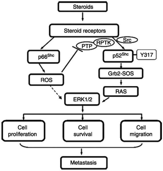
Tyrosine phosphorylation plays a critical role in growth regulation, and its aberrant regulation can be involved in carcinogenesis. The association of Shc (Src homolog and collagen homolog) adaptor protein family members in tyrosine phosphorylation signaling pathway is well recognized. Shc adaptor proteins transmit activated tyrosine phosphorylation signaling that suggest their plausible role in growth regulation including carcinogenesis and metastasis. In parallel, by sharing a similar mechanism of carcinogenesis, the steroids are involved in the early stage of carcinogenesis as well as the regulation of cancer progression and metastatic processes. Recent evidence indicates a cross-talk between tyrosine phosphorylation signaling and steroid hormone action in epithelial cells, including prostate and breast cancer cells. Therefore, the members of Shc proteins may function as mediators between tyrosine phosphorylation and steroid signaling in steroid-regulated cell proliferation and carcinogenesis. In this communication, we discuss the novel roles of Shc proteins, specifically p52(Shc) and p66(Shc), in steroid hormone-regulated cancers and a novel molecular mechanism by which redox signaling induced by p66(Shc) mediates steroid action via a non-genomic pathway. The p66(Shc) protein may serve as an effective biomarker for predicting cancer prognosis as well as a useful target for treatment.


Conflict of interest statement

Declaration of interest

All authors declare that there is no conflict of interest that could be perceived as prejudicing the impartiality of the research reported.

Figures

Figure 1
Figure 1
A schematic organization of Shc isoforms. The Shc proteins include three isoforms that are encoded by the same gene. They contain overlapping sequences. Three major tyrosine phosphorylation sites have been identified within the CH1 domain in all Shc isoforms. The unique CH2 domain of p66Shc isoform consists of 110 amino acids and contains a serine phosphorylation site (Ser-36). The PTB domain of p46Shc is deficient of the first 46 amino acids.
Figure 2
Figure 2
A schematic representation of Shc proteins activating the Ras and MAP kinase cascade. Upon stimulation by growth factors, RTK undergoes dimerization and tyrosine phosphorylation. Subsequently, Shc proteins are recruited and phosphorylated on tyrosine residue via forming a complex with RTK through the SH2 and/or PTB domain of Shc. The phosphorylated Shc proteins then associate with Grb2 adaptor protein through its tyrosine phosphorylation site to the SH2 domain of Grb2; the latter is constitutively complexed with SOS through its SH3 domains. These events result in the translocation of SOS to the plasma membrane and subsequently activate membrane-bound Ras in the exchange of GDP for GTP and trigger the activation of MAP kinase cascade, resulting in cell proliferation, differentiation, migration, invasion, and survival.
Figure 3
Figure 3
p66Shc acting as a stress sensor and increasing intracellular ROS level. Increased ROS induces the phosphorylation of serine 36 at p66Shc protein that promotes the generation of more ROS, leading to cell proliferation, adhesion, migration, or apoptosis.
Figure 4
Figure 4
Involvement of p66Shc and p52Shc in tumor metastasis. Growing primary tumors attract new blood vessels, i.e., angiogenesis, and lymphatic vessels, i.e., lymphangiogenesis, to promote local tumor growth, involvement of regional lymph nodes, and finally distant metastasis. In the process of metastasis after degrading or remodeling the basement membrane, metastatic cells detach from the primary tumor mass, intravasate, survive the stress of vascular transportation, and then evade host defense mechanism, which are in part regulated by the adaptor proteins p52Shc and p66Shc. Furthermore, targeting via microvessels and cell adhesion molecules, the increased phosphorylation of p52Shc and p66Shc proteins mediate the proliferation of tumor cells that metastasize to their preferred sites after extravasation into the target organ parenchyma, and are permitted to reside in the target tissue in which cancer cells respond to transendothelial growth factors from that specific organ. (The figure is adapted from La Porta 2000).
Figure 5
Figure 5
A proposed scheme of steroid-regulated cancer progression via Shc proteins. Upon steroid activation, p52Shc undergoes phosphorylation at Y317 and promotes tumor growth at least in part via transducing signals through the Grb2–Ras–MAPK pathway. Like p52Shc, p66Shc protein also promotes tumor progression, nevertheless, via mediating oxidative stress signals through the generation of ROS. Upon stimulation by steroids, p66Shc is translocated to mitochondria via Ser-36 phosphorylation-independent manner resulting in the generation of ROS. ROS may then inhibit PTP, resulting in RPTK activation, ERK/MAPK activation, and promote cell proliferation, survival, and migration, which collectively lead to metastasis.

Similar articles

Cited by

References

    1. Abdollahi A, Gruver BN, Patriotis C, Hamilton TC. Identification of epidermal growth factor-responsive genes in normal rat ovarian surface epithelial cells. Biochemical and Biophysical Research Communications. 2003;18:188–197. - PubMed
    1. Alzamora R, Harvey BJ. Direct binding and activation of protein kinase C isoforms by steroid hormones. Steroids. 2008;73:885–888. - PubMed
    1. Anand-Apte B, Zetter BR, Viswanathan A, Qiu RG, Chen J, Ruggieri R, Symons M. Platelet-derived growth factor and fibronectin-stimulated migration are differentially regulated by the Rac and extracellular signal-regulated kinase pathways. Journal of Biological Chemistry. 1997;272:30688–30692. - PubMed
    1. Armen TA, Gay CV. Simultaneous detection and functional response of testosterone and estradiol receptors in osteoblast plasma membranes. Journal of Cellular Biochemistry. 2000;79:620–627. - PubMed
    1. Benten WP, Lieberherr M, Giese G, Wunderlich F. Estradiol binding to cell surface raises cytosolic free calcium in T cells. FEBS Letters. 1998;422:349–353. - PubMed

Publication types

MeSH terms