

Temporal and spatial localization of the dentin matrix proteins during dentin biomineralization
- PMID: 19001636
- PMCID: PMC2664930
- DOI: 10.1369/jhc.2008.952119
Temporal and spatial localization of the dentin matrix proteins during dentin biomineralization
Abstract
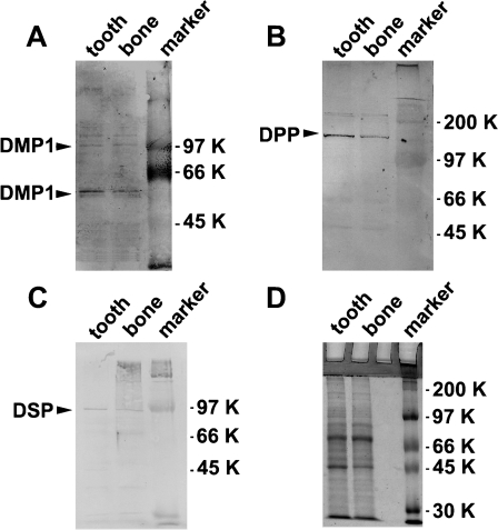
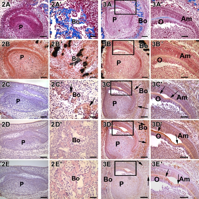
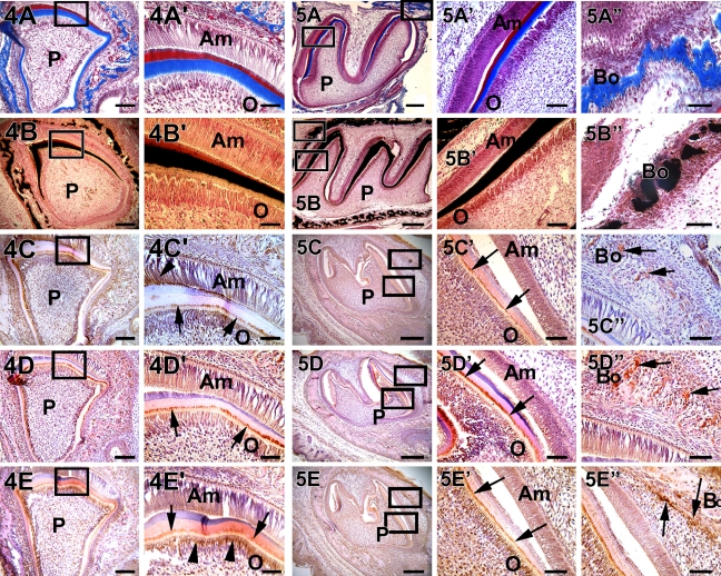
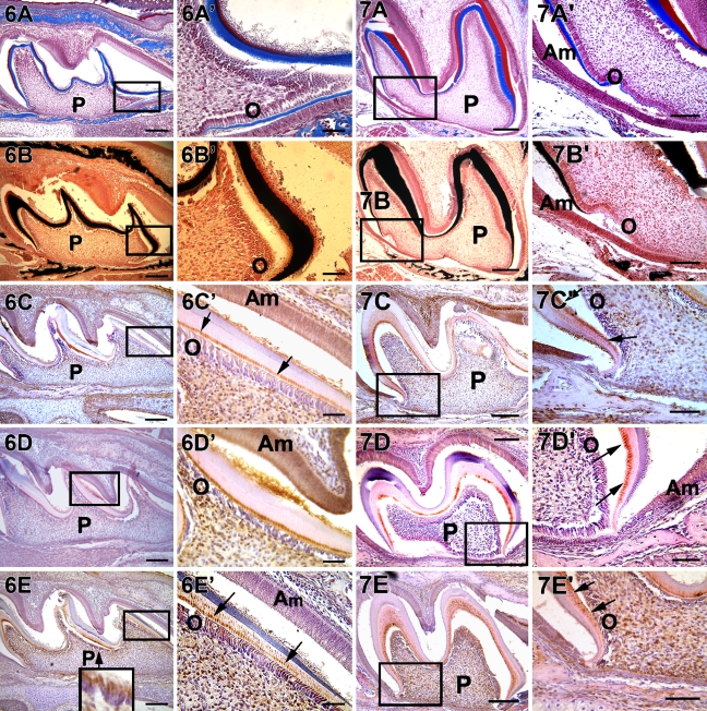
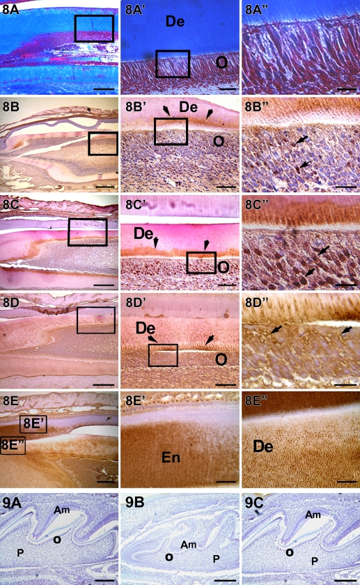
Formation of bone and dentin are classical examples of matrix-mediated mineralization. The mineral phase is essentially the same in these two tissues and primarily consists of a carbonated hydroxyapatite, but the difference lies in the crystal size and shape. There are three components that are necessary for proper mineralization, namely the proper synthesis and secretion of the non-collagenous proteins (NCPs), self-assembly of the collagenous matrix, and delivery of calcium and phosphate ions to the extracellular matrix. Three major NCPs present in the dentin matrix are dentin matrix protein 1 (DMP1), dentin phosphophorin (DPP), and dentin sialoprotein (DSP). In this study, we show the temporal and spatial localization of these NCPs and correlate their expression with the presence of collagenous matrix and calcified deposits in developing mouse incisors and molars. DMP1, an acidic protein, is present predominantly at the mineralization front and in the nucleus of undifferentiated preodontoblast cells. DPP, the major NCP, is present in large amounts at the mineralization front and might function to regulate the size of the growing hydroxyapatite crystals. For the first time, we report the localization of DPP in the nucleus of preodontoblast cells, suggesting a signaling function during the odontoblast differentiation process. DSP is localized predominantly in the dentinal tubules at the site of peritubular dentin, which is highly mineralized in nature. Thus, the precise localization of DMP1, DPP, and DSP in the dentin tissue suggests that a concerted effort between several NCPs is necessary for dentin formation.
Figures
Similar articles
-
Differential expression patterns of the dentin matrix proteins during mineralized tissue formation.Bone. 2004 Jun;34(6):921-32. doi: 10.1016/j.bone.2004.01.020. Bone. 2004. PMID: 15193538
-
The nature and functional significance of dentin extracellular matrix proteins.Int J Dev Biol. 1995 Feb;39(1):169-79. Int J Dev Biol. 1995. PMID: 7626404 Review.
-
The colocalizations of pulp neural stem cells markers with dentin matrix protein-1, dentin sialoprotein and dentin phosphoprotein in human denticle (pulp stone) lining cells.Ann Anat. 2022 Jan;239:151815. doi: 10.1016/j.aanat.2021.151815. Epub 2021 Aug 13. Ann Anat. 2022. PMID: 34400302
-
Extracellular matrix proteins of dentine.Ciba Found Symp. 1997;205:107-15; discussion 115-7. doi: 10.1002/9780470515303.ch8. Ciba Found Symp. 1997. PMID: 9189620 Review.
-
Dentin matrix proteins and dentinogenesis.Connect Tissue Res. 1995;33(1-3):59-65. doi: 10.3109/03008209509016983. Connect Tissue Res. 1995. PMID: 7554963 Review.
Cited by
-
CD146 positive human dental pulp stem cells promote regeneration of dentin/pulp-like structures.Hum Cell. 2018 Apr;31(2):127-138. doi: 10.1007/s13577-017-0198-2. Epub 2018 Jan 8. Hum Cell. 2018. PMID: 29313241 Free PMC article.
-
Effects of D-galactose Induction on Aging Characteristics of the Human Dental Pulp Cell Culture Model: An In Vitro Study.Eur Endod J. 2025 Mar;10(2):142-150. doi: 10.14744/eej.2024.15010. Eur Endod J. 2025. PMID: 40143562 Free PMC article.
-
Immunohistochemical localization of the NH(2)-terminal and COOH-terminal fragments of dentin sialoprotein in mouse teeth.Cell Tissue Res. 2012 Aug;349(2):605-14. doi: 10.1007/s00441-012-1418-4. Epub 2012 May 13. Cell Tissue Res. 2012. PMID: 22581382 Free PMC article.
-
Dentin Sialophosphoprotein Expression Profile in Developing Human Primary Teeth: An Experimental Study.J Dent (Shiraz). 2025 Mar 1;26(1):55-60. doi: 10.30476/dentjods.2024.100219.2202. eCollection 2025 Mar. J Dent (Shiraz). 2025. PMID: 40092544 Free PMC article.
-
Transcriptome profiling of DPP stimulated DPSCs identifies the role of autophagy in odontogenic differentiation.J Struct Biol. 2024 Dec;216(4):108134. doi: 10.1016/j.jsb.2024.108134. Epub 2024 Oct 9. J Struct Biol. 2024. PMID: 39389242
References
-
- Baba O, Qin C, Brunn JC, Wygant JN, McIntyre BW, Butler WT (2004) Colocalization of dentin matrix protein 1 and dentin sialoprotein at late stages of rat molar development. Matrix Biology 23:371–379 - PubMed
-
- Begue-Kirn C, Krebsbach PH, Bartlett JD, Butler WT (1998a) Dentin sialoprotein, dentin phosphoprotein, enamelysin and ameloblastin: tooth-specific molecules that are distinctively expressed during murine dental differentiation. Eur J Oral Sci 106:963–970 - PubMed
-
- Begue-Kirn C, Ruch JV, Ridall AL, Butler WT (1998b) Comparative analysis of mouse DSP and DPP expression in odontoblasts, preameloblasts, and experimentally induced odontoblast-like cells. Eur J Oral Sci 106(suppl 1):254–259 - PubMed
-
- Bleicher F, Couble ML, Farges JC, Couble P, Magloire H (1999) Sequential expression of matrix protein genes in developing rat teeth. Matrix Biol 18:133–143 - PubMed
-
- Boskey A, Spevak L, Tan M, Doty SB, Butler WT (2000) Dentin sialoprotein (DSP) has limited effects on in vitro apatite formation and growth. Calcif Tissue Int 67:472–478 - PubMed
Publication types
MeSH terms
Substances
Grants and funding
LinkOut - more resources
Full Text Sources
Other Literature Sources
Molecular Biology Databases
Research Materials
Miscellaneous
