

Altered expression of fibronectin and collagens I and IV in multiple myeloma and monoclonal gammopathy of undetermined significance
- PMID: 19001640
- PMCID: PMC2664936
- DOI: 10.1369/jhc.2008.952200
Altered expression of fibronectin and collagens I and IV in multiple myeloma and monoclonal gammopathy of undetermined significance
Abstract
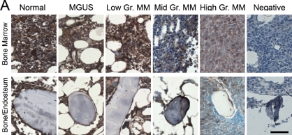
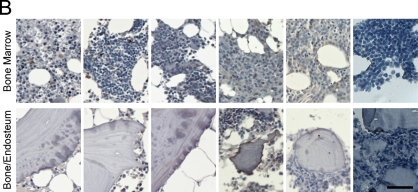
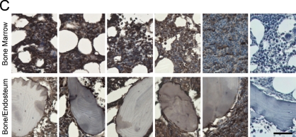
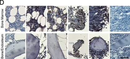
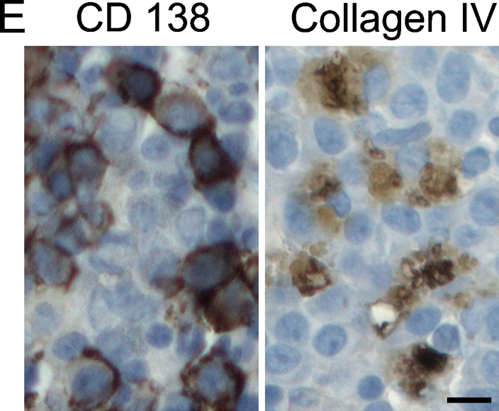
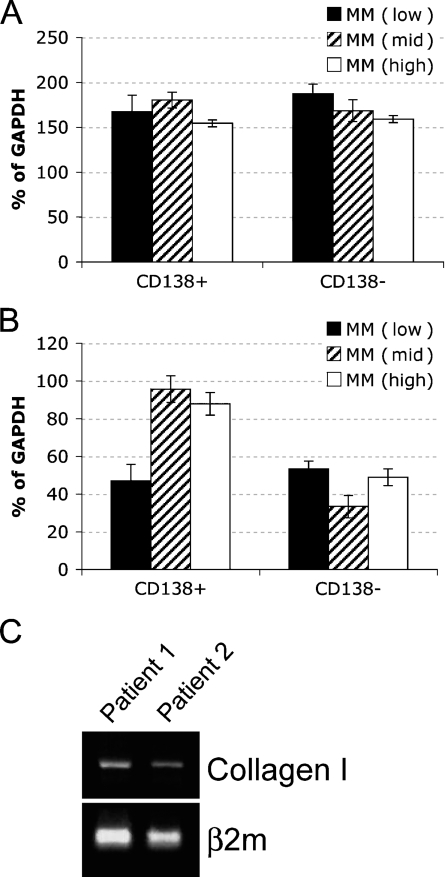
Multiple myeloma (MM) is an incurable B-cell malignancy that arises in the bone marrow (BM). The malignant cells within the BM have extensive interaction with the structural components of their microenvironment. It has been previously shown that the interactions between MM cells and the BM extracellular matrix (ECM) proteins contribute to drug resistance. To understand the underlying causes of adhesion-mediated drug resistance in MM, the components of human BM ECM available for interactions with MM cells must be characterized. We analyzed the expression and localization of fibronectin, laminin, and collagens I and IV in the core biopsies of normal donors and patients with monoclonal gammopathy of undetermined significance (MGUS) or MM. In addition, we compared the patterns of ECM expression in MM patients with low-, mid-, and high-level plasmacytosis of the BM. Although expression of laminin was the same for all groups tested, levels of fibronectin and collagen I were reduced in MM patients with high-level plasmacytosis. Expression of collagen IV in the BM of MGUS and MM patients was higher than in the BM from normal donors. Compared with the plasma cells isolated from the patients with low- and mid-level plasmacytosis, sorted CD138(+) plasma cells from MM patients with high-level plasmacytosis overexpressed collagen IV. Our findings show that, compared with normal controls, the ECM composition of the bone, endosteum, and BM is aberrant in patients with MM, further establishing ECM as a key player in the MM disease process.
Figures
Similar articles
-
Neural cell adhesion molecule expression in plasma cells in bone marrow biopsies and aspirates allows discrimination between multiple myeloma, monoclonal gammopathy of uncertain significance and polyclonal plasmacytosis.Histopathology. 2004 Apr;44(4):375-80. doi: 10.1111/j.1365-2559.2004.01834.x. Histopathology. 2004. PMID: 15049904
-
Monoclonal gammopathy of undetermined significance and smoldering multiple myeloma.Hematol Oncol Clin North Am. 2014 Oct;28(5):775-90. doi: 10.1016/j.hoc.2014.06.005. Epub 2014 Jul 22. Hematol Oncol Clin North Am. 2014. PMID: 25212882 Review.
-
Proteomic characterization of human multiple myeloma bone marrow extracellular matrix.Leukemia. 2017 Nov;31(11):2426-2434. doi: 10.1038/leu.2017.102. Epub 2017 Mar 27. Leukemia. 2017. PMID: 28344315
-
Competition between clonal plasma cells and normal cells for potentially overlapping bone marrow niches is associated with a progressively altered cellular distribution in MGUS vs myeloma.Leukemia. 2011 Apr;25(4):697-706. doi: 10.1038/leu.2010.320. Epub 2011 Jan 21. Leukemia. 2011. PMID: 21252988
-
Molecular Features of the Mesenchymal and Osteoblastic Cells in Multiple Myeloma.Int J Mol Sci. 2022 Dec 7;23(24):15448. doi: 10.3390/ijms232415448. Int J Mol Sci. 2022. PMID: 36555090 Free PMC article. Review.
Cited by
-
Good Cop, Bad Cop: Profiling the Immune Landscape in Multiple Myeloma.Biomolecules. 2023 Nov 7;13(11):1629. doi: 10.3390/biom13111629. Biomolecules. 2023. PMID: 38002311 Free PMC article. Review.
-
Overcoming drug resistance and treating advanced prostate cancer.Curr Drug Targets. 2012 Sep 1;13(10):1308-23. doi: 10.2174/138945012802429615. Curr Drug Targets. 2012. PMID: 22746994 Free PMC article. Review.
-
A three-dimensional tissue culture model to study primary human bone marrow and its malignancies.J Vis Exp. 2014 Mar 8;(85):50947. doi: 10.3791/50947. J Vis Exp. 2014. PMID: 24637629 Free PMC article.
-
MT1-MMP-dependent control of skeletal stem cell commitment via a β1-integrin/YAP/TAZ signaling axis.Dev Cell. 2013 May 28;25(4):402-16. doi: 10.1016/j.devcel.2013.04.011. Epub 2013 May 16. Dev Cell. 2013. PMID: 23685250 Free PMC article.
-
Switching between individual and collective motility in B lymphocytes is controlled by cell-matrix adhesion and inter-cellular interactions.Sci Rep. 2018 Apr 11;8(1):5800. doi: 10.1038/s41598-018-24222-4. Sci Rep. 2018. PMID: 29643414 Free PMC article.
References
-
- Adams GB, Scadden DT (2006) The hematopoietic stem cell in its place. Nat Immunol 7:333–337 - PubMed
-
- Barcellos-Hoff MH, Ravani SA (2000) Irradiated mammary gland stroma promotes the expression of tumorigenic potential by unirradiated epithelial cells. Cancer Res 60:1254–1260 - PubMed
-
- Bataille R, Manolagas SC, Berenson JR (1997) Pathogenesis and management of bone lesions in multiple myeloma. Hematol Oncol Clin North Am 11:349–361 - PubMed
-
- Billadeau D, Van Ness B, Kimlinger T, Kyle RA, Therneau TM, Greipp PR, Witzig TE (1996) Clonal circulating cells are common in plasma cell proliferative disorders: a comparison of monoclonal gammopathy of undetermined significance, smoldering multiple myeloma, and active myeloma. Blood 88:289–296 - PubMed
-
- Bouterfa H, Darlapp AR, Klein E, Pietsch T, Roosen K, Tonn JC (1999) Expression of different extracellular matrix components in human brain tumor and melanoma cells in respect to variant culture conditions. J Neurooncol 44:23–33 - PubMed
Publication types
MeSH terms
Substances
LinkOut - more resources
Full Text Sources
Other Literature Sources
Medical
