. 2008 Nov;155(6):884-95.
doi: 10.1038/bjp.2008.328. Epub 2008 Aug 18.

Effects of levo- and dextrosimendan on NF-kappaB-mediated transcription, iNOS expression and NO production in response to inflammatory stimuli

Affiliations

Effects of levo- and dextrosimendan on NF-kappaB-mediated transcription, iNOS expression and NO production in response to inflammatory stimuli

O Sareila et al. Br J Pharmacol. 2008 Nov.

Abstract

Background and purpose: Levosimendan is used in the treatment of decompensated heart failure. It increases the contractility of the myocardium by sensitizing troponin C to calcium. In addition, levosimendan has been reported to have beneficial effects in experimental models of septic shock. Because heart failure and sepsis have been associated with excessive nitric oxide (NO) production through inducible NOS (iNOS), we investigated the effects of the simendans on NO production and iNOS expression and on generation of pro-inflammatory cytokines.

Experimental approach: Macrophages and fibroblasts were exposed to inflammatory stimuli to induce iNOS expression. Proteins were measured by western blot and mRNA expression was determined by quantitative RT-PCR. Promoter activity and nuclear factor-kappaB (NF-kappaB) and the gamma-activated site (GAS; binding site for signal transducer and activator of transcription 1; STAT1)-mediated transcription were investigated using luciferase reporter constructs. Cytokines tumour necrosis factor-alpha (TNF-alpha) and interleukin-6 (IL-6) were measured by ELISA.

Key results: Levosimendan and dextrosimendan decreased NO production in a dose-dependent manner in cells exposed to inflammatory stimuli. The simendans decreased iNOS protein and mRNA expression but did not affect iNOS mRNA decay. These compounds decreased iNOS promoter activity and inhibited NF-kappaB-mediated transcription but not that mediated by STAT1/GAS. The simendans reduced IL-6 production slightly but they had no effect on TNF-alpha synthesis.

Conclusions and implications: The simendans downregulated NF-kappaB-dependent transcription and decreased iNOS promoter activity, iNOS expression and NO production. These mechanisms may contribute to their beneficial clinical effects.


Figures

Figure 1
Figure 1
The chemical structures of levosimendan and dextrosimendan.
Figure 2
Figure 2
The effects of simendans on the production of inflammatory cytokines tumour necrosis factor-α (TNF-α) (a) and interleukin-6 (IL-6) (b) and nitric oxide (NO) (c) in lipopolysaccharide (LPS)-stimulated J774 macrophages. Cells were incubated with the indicated combinations of LPS (10 ng mL−1) and simendans (10 μM) for 24 h. TNF-α and IL-6 levels were measured by ELISA. NO production was measured as nitrite accumulated in the culture medium by the Griess reaction. (d) The effect of simendans on LPS-induced NO production in J774 macrophages when the simendans were added simultaneously with LPS or 4 or 8 h after LPS. Cells were incubated with the indicated combinations of LPS (100 ng mL−1) and simendans (10 μM) for 24 h, and NO production was measured as nitrite accumulated in the culture medium by Griess reaction. The results are expressed as mean+s.e.mean (n=4 in a, b and n=6 in c, d). *P<0.05 and **P<0.01 when compared with cells treated with LPS alone.
Figure 3
Figure 3
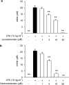
The concentration-dependent effect of levosimendan (a) and dextrosimendan (b) on lipopolysaccharide (LPS)-induced nitric oxide (NO) production in J774 macrophages. Cells were incubated with LPS (10 ng mL−1) and levosimendan or dextrosimendan (1–30 μM) for 24 h. NO production was measured as nitrite accumulated in the culture medium by Griess reaction. The results are expressed as mean+s.e.mean (n=6). **P<0.01 when compared with cells treated with LPS alone.
Figure 4
Figure 4
The effects of levosimendan (a) and dextrosimendan (b) on lipopolysaccharide (LPS)-induced iNOS protein expression in J774 macrophages. Cells were incubated with LPS (10 ng mL−1) in the presence or in the absence of levosimendan or dextrosimendan (10 μM) for 24 h, and iNOS expression was detected by western blot. LPS-induced iNOS protein expression was set as 100% and the other values were related to that value. Actin was used as a loading control. The results are expressed as mean+s.e.mean (n=6 in a, n=3–6 in b). **P<0.01 when compared with cells treated with LPS alone.
Figure 5
Figure 5
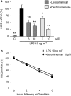
The effects of the simendans on levels of iNOS mRNA and its decay in lipopolysaccharide (LPS)-stimulated J774 macrophages. (a) Cells were incubated with LPS (10 ng mL−1) in the presence or absence of levosimendan or dextrosimendan (3 and 10 μM) for 3 h and iNOS mRNA levels were measured by quantitative RT-PCR. LPS-induced iNOS mRNA expression was set as 100% and the other values were related to that value. The results are expressed as mean+s.e.mean (n=3–6). *P<0.05 and **P<0.01 when compared with cells treated with LPS alone. (b) The effects of levosimendan on iNOS mRNA decay in LPS-stimulated J774 macrophages as measured by actinomycin D assay. Cells were incubated with LPS (10 ng mL−1) in the presence or absence of levosimendan (10 μM) for 6 h, and actinomycin D (actD; 0.5 μg mL−1) was then added to stop transcription. Cells were further incubated for 2, 4 or 6 h before RNA was extracted and iNOS mRNA levels were measured by quantitative RT-PCR. iNOS mRNA levels at the time of addition of actinomycin D were set as 100% and the other values were related to that value. The results are expressed as mean+s.e.mean (n=3).
Figure 6
Figure 6
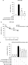
The effects of the simendans on iNOS mRNA expression and decay and on iNOS promoter activity in L929 fibroblasts stimulated with pro-inflammatory cytokines. (a) Cells were incubated with a combination of pro-inflammatory cytokines (cytokine mixture (CM 10), tumour necrosis factor-α, interferon-γ and interleukin-1β, 10 ng mL−1 each) and levo- or dextrosimendan (10 μM) for 6 h, and iNOS mRNA levels were measured by quantitative RT-PCR. Cytokine-induced iNOS mRNA expression was set as 100% and the other values were related to that value. The results are expressed as mean+s.e.mean (n=6). **P<0.01 when compared with cells treated with cytokine mixture alone. (b) Cells were incubated with a combination of pro-inflammatory cytokines (CM 10) in the presence or absence of dextrosimendan (10 μM) for 3 h, and actinomycin D (ActD; 5 μg mL−1) was then added to stop transcription. Cells were further incubated for 2, 4 of 6 h before RNA was extracted, and iNOS mRNA levels were measured by quantitative RT-PCR. iNOS mRNA levels at the time of addition of ActD were set as 100% and the other values were related to that value. The results are expressed as mean+s.e.mean (n=4). (c) The effects of levosimendan and dextrosimendan on iNOS promoter activity in L929 pGL4(miNOS-prom)neo cells. L929 cells stably transfected with pGL4(miNOS-prom)neo were stimulated with a combination of pro-inflammatory cytokines (CM 10) alone or in combination with levosimendan, dextrosimendan, Janus kinase inhibitor WHI-P154 or NF-κB inhibitor MG 132 for 6 h, and luciferase mRNA levels were measured by quantitative RT-PCR. Luciferase mRNA levels in cytokine-treated cells were set as 100% and the other values were related to that value. The results are expressed as mean+s.e.mean (n=4). *P<0.05 and **P<0.01 when compared with luciferase mRNA levels in cells treated with CM alone.
Figure 7
Figure 7
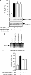
The effects of simendans on nuclear translocation, DNA binding and transcriptional activity of nuclear factor-κB (NF-κB). (a) J774 macrophages were incubated with lipopolysaccharide (LPS) (10 ng mL−1), levosimendan (10 μM) or dextrosimendan (3 μM) for 30 min. Nuclear proteins were then extracted and NF-κB p65 was detected by western blot. Lamin A/C was used as a loading control. The nuclear level of NF-κB p65 in LPS-treated cells was set as 100% and the other values were related to that value. The results are expressed as mean+s.e.mean (n=6). **P<0.01 when compared with cells treated with LPS alone. (b) J774 macrophages were treated as in (a), and NF-κB-binding activity in nuclear extracts was analysed by electrophoretic mobility shift assay. One representative blot of three with similar results is shown. (c) L929-pNF-κB(luc)neo cells, that contained NF-κB luciferase reporter construct, were incubated with a combination of pro-inflammatory cytokines (cytokine mixture (CM 10); tumour necrosis factor-α, interferon-γ and interleukin-1β, 10 ng mL−1 each) and dextro- or levosimendan for 6 h, and luciferase mRNA levels were measured by quantitative RT-PCR. Cytokine-induced luciferase expression was set as 100% and the other values were related to that value. The results are expressed as mean+s.e.mean (n=3). **P<0.01 when compared with cells treated with cytokine mixture alone.
Figure 8
Figure 8
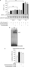
The effects of simendans on nuclear translocation, DNA binding and transcriptional activity of signal transducer and activator of transcription 1 (STAT1). (a) J774 macrophages were incubated with lipopolysaccharide (LPS) and levosimendan or dextrosimendan. Nuclear proteins were extracted at the indicated time points and STAT1α was detected by western blot. Lamin A/C was used as a loading control. The nuclear level of STAT1α in LPS-treated cells was set as 100% and the other values were related to that value. The results are expressed as mean+s.e.mean (n=3). **P<0.01. (b) J774 macrophages were incubated with (1) vehicle, (2) LPS (10 ng mL−1)+interferon-γ (IFN-γ) (5 ng mL−1) or (3) LPS (10 ng mL−1)+IFN-γ (5 ng mL−1)+dextrosimendan (10 μM) for 1 h. Nuclear extracts were then prepared and STAT1α DNA-binding activity was analysed by electrophoretic mobility shift assay. One representative blot of three with similar results is shown. (c) L929-pGAS(luc)neo cells, which contain the GAS-motif (binding site for STAT1)-luciferase reporter construct, were incubated with a combination of pro-inflammatory cytokines (cytokine mixture (CM 10); tumour necrosis factor-α, IFN-γ and interlekin-1β, 10 ng mL−1 each) and dextrosimendan or levosimendan for 6 h and luciferase mRNA levels were measured by quantitative RT-PCR. Cytokine-induced luciferase mRNA expression was set as 100% and the other values were related to that value. The results are expressed as mean+s.e.mean (n=3). *P<0.05 and **P<0.01 when compared with cells treated with cytokine mixture alone.

Similar articles

Cited by

References

    1. Alderton WK, Cooper CE, Knowles RG. Nitric oxide synthases: structure, function and inhibition. Biochem J. 2001;357:593–615. - PMC - PubMed
    1. Avgeropoulou C, Andreadou I, Markantonis-Kyroudis S, Demopoulou M, Missovoulos P, Androulakis A, et al. The Ca(2+)-sensitizer levosimendan improves oxidative damage, BNP and pro-inflammatory cytokine levels in patients with advanced decompensated heart failure in comparison to dobutamine. Eur J Heart Fail. 2005;7:882–887. - PubMed
    1. Becker C, Barbulescu K, Wirtz S, Meyer zum Büschenfelde KH, Pettersson S, Neurath MF. Constitutive and inducible in vivo protein–DNA interactions at the tumor necrosis factor-alpha promoter in primary human T lymphocytes. Gene Expr. 1999;8:115–127. - PMC - PubMed
    1. Bradford MM. A rapid and sensitive method for the quantitation of microgram quantities of protein utilizing the principle of protein-dye binding. Anal Biochem. 1976;72:248–254. - PubMed
    1. Brown RD, Ambler SK, Mitchell MD, Long CS. The cardiac fibroblast: therapeutic target in myocardial remodeling and failure. Annu Rev Pharmacol Toxicol. 2005;45:657–687. - PubMed

Publication types

MeSH terms