. 2009 Jan;17(1):169-75.
doi: 10.1038/mt.2008.231. Epub 2008 Nov 11.

Artificial microRNAs as siRNA shuttles: improved safety as compared to shRNAs in vitro and in vivo

Affiliations

Artificial microRNAs as siRNA shuttles: improved safety as compared to shRNAs in vitro and in vivo

Ryan L Boudreau et al. Mol Ther. 2009 Jan.

Abstract

RNA interference (RNAi) provides a promising therapeutic approach to human diseases. However, data from recent reports demonstrate that short-hairpin RNAs (shRNAs) may cause cellular toxicity, and this warrants further investigation of the safety of using RNAi vectors. Earlier, in comparing hairpin-based RNAi vectors, we noted that shRNAs are highly expressed and yield an abundance of unprocessed precursors, whereas artificial microRNAs (miRNAs) are expressed at lower levels and are processed efficiently. We hypothesized that unprocessed shRNAs arise from the saturation of endogenous RNAi machinery, which poses likely a burden to cells. In this study, we tested that hypothesis by assessing the relative effects of shRNAs and artificial miRNAs on the processing and function of miRNAs. In competition assays, shRNAs disrupted miRNA biogenesis and function, whereas artificial miRNAs avoided this interference even when dosed to silence as effectively as shRNAs. We next compared the safety of these vectors in mouse cerebella, and found that shRNAs cause Purkinje cell neurotoxicity. By contrast, artificial miRNA expression was well tolerated, resulting in effective target gene silencing in Purkinje cells. These findings, together with data from earlier work in mouse striata, suggest that miRNA-based platforms are better suited for therapeutic silencing in the mammalian brain.


Figures

<b>Figure 1</b>
Figure 1
Design of hairpin-based RNAi vectors for fair comparison studies. shRNA and artificial miRNA vectors were designed to release the same siRNA sequences [either targeting SCA1 (shown) or eGFP] after processing by Drosha and/or Dicer. Processing sites (arrows) and proper loading of the intended antisense strand (bottom) as opposed to the unintended sense strand (top) were previously confirmed.10
<b>Figure 2</b>
Figure 2
Effects of shRNA- and miRNA-based vectors on artificial miRNA biogenesis and function. (a) Cartoon of RNAi and RNAi luciferase reporter vectors. “TTTTT” designates the Pol-III termination signal. (b) HEK293 cells were transfected with miGFP and GFP RNAi luciferase reporter expression plasmids to establish baseline silencing levels (dotted line). To assess for disruptions in miGFP biogenesis and function, shSCA1 or miSCA1 competitors were added to the transfections at varying doses of RNAi:target. U6 serves as the promoter-only control. (c) Gene silencing assays were performed by co-transfecting HEK293 cells with SCA1 RNAi and RNAi reporter plasmids at varying doses of RNAi:target. (d,e) Reciprocal experiments evaluating the effects of GFP RNAi competitors (shGFP or miGFP) on miSCA1 activity, in parallel with GFP RNAi efficacy studies. All bar graphs represent mean values ± SEM (n = 3; ***, ** and NS indicate P < 0.001, P < 0.01, and no significance, P > 0.05, respectively). (f) Northern blot analyses assessing the processing of miGFP in the presence of SCA1 RNAi competitors. Blots were probed for either GFP (top blot-pair) or SCA1 (bottom blot-pair) RNAi transcripts.
<b>Figure 3</b>
Figure 3
Effects of shSCA1 and miSCA1 on miR-1 function in differentiating myoblasts. (a) Cartoons depicting the miR-1 luciferase reporter (contains a perfect complementary miR-1 target site in the 3′UTR of Firefly luciferase) and the RNAi-hrGFP dual expression vector used in C2C12 studies. (b) Disruption of endogenous miRNA biogenesis and function was assessed in C2C12 cells which show induced miR-1 expression after differentiation. Cells were co-transfected with RNAi and miR-1 luciferase reporter plasmids, and then differentiated. Each group (n = 4) was normalized to cells treated with siCheck2-alone (i.e., no miR-1 target), and the results are shown as mean values ± SEM. No RNAi (---) served as the empty-vector control. (c) RNAi plasmids co-expressing hrGFP were co-transfected into C2C12 cells, and these were differentiated and stained for myosin heavy-chain (MHC) to identify transfected differentiating myotubes (i.e., hrGFP+/MHC+). (d) The lengths of hrGFP+/MHC+ cells were measured, and the results (mean values ± SEM) are shown as fold-elongation relative to undifferentiating cells (n indicated in parentheses, **P < 0.01).
<b>Figure 4</b>
Figure 4
Effects of shSCA1 and miSCA1 on cell viability in vitro. C2C12 cells were transfected with RNAi plasmids co-expressing hrGFP, and differentiated. Photomicrographs depicting hrGFP expression at 24 and 72 h after treatment are shown. Cell viability was measured by CellTiter-96 AQueous MTS assay (Promega, n = 3, mean values ± SEM).
<b>Figure 5</b>
Figure 5
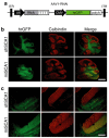
Effects of shSCA1 and miSCA1 on cerebellar Purkinje cell viability in vivo. (a) Diagram of the recombinant AAV2/1 viral vectors containing SCA1 RNAi and hrGFP expression cassettes. (b,c) Wild-type mice were injected with either AAV1-shSCA1 or AAV1-miSCA1 into the cerebellum, and histological analyses were performed 10 weeks later. Representative photomicrographs, captured at (b) low magnification (Bar = 500 µm) and (c) high magnification (Bar = 100 µm), depicting hrGFP autofluorescence and immunohistochemical staining of calbindin-positive Purkinje cells, are shown for each treatment group.
<b>Figure 6</b>
Figure 6
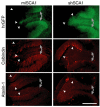
Silencing of mutant ataxin-1 in cerebellar Purkinje cells. SCA1 mice, expressing mutant human ataxin-1 in cerebellar Purkinje cells, were injected with either AAV1-miSCA1 or AAV1-shSCA1 into the cerebellum, and histological analyses were performed 7 weeks later. Representative photomicrographs depicting hrGFP autofluorescence and immunohistochemical staining for calbindin-positive Purkinje cells and mutant human ataxin-1 are shown. Silencing of nuclear localized mutant ataxin-1 is observed in hrGFP-positive regions (gray arrows) of miSCA1-treated cerebella relative to nontransduced areas (white arrowheads). By contrast, there is a lack of ataxin-1 staining in shSCA1-treated regions, resulting from the loss of Purkinje cells as observed by the overlapping loss of calbindin staining (gray arrows). Bar = 200 µm for all photomicrographs.

Similar articles

Cited by

References

    1. McManus MT., and , Sharp PA. Gene silencing in mammals by small interfering RNAs. Nat Rev Genet. 2002;3:737–747. - PubMed
    1. Fire A, Xu SQ, Montgomery MK, Kostas SA, Driver SE., and , Mello CC. Potent and specific genetic interference by double-stranded RNA in Caenorhabditis elegans. Nature. 1998;391:806–811. - PubMed
    1. Provost P, Dishart D, Doucet J, Frendewey D, Samuelsson B., and , Radmark O. Ribonuclease activity and RNA binding of recombinant human Dicer. EMBO J. 2002;21:5864–5874. - PMC - PubMed
    1. Lee YS, Nakahara K, Pham JW, Kim K, He Z, Sontheimer EJ, et al. Distinct roles for Drosophila Dicer-1 and Dicer-2 in the siRNA/miRNA silencing pathways. Cell. 2004;117:69–81. - PubMed
    1. Elbashir SM, Harborth J, Lendeckel W, Yalcin A, Weber K., and , Tuschl T. Duplexes of 21-nucleotide RNAs mediate RNA interference in cultured mammalian cells. Nature. 2001;411:494–498. - PubMed

Publication types

LinkOut - more resources