Review
. 2008 Dec;9(12):944-57.
doi: 10.1038/nrm2546. Epub 2008 Nov 12.

One step at a time: endoplasmic reticulum-associated degradation

Affiliations
Review

One step at a time: endoplasmic reticulum-associated degradation

Shruthi S Vembar et al. Nat Rev Mol Cell Biol. 2008 Dec.

Abstract

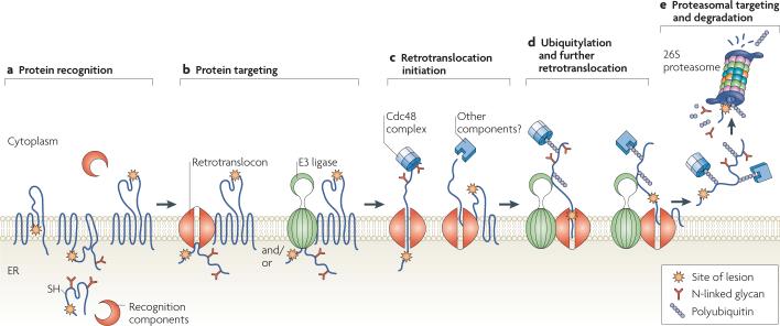
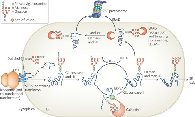
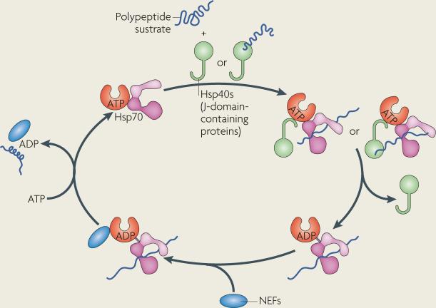
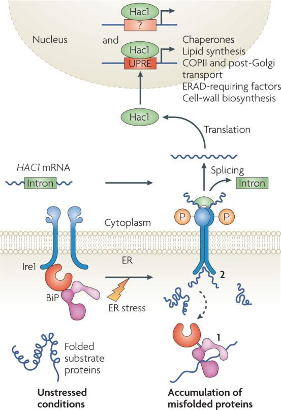
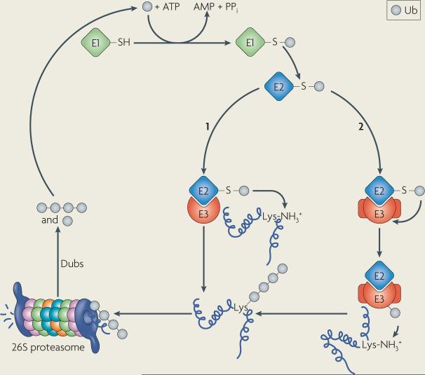
Protein folding in the endoplasmic reticulum (ER) is monitored by ER quality control (ERQC) mechanisms. Proteins that pass ERQC criteria traffic to their final destinations through the secretory pathway, whereas non-native and unassembled subunits of multimeric proteins are degraded by the ER-associated degradation (ERAD) pathway. During ERAD, molecular chaperones and associated factors recognize and target substrates for retrotranslocation to the cytoplasm, where they are degraded by the ubiquitin-proteasome machinery. The discovery of diseases that are associated with ERAD substrates highlights the importance of this pathway. Here, we summarize our current understanding of each step during ERAD, with emphasis on the factors that catalyse distinct activities.


Figures

Figure 1
Figure 1. A step-by-step illustration of endoplasmic reticulum-associated degradation
a | Protein recognition. Misfolded proteins containing cytoplasmic, intramembrane or endoplasmic reticulum (ER)-luminal lesions are recognized by cytoplasmic and luminal chaperones and associated factors, such as 70 kDa heat-shock protein (Hsp70)-family members, calnexin and calreticulin, and protein disulphide isomerases. b | Protein targeting. ER-associated degradation (ERAD) substrates are targeted to the retrotranslocation machinery (the retrotranslocon) and/or to E3 ligases. c | Retrotranslocation initiation. Substrate retrotranslocation into the cytoplasm might be initiated in part by the cell-division cycle-48 (Cdc48) complex; other components, such as molecular chaperones or the proteasome, might also be required for this step. The energy derived from ATP hydrolysis by Cdc48, which is a AAA+ATPase, is coupled to retrotranslocation. d | Ubiquitylation and further retrotranslocation. As proteins exit the retrotranslocon they are polyubiquitylated by E3 ubiquitin ligases. This promotes further retrotranslocation and is aided by cytoplasmic ubiquitin-binding protein complexes. e | Proteasomal targeting and degradation. Once a polyubiquitylated substrate is displaced into the cytoplasm, it is recognized by receptors in the 19S cap of the 26S proteasome. De-ubiquitylating enzymes (not shown) remove the polyubiquitin tag, and peptide N-glycanase (not shown) might also be required for efficient degradation. The substrate is then threaded into the 20S catalytic core of the proteasome where it is broken down into peptide fragments. Ubiquitin that is generated by this process can be recycled for subsequent rounds of modification.
Figure 2
Figure 2. N-linked glycosylation and the degradation of glycosylated proteins
Proteins that enter the endoplasmic reticulum (ER) are often modified by the addition of a GlcNAc2-Man9-Glc3 glycan to the side-chain nitrogen of Asn residues in the consensus Asn-X-Ser/Thr motif. First, the translocon-associated oligosaccharyl transferase (OST) complex co-translationally transfers GlcNAc2-Man9-Glc3 glycans from dolichol to substrate proteins. Next, glucosidase-I and glucosidase-II sequentially remove two terminal glucoses, generating monoglucosylated substrates that are recognized by calnexin and calreticulin through their carbohydrate-binding globular domains (calreticulin is a soluble protein and is not shown). The interaction with calnexin and calreticulin facilitates folding. ERP57, a protein disulphide isomerase homologue that is associated with the arm domain of calnexin and calreticulin, catalyses disulphide bond formation. Following release from the calnexin–calreticulin cycle, the final glucose is trimmed by glucosidase-II. If glycoproteins have adopted their native conformations, they can be demannosylated (denoted by the use of parentheses around the mannoses) by ER mannosidases I and II (ER man-I and man-II) and exit the ER through coatomer protein complex-II vesicles. However, the folding of some glycoproteins requires multiple rounds of association with calnexin–calreticulin. Such proteins are reglucosylated by UDP-glucose:glycoprotein glucosyltransferase (UGGT), which recognizes non-native states and transfers a glucose from UDP-glucose to the N-linked GlcNAc2-Man9 glycan. Re-monoglucosylation promotes re-entry into the folding cycle. Terminally misfolded glycoproteins might also be targeted for ER-associated degradation (ERAD) by calnexin and calreticulin or by other ERAD-requiring components. EDEM, ER degradation-enhancing α-mannosidase-like lectins; Glc, glucose; GlcNAc, N-acetylglucosamine; Man, mannose.
None
None
None

Similar articles

Cited by

References

    1. Ellis RJ. Macromolecular crowding: obvious but underappreciated. Trends Biochem. Sci. 2001;26:597–604. - PubMed
    1. Despa F, Orgill DP, Lee RC. Molecular crowding effects on protein stability. Ann. NY Acad. Sci. 2005;1066:54–66. - PubMed
    1. Jahn TR, Radford SE. The yin and yang of protein folding. FEBS J. 2005;272:5962–5970. - PubMed
    1. Bukau B, Weissman J, Horwich A. Molecular chaperones and protein quality control. Cell. 2006;125:443–451. - PubMed
    1. Ghaemmaghami S, et al. Global analysis of protein expression in yeast. Nature. 2003;425:737–741. - PubMed

Publication types

LinkOut - more resources