. 2008 Nov;4(11):e1000204.
doi: 10.1371/journal.ppat.1000204. Epub 2008 Nov 11.

Foamy macrophages from tuberculous patients' granulomas constitute a nutrient-rich reservoir for M. tuberculosis persistence

Affiliations

Foamy macrophages from tuberculous patients' granulomas constitute a nutrient-rich reservoir for M. tuberculosis persistence

Pascale Peyron et al. PLoS Pathog. 2008 Nov.

Abstract

Tuberculosis (TB) is characterized by a tight interplay between Mycobacterium tuberculosis and host cells within granulomas. These cellular aggregates restrict bacterial spreading, but do not kill all the bacilli, which can persist for years. In-depth investigation of M. tuberculosis interactions with granuloma-specific cell populations are needed to gain insight into mycobacterial persistence, and to better understand the physiopathology of the disease. We have analyzed the formation of foamy macrophages (FMs), a granuloma-specific cell population characterized by its high lipid content, and studied their interaction with the tubercle bacillus. Within our in vitro human granuloma model, M. tuberculosis long chain fatty acids, namely oxygenated mycolic acids (MA), triggered the differentiation of human monocyte-derived macrophages into FMs. In these cells, mycobacteria no longer replicated and switched to a dormant non-replicative state. Electron microscopy observation of M. tuberculosis-infected FMs showed that the mycobacteria-containing phagosomes migrate towards host cell lipid bodies (LB), a process which culminates with the engulfment of the bacillus into the lipid droplets and with the accumulation of lipids within the microbe. Altogether, our results suggest that oxygenated mycolic acids from M. tuberculosis play a crucial role in the differentiation of macrophages into FMs. These cells might constitute a reservoir used by the tubercle bacillus for long-term persistence within its human host, and could provide a relevant model for the screening of new antimicrobials against non-replicating persistent mycobacteria.


Conflict of interest statement

The authors have declared that no competing interests exist.

Figures

Figure 1
Figure 1. Granulomas from TB patients display many M.tb-containing FMs surrounding the necrotic center.
Thin sections of lymph node biopsy samples from 10 tuberculous patients were stained and analyzed. A) Haematoxylin and Eosin staining, original magnification ×12. B, C) Oil red-O staining, original magnification ×200 (B) and ×1000 (C). D) Ziehl-Nielsen staining (×1000). G: granuloma, N: necrosis, I: interface. Arrow: Giant Langhans cell (B), M.tb (D).
Figure 2
Figure 2. M.tb but not M. smegmatis induces FM formation within in vitro granulomas.
PBMCs from a healthy donor were infected with M.tb or M. smegmatis. Granuloma cells were collected 3 (A, B) and 11 (C) days later and stained with Oil red-O. The percentage of FMs within the whole macrophage population is indicated in panel D. The original magnification for the general views is ×100 (A, B), and ×200 (C), ×1000 for the enlarged views. These pictures are representative of 5 independent experiments with 5 unrelated controls.
Figure 3
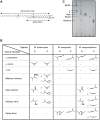
Figure 3. Structure of the mycolic acids found in M.tb, M. smegmatis and M. smegmatis/hma respectively.
A) Dicyclopropanated mycolic acid (α-mycolate) from M. tuberculosis. The chemical functions introduced by methyltransferases occur at the proximal (P) and distal (D) positions. B) TLC profile of the mycolic acid methyl esters from: lane1, M. smegmatis; lane 2, M. smegmatis/hma; lane 3, ketomycolate; lane 4, epoxymycolate; lane 5, hydromycolate. NHFE, non-hydroxylated fatty esters. C) Types of functional groups present at both the proximal and distal positions of the different species of mycolates found in M. tuberculosis, M. smegmatis and M. smegmatis/hma.
Figure 4
Figure 4. Role of hma-controlled mycolic acids in FM formation.
PBMCs from a healthy donor were infected with either M. smegmatis or M. smegmatis/hma for 3 days. The granuloma cells were then collected and stained with Oil red-O and May-Grünwald Giemsa (A, B). In parallel experiments, isolated macrophages were infected with either M. smegmatis (D) or M. smegmatis-hma (E), or incubated with mycolic acids extracted from both species (M. smegmatis G, M. smegmatis-hma H). The cells were then stained with Nile red. The pictures are representative of 3 independent experiments with 3 unrelated controls. Original magnification: A, B: ×100; D, E, G, H: ×400. The percentage of FMs in the different macrophage populations are indicated in panels C, F, I, respectively.
Figure 5
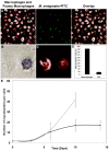
Figure 5. Phagocytic uptake and survival of mycobacteria within FMs.
A) Isolated macrophages from a control donor were incubated for 2 days with M. smegmatis/hma-isolated mycolic acids, and then infected with FITC-labelled M. smegmatis for 1 hour. The cells were then stained with Nile red. Original magnification ×400. Isolated macrophages from a control donor were infected with M.tb for 2 days, and then stained with Nile red (B, ×1000) and subsequently analyzed for NBT reduction ability (C, ×1000). The proportion of FMs (Nile red positive cells) able to reduce NBT was then evaluated (D). E) Macrophages from a healthy donor were infected with GFP-expressing M.tb for 1 hour, washed and re-incubated in fresh medium. At selected times after infection, the number of bacteria per cell was determined under a confocal microscope for both macrophages (Nile red-negative cells) and FMs (Nile red-positive cells). The data are representative of 3 independent experiments. In order to distinguish the lipids contained within lipid bodies, from those of the cell membrane, we made use of the fluorescent emission spectrum properties of Nile red which depend upon the lipid which is associated with Nile red, i.e. for triacylglycerol (λmax em = 590 nm), for phospholipids (λmax em = 640 nm) (Molecular Probes handbook). On confocal microscopy pictures, the phospholipids background of both macrophages and FMs appears in red, and the triacylglycerol-rich lipid bodies in white. Cells were regarded as positively stained by Nile red when more than 50% of the cell surface was stained (see Figure S2).
Figure 6
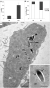
Figure 6. Morphological appearance of foamy macrophages and bacteria within granulomas.
PBMCs were infected with M.tb H37Rv, fixed and processed for electron microscopy at days 3 and 11 post-infection. A) The percentage of FM, with respect to the total amount of macrophages encountered on thin sections of granulomas, was determined. Care was taken to avoid serial sections. B) The number of LBs encountered in the above FMs was next determined. Results are expressed as the percentage of FMs, with respect to the total number of FMs, displaying either 2 to 5 LBs (□) or more than 5 LBs (▪). C) General view of a FM containing a large number of lipid bodies (LB) at day 11 post-infection. In this cell, some of the M.tb-containing phagosomes are in the close vicinity of a LB (large arrow). Enlarged view of one bacterium within a membrane-bound phagosome (small arrows) showing the ultrastructural integrity of bacteria within foamy macrophages and the lack of bacterial intracytoplasmic lipid bodies (ILI). Bar: C) 1 µm; D) 0.5 µm.
Figure 7
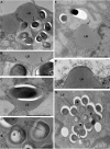
Figure 7. Interaction between cellular lipid bodies (LB) of foamy macrophages and M.tb-containing phagosomes.
PBMCs were infected with M.tb H37Rv, fixed and processed for electron microscopy at day 11 post-infection. A) shows 3 M.tb-containing phagosomes which are closely apposed to a LB (arrows); B) enlarged view of part of the phagosomes in A, showing the tight contact between the phagosome membrane and the LB (arrows); C) M.tb in a LB. This bacterium displays no intracytoplasmic lipid bodies (ILI); D) enlarged view of M.tb within the LB depicted in G. These bacteria contain large ILI. E) shows a M.tb-containing phagosome, tightly apposed to a LB, and which is starting to engulf this LB; F) Enlarged view of part of E showing the tight apposition of the phagosome membrane to the LB; G) LB containing several M.tb. Some of the bacteria now display ILIs (see D for detailed view of these bacteria). Bar: A, C, E, G) 0.5 µm; B, D, F) 0.25 µm.

Similar articles

Cited by

References

    1. WHO. Global Tuberculosis Control, WHO Report. 2006. http://www.who.int/mediacentre/factsheets/fs104/en/index.html.
    1. Glickman MS, Jacobs WR., Jr Microbial pathogenesis of Mycobacterium tuberculosis: dawn of a discipline. Cell. 2001;104:477–485. - PubMed
    1. Tufariello JM, Chan J, Flynn JL. Latent tuberculosis: mechanisms of host and bacillus that contribute to persistent infection. Lancet Infect Dis. 2003;3:578–590. - PubMed
    1. Cardona PJ, Llatjos R, Gordillo S, Diaz J, Ojanguren I, et al. Evolution of granulomas in lungs of mice infected aerogenially with Mycobacterium tuberculosis. Scand J Immunol. 2000;52:156–163. - PubMed
    1. Ridley DS, Ridley MJ. Rationale for the histological spectrum of tuberculosis: a basis for classification. Pathology. 1987;19:186–192. - PubMed

Publication types