. 2008;3(11):e3692.
doi: 10.1371/journal.pone.0003692. Epub 2008 Nov 12.

Astrocytic Ca(2+) waves guide CNS growth cones to remote regions of neuronal activity

Affiliations

Astrocytic Ca(2+) waves guide CNS growth cones to remote regions of neuronal activity

Johanna Hung et al. PLoS One. 2008.

Abstract

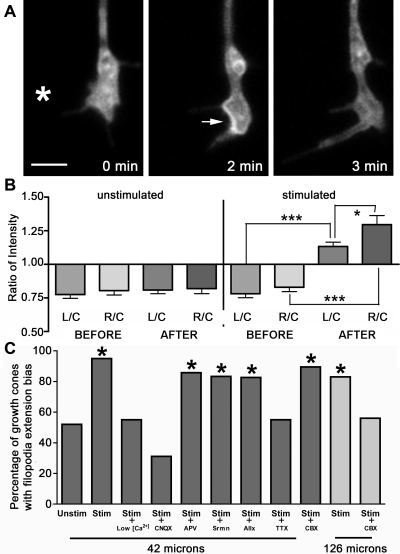
Activity plays a critical role in network formation during developmental, experience-dependent, and injury related remodeling. Here we report a mechanism by which axon trajectory can be altered in response to remote neuronal activity. Using photoconductive stimulation to trigger high frequency action potentials in rat hippocampal neurons in vitro, we find that activity functions as an attractive cue for growth cones in the local environment. The underlying guidance mechanism involves astrocyte Ca(2+) waves, as the connexin-43 antagonist carbenoxolone abolishes the attraction when activity is initiated at a distance greater than 120 microm. The asymmetric growth cone filopodia extension that precedes turning can be blocked with CNQX (10 microM), but not with the ATP and adenosine receptor antagonists suramin (100 microM) and alloxazine (4 microM), suggesting non-NMDA glutamate receptors on the growth cone mediate the interaction with astrocytes. These results define a potential long-range signalling pathway for activity-dependent axon guidance in which growth cones turn towards directional, temporally coordinated astrocyte Ca(2+) waves that are triggered by neuronal activity. To assess the viability of the guidance effect in an injury paradigm, we performed the assay in the presence of conditioned media from lipopolysaccharide (LPS) activated purified microglial cultures, as well as directly activating the glia present in our co-cultures. Growth cone attraction was not inhibited under these conditions, suggesting this mechanism could be used to guide regeneration following axonal injury.


Conflict of interest statement

Competing Interests: The authors have declared that no competing interests exist.

Figures

Figure 1
Figure 1. Local calcium elevation in astrocytes leads to the propagation of an intercellular Ca2+ wave.
Calcium levels were monitored using fluo-4 AM. (a) Illustration of the experimental protocol, which entailed the depolarization of a 42 µm diameter area either 42 µm or 126 µm to the right of an axonal growth cone. (b,c) Representative images of a glial wave. Panel c shows the progression of the wave front after image subtraction in ImageJ (W. Rasband, NIH). Scale bars, 50 µm.
Figure 2
Figure 2. Activity induces attractive growth cone turning and filopodia sprouting.
(a) An example of a growth cone that turned when the stimulation was repeated once every 5 minutes, as visualized by YFP-actin. (b) Superimposed traces of the trajectories of growth cones (n = 5) that extended more than 5 µm during long term experiments wherein neurons to the right side of the growth cone were stimulated every 5 minutes for 0.5 to 1 hour. The origin shows the center of the growth cone at t = 0, when its direction of growth was vertical. The axes show the distance in microns. (c) More filopodia protrude on the side of the growth cone that faces the area of stimulation, as visualized by YFP-actin. (d) A representative time series of the total perimeter of the left and right side of a growth cone during an experiment, measured from images taken 20 seconds apart. The vertical bar marks the first set of images taken after the stimulation. (e) The ratio of the total perimeter of the right side to that of the left (R/L) after a real stimulation is greater compared with that of a mock stimulation (unstimulated, n = 25; stimulated, n = 20). Bars show the mean, error bars are the SEM. Scale bars, 2 µm.
Figure 3
Figure 3. A role for VAMP-2, calcium, non-NMDA receptors and gap junctions in activity-induced attractive filopodia extension.
(a) 2 minutes after stimulation GFP-VAMP-2 clusters near the membrane, mostly on the right side. (b) The mean grey value (intensity) of a region of interest was measured from the left side (L), right side (R) and cytoplasm (C) of growth cones from GFP-VAMP-2 neurons before and after stimulation. The intensity ratios L/C and R/C increase after a stimulation (n = 20; p<0.0001, two-sided unpaired student's t-test). R/C is also significantly higher then L/C after stimulation (p = 0.0344). The intensity ratios (L/C and R/C) do not change when cells are monitored for the same amount of time without a voltage pulse (n = 21, p>0.05). Error bars represent standard deviations. (c) The attractive filopodia extension is calcium dependent and is blocked by CNQX. The effect is blocked by CBX when depolarization is performed at 126 µm but not 42 µm. Control, n = 25; stimulated (42 µm), n = 20; low Ca2+ (42 µm), n = 20; 10 µM CNQX(42 µm), n = 22, 50 µM APV(42 µm), n = 21; 100 µM suramin (Srmn) (42 µm), n = 18; 4 µM alloxazine (Allx) (42 µm), n = 23; 1 µM TTX (42 µm), n = 20; 50 µM CBX (42 µm), n = 19; stimulated (126 µm), n = 24; 50 µM CBX(126 um), n = 27. Asterisk indicates p<0.05. Scale bars, 2 µm.
Figure 4
Figure 4. Activity-driven axon guidance in the presence of activated microglia and astrocytes.
(a) Astrocytes and microglia in rat hippocampal cultures. Cultures were labeled using anti-Iba1 as a microglia marker (green), GFAP as an astrocyte marker (red), and were counterstained for nuclei with DAPI (blue). There were very few microglia in the cultures (∼1 in 300, see text). (b) shows an enlarged panel of (a). (c) The few microglia that were present in our cultures sometimes formed clusters. (d) The attractive filopodia extension bias occurs in the presence of conditioned media from minocycline treated rat hippocampal cultures (RHC-MIN) (n = 17) and LPS activated human adult microglia cultures (HAM-LPS) (n = 18). This was also the case for conditioned media from LPS (RHC-LPS) activated rat hippocampal microglia and astrocytes(n = 22). Asterisk indicates p<0.05, cross indicates p<0.06. Scale bars, 25 µm.

Similar articles

Cited by

References

    1. Knowles WD, Schwartzkroin PA. Axonal ramifications of Hippocampal CA1 pyramidal cells. J Neurosci. 1981;1(11):1236–1241. - PMC - PubMed
    1. De Paola V, Holtmaat A, Knott G, Song S, Wilbrecht L, et al. Cell type-specific structural plasticity of axonal branches and boutons in the adult neocortex. Neuron. 2006;49:861–875. - PubMed
    1. Gogolla N, Galimberti I, Caroni P. Structural plasticity of axon terminals in the adult. Curr Opin Neuro. 2007;17:516–524. - PubMed
    1. Chen R, Cohen LG, Hallett M. Nervous system reorganization following injury. Neuroscience. 2002;111(4):761–773. - PubMed
    1. Neumann H. Molecular mechanisms of axonal damage in inflammatory central nervous system diseases. Curr Opin Neurol. 2003;16(3):267–73. - PubMed

Publication types

MeSH terms