. 2008;3(11):e3706.
doi: 10.1371/journal.pone.0003706. Epub 2008 Nov 12.

Bacillus anthracis peptidoglycan stimulates an inflammatory response in monocytes through the p38 mitogen-activated protein kinase pathway

Affiliations

Bacillus anthracis peptidoglycan stimulates an inflammatory response in monocytes through the p38 mitogen-activated protein kinase pathway

Marybeth Langer et al. PLoS One. 2008.

Abstract

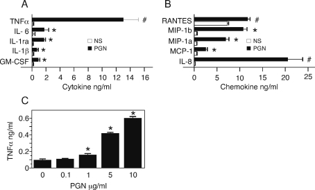
We hypothesized that the peptidoglycan component of B. anthracis may play a critical role in morbidity and mortality associated with inhalation anthrax. To explore this issue, we purified the peptidoglycan component of the bacterial cell wall and studied the response of human peripheral blood cells. The purified B. anthracis peptidoglycan was free of non-covalently bound protein but contained a complex set of amino acids probably arising from the stem peptide. The peptidoglycan contained a polysaccharide that was removed by mild acid treatment, and the biological activity remained with the peptidoglycan and not the polysaccharide. The biological activity of the peptidoglycan was sensitive to lysozyme but not other hydrolytic enzymes, showing that the activity resides in the peptidoglycan component and not bacterial DNA, RNA or protein. B. anthracis peptidoglycan stimulated monocytes to produce primarily TNFalpha; neutrophils and lymphocytes did not respond. Peptidoglycan stimulated monocyte p38 mitogen-activated protein kinase and p38 activity was required for TNFalpha production by the cells. We conclude that peptidoglycan in B. anthracis is biologically active, that it stimulates a proinflammatory response in monocytes, and uses the p38 kinase signal transduction pathway to do so. Given the high bacterial burden in pulmonary anthrax, these findings suggest that the inflammatory events associated with peptidoglycan may play an important role in anthrax pathogenesis.


Conflict of interest statement

Competing Interests: The authors have declared that no competing interests exist.

Figures

Figure 1
Figure 1. Purified peptidoglycan from B. anthracis lacks noncovalently bound proteins.
(A) Silver-stained SDS PAGE showing with samples from purification steps and comparison to commercial preparation. Lane 1: bacteria after harvest, lane 2: equivalent sample of extracted PGN, lane 3: molecular weight marker. (B) Amino acid content of B. anthracis PGN.
Figure 2
Figure 2. TNFα is the major cytokine in PB produced in response to B. anthracis PGN.
PB was stimulated with either 10 µg/ml or increasing doses of PGN for 6 hours. (A, B). Secreted cytokines (A) and chemokines (B) in PB supernatants were measured by human multiplex bead immunoassay. Data are expressed as mean±SEM of 3 replicate wells. Statistical significance was determined for stimulated PB (PGN; 10 µg/ml) versus unstimulated (NS) by unpaired, one-tailed t test; *, p<0.001 versus 0 PGN; #, p<0.05 versus 0 PGN (C) Dose-response of secreted TNFα in supernatant measured by ELISA. The data are expressed as mean±SEM of 3 replicate wells. Statistical significance was determined by ANOVA with Bonferroni post test. *, p<0.001 versus 0 PGN.
Figure 3
Figure 3. Monocytes produce TNFα in response to PGN.
(A) Blood cells were sorted by SSC/FSC properties and staining intensity of fluorescent surface markers CD14, CD16b, CD3, CD19. Cells were separated as CD14+/CD16b− (monocytes), CD14−/CD16b+ (neutrophils) (A), CD19+ (B lymphocytes), CD3+ (T lymphocytes) (B). Slides were prepared of the sorted subpopulations. Leukocyte type was determined based on nuclear and cytoplasmic stain and morphology. (C) TNFα positive cells were identified using surface markers as above, in combination with intracellular staining for TNFα. (D) PB was stimulated with PGN (10 µg/ml) for 2, 4 and 6 hours. Cell populations were identified by surface markers and TNFα was measured by intracellular staining and flow cytometry. Two separate preparations from two donors were used; for the 6 hour point, five preparations were used. (E) PB was stimulated with PGN and the percent TNFα+monocytes (CD14+/CD16b−) was measured after 6 hours at each dose by intracellular staining and flow cytometry. Three separate preparations from two donors were used. For PGN (10 µg/ml), five preparations were used. The data for panel D and E are expressed as the mean±SEM. Statistical significance was determined by ANOVA with Bonferroni post test. *, p<0.001 versus 0 PGN.
Figure 4
Figure 4. Production of TNFα is not due to endotoxin, bacterial DNA, RNA or contaminating proteins in B. anthracis peptidoglycan.
A) The Limulus amebocyte lysate assay was used to test PGN (10 µg/ml), LPS (1 pg/ml, 10 pg/ml, 100 pg/ml) and endotoxin-free water for endotoxin activity, Endotoxin units (EU)/ml. Data are expressed as mean±SEM of 3 replicates. Statistical significance was determined for LPS (10 pg/ml) compared with PGN (10 µg/ml) by unpaired, two-tailed t test; p = 0.49, indicating no significant difference in level of endotoxin activity. (B) PB was stimulated with 10 pg/ml LPS 6 hours and TNFα positive monocytes were measured by flow cytometry. (C) PB was stimulated with PGN or LPS with and without polymixin B (PMB) (10 µg/ml) to bind endotoxin. TNFα production is expressed as fold difference±SEM of MFI of TNFα positive monocytes for 2 separate preparations on two individuals, 2 replicates each. Statistical significance was determined by ANOVA with Bonferroni post test. *, p<0.001 versus 1 mg/ml LPS without PMB. (D) B. anthracis PGN was digested with the indicted hydrolytic enzymes for 24 hours. The preparations were heat inactivated and used to stimulate PB at 10 µg/ml PGN along with an equivalent preparation of untreated PGN. The percent of TNFα positive monocytes was measured by intracellular cytokine staining. Data are expressed as the mean±SEM of three separate preparations. Statistical significance was performed on results from the enzyme treated PGN stimulations versus stimulation with untreated PGN by ANOVA with Bonferroni post test. *, p<0.001 versus untreated PGN.
Figure 5
Figure 5. The PGN-associated polysaccharide of B. anthracis is not necessary for TNFα activity.
B. anthracis PGN was sonicated and hydrolyzed with HF for 24 hours. The HF-soluble polysaccharide was separated from the insoluble peptidoglycan backbone by centrifugation. The original PGN, sonicated PGN, and HF-soluble polysaccharide and HF-insoluble PGN fractions were used to stimulate PB. The percent TNFα positive monocytes was calculated per µg of PGN (OD600). Data is expressed as mean±SEM of three separate preparations from 2 donors including two separate HF hydrolysis reactions of PGN. Statistical analysis was by ANOVA with Bonferroni post test of the stimulated samples versus PGN. *, p<0.001 versus PGN.
Figure 6
Figure 6. PGN stimulates the p38 MAP kinase and ERK pathways and TNFα is blocked by inhibition of p38 MAP kinase.
(A) PB was stimulated with PGN (10 µg/ml) or LPS (1 µg/ml) and phospho-MAP kinases were measured by intracellular staining in CD14+ monocytes. (B) Phospho-p38 MAP kinase was measured after 15 min for the unstimulated (thin solid line), PGN (thick solid line), and LPS (dashed line). (C) Phospho p44/42 MAP kinase, 15 minutes and (D) phospho-JnK, 15 minutes. (E) Fold increase in phospho-MAP kinases for unstimulated (NS), PGN and LPS stimulation. Data are expressed as mean±SEM of three separate PB preparations. Statistical analysis was performed on MFI of stimulated PB versus MFI unstimulated PB. *, p<0.03; # p<0.005 by paired one-tailed t test. (F) PB was co-stimulated for 2 hours with PGN (10 µg/ml) with the MAP kinase inhibitors SB202190 (p38 MAP kinase inhibitor), SP600125 (JnK inhibitor) and PD 98095 (ERK inhibitor). TNFα in monocytes was measured by intracellular staining and flow cytometry. Data are expressed as mean±SEM of three separate preparations from two donors. Statistical analysis was by ANOVA with Bonferroni post test. *, p<0.01 versus PGN (10 µg/ml).
Figure 7
Figure 7. TNFα protein and mRNA is upregulated by PGN.
(A) Time course of secreted TNFα induced by PGN in whole blood. PB was stimulated with PGN (10 µg/ml) and the supernatant was sampled at the indicated times. TNFα concentration was measured by ELISA. Data are expressed as mean±SEM for 3 replicates. Statistical analysis was by ANOVA with Bonferroni post test. *, p<0.001 versus 0 PGN; #, p<0.01 versus 0 PGN. (B) PB was stimulated with PGN (10 µg/ml)±SB202190 for 2 hours. RNA was isolated from leukocytes, converted to cDNA which was subjected to real-time quantitative PCR. Gene specific primers used were for TNFα and β-actin in a real-time PCR assay. Data presented are the mean±SEM of separate preparations of the TNFα/β-actin ratio from 4 individuals, 3 replicates each and normalized to the unstimulated ratio. Statistical analysis was by t test. *, p<0.01 for TNFα, PGN versus unstimulated; # p>0.3 for β-actin versus unstimulated. (C) PB was stimulated with PGN (10 µg/ml) for 3 hours. Cells were harvested to represent 100% RNA at 3 hours. To equivalent samples, actinomycin D was added immediately to block further RNA synthesis, or actinomycin+SB202190 were added to block RNA synthesis and the p38 MAP kinase pathway. Cells were harvested at 10, 20, 30, 60 minutes after these additions. RNA was isolated, cDNA was prepared and subjected to real-time quantitative PCR for TNFα as in (B). Quantity of RNA was expressed as % of RNA after 3 hours of stimulation. Data presented are the mean±SEM from 3 replicate PCR reactions from 1 individual. Statistical analysis was by ANOVA with Bonferroni post test of *, p<0.001 for PGN+ActD versus PGN+ActD+SB; #, p<0.01.

Similar articles

Cited by

References

    1. Cote CK, Van Rooijen N, Welkos SL. Roles of macrophages and neutrophils in the early host response to Bacillus anthracis spores in a mouse model of infection. Infect Immun. 2006;74:469–480. - PMC - PubMed
    1. Rhie GE, Roehrl MH, Mourez M, Collier RJ, Mekalanos JJ, et al. A dually active anthrax vaccine that confers protection against both bacilli and toxins. Proc Natl Acad Sci U S A. 2003;100:10925–10930. - PMC - PubMed
    1. Dixon TC, Meselson M, Guillemin J, Hanna PC. Anthrax. N Engl J Med. 1999;341:815–826. - PubMed
    1. Guarner J, Jernigan JA, Shieh WJ, Tatti K, Flannagan LM, et al. Pathology and pathogenesis of bioterrorism-related inhalational anthrax. Am J Pathol. 2003;163:701–709. - PMC - PubMed
    1. Jernigan JA, Stephens DS, Ashford DA, Omenaca C, Topiel MS, et al. Bioterrorism-related inhalational anthrax: the first 10 cases reported in the United States. Emerg Infect Dis. 2001;7:933–944. - PMC - PubMed

Publication types

MeSH terms

LinkOut - more resources