. 2009 Feb;52(2):262-70.
doi: 10.1007/s00125-008-1200-z. Epub 2008 Nov 11.

Pancreatic adenocarcinoma patients with localised chronic severe pancreatitis show an increased number of single beta cells, without alterations in fractional insulin area

Affiliations

Pancreatic adenocarcinoma patients with localised chronic severe pancreatitis show an increased number of single beta cells, without alterations in fractional insulin area

M Campbell-Thompson et al. Diabetologia. 2009 Feb.

Abstract

Aims/hypothesis: Recent histological analysis of pancreases obtained from patients with long-standing type 1 diabetes identified chronic islet inflammation and limited evidence suggestive of beta cell replication. Studies in rodent models also suggest that beta cell replication can be induced by certain inflammatory cytokines and by gastrin. We therefore tested the hypothesis that beta cell replication is observed in non-autoimmune human pancreatic disorders in which localised inflammation or elevated gastrin levels are present.

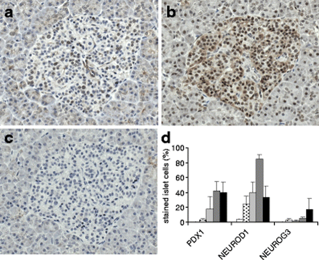
Methods: Resected operative pancreatic specimens were obtained from patients diagnosed with primary adenocarcinoma (with or without chronic severe pancreatitis) or gastrinoma. Additional pancreatic tissue was obtained from autopsy control patients. Immunohistochemistry was used to assess fractional insulin area, beta cell number and replication rate and differentiation factors relevant to beta cell development.

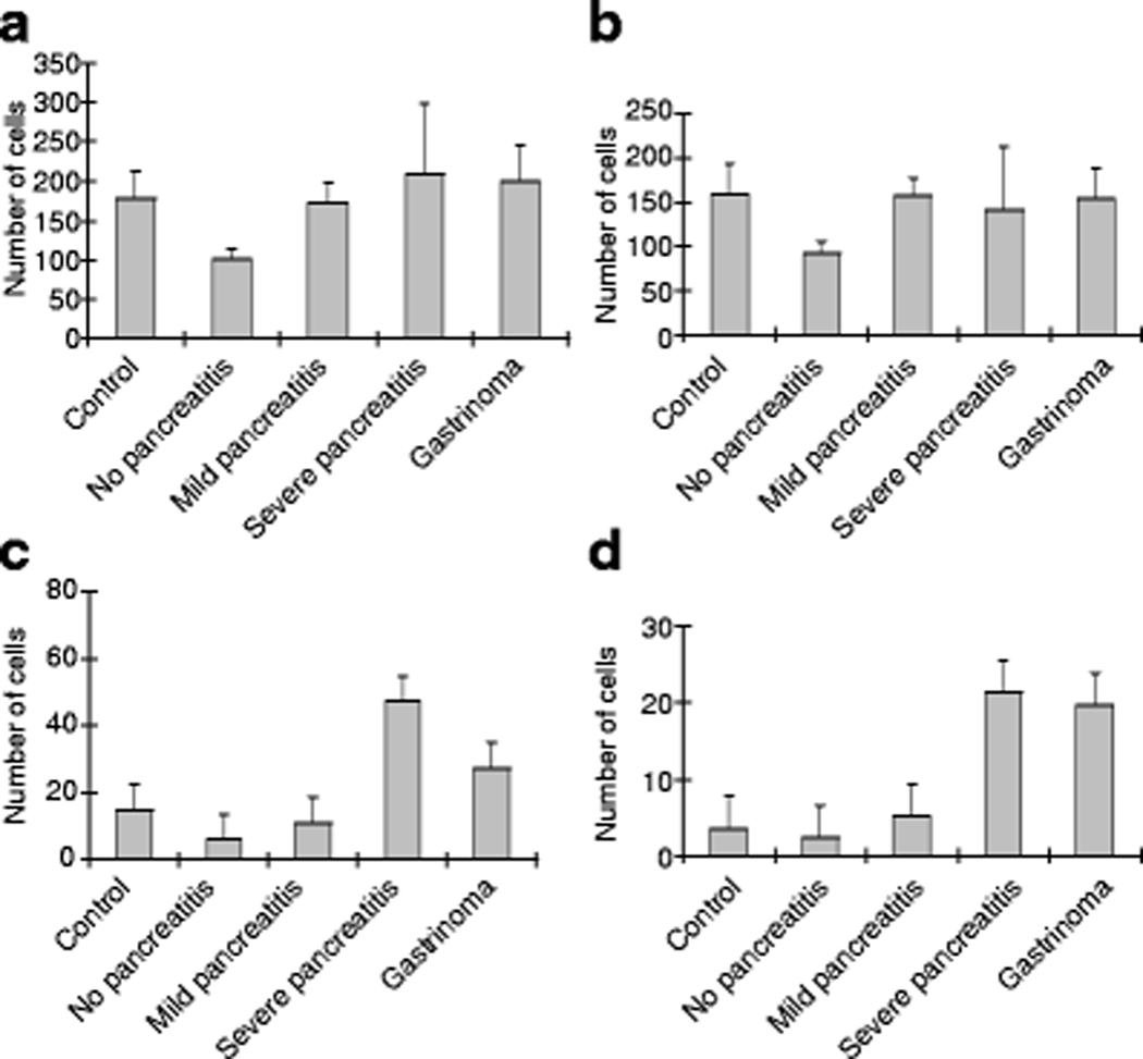
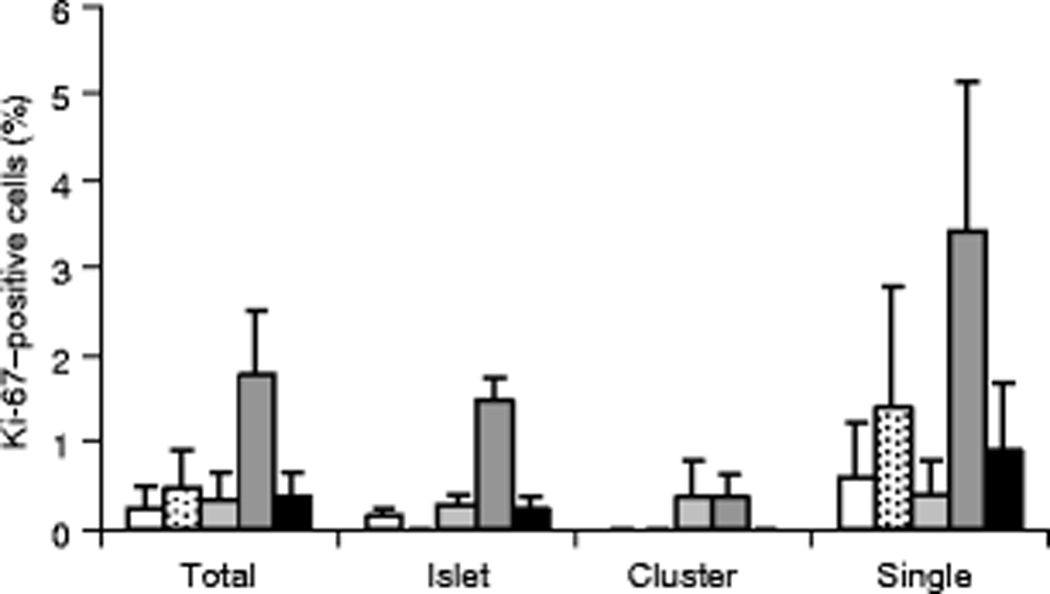
Results: Fractional insulin area was similar among groups. Patients with pancreatic adenocarcinoma and localised chronic severe pancreatitis displayed significant increases in the number of single beta cells, as well as increased beta cell replication rate and levels of neurogenic differentiation 1 in islets. Patients with gastrinoma demonstrated significant increases in the number of single beta cells, but the beta cell replication rate and islet differentiation factor levels were similar to those in the control group.

Conclusions/interpretation: These findings indicate that chronic severe pancreatic inflammation can be associated with significant effects on beta cell number or replication rate, depending on the distribution of the cells. This information may prove useful for attempts seeking to design therapies aimed at inducing beta cell replication as a means of reversing diabetes.


Figures

Fig. 1.
Fig. 1.
Pancreatic histopathology in uninvolved regions from patients with primary adenocarcinoma. Paraffin sections were stained with AF-H&E to delineate insulin-positive beta cells (dark purple). a Section from a patient with adenocarcinoma and no pancreatitis (1.25x). Red dye at lower margin delineates orientation towards the adenocarcinoma. Mild intralobular adipose tissue was present. b Focal ductular proliferation in a patient with no pancreatitis. Cross sections of ducts are visible (arrows) in addition to a large group of cells that are negative by AF-H&E staining. Elastic fibers in a large blood vessel stain dark purple with AF-H&E. c Pancreatic morphology in a patient with chronic mild pancreatitis. Islets of varying sizes are observed in different lobules. Focal ductular hyperplasia (arrow) is observed in a large duct with multifocal mild adipose tissue (clear spaces) in the acinar regions. d Pancreatic morphology in a patient with chronic severe pancreatitis. Focal, mild acinar atrophy was observed (white star) with focal, moderate ductular proliferation and hyperplasia (arrows).
Fig. 2
Fig. 2
Pancreatic immunohistochemistry for pancreatic ductular epithelium and insulin. Pancreatic sections were double stained for cytokeratin 7 (CK7, brown) and insulin (red) as described in Methods. CK7 immunopositivity was found in ducts and centroacinar cells (a-c, black arrows) adjacent to unstained acinar cells. Insulin positive cells were identified in close approximation to or within ductular epithelium in all patients groups. Insulin positive cells were found as single cells (a, d, white arrows) or clusters (a-d) and were only rarely observed in ducts with hyperplasia (d).
Fig. 3
Fig. 3
Beta cell numbers and distribution within islets or clusters, or as single cells. Beta cells were identified by anti-insulin immunofluorescence, images acquired using a 20x objective, and positive cells counted per field of view. a Mild pancreatitis and gastrinoma subjects had significantly higher total beta cell numbers compared to patients with no pancreatitis. b Beta cell numbers within islets were lowest in the no pancreatitis group when compared to patients with mild pancreatitis (*p=0.02). c Beta cell numbers within clusters were significantly higher in severe pancreatitis and gastrinoma patients compared to no pancreatitis patients (*p<0.01). d Patients with severe pancreatitis and gastrinoma had significantly higher beta cell counts as single cells compared to controls and both no and mild pancreatitis groups (***p<0.005).
Fig. 4
Fig. 4
Beta cell proliferation rates determined by Ki-67 levels. Proportion of Ki-67–positive beta cells are shown for each group expressed as total cells or beta cells within islets, clusters, and single cells. Despite variability, the severe pancreatitis group showed significantly higher proportions of total Ki-67-positive beta cells (***p<0.005) compared to all other groups. The average count of Ki-67-positive beta cells in islets was also significantly higher in the severe pancreatitis patients compared to all other groups (***p<0.005). Ki-67 positivity was most variable within single beta cells. Legend: Control (white); No pancreatitis (stippled); Mild pancreatitis (light grey); Severe pancreatitis (dark grey); Gastrinoma (black).
Fig. 5
Fig. 5
PDX1, NEUROD1, and NEUROG3 levels in pancreatic islets. Representative islet images showing immunostaining for PDX1 (a), NEUROD1 (b), and NEUROG3 (c) in serial sections from a patient with mild pancreatitis. d Semi-quantitative analysis of percentage nuclear staining for PDX1, NEUROD1, and NEUROG3 staining in islets. The level of PDX1 was significantly higher in severe pancreatitis and gastrinoma patients compared to both autopsy control and no pancreatitis patients (**p<0.03). NEUROD1 was significantly higher in severe pancreatitis patients compared to all other groups (***p<0.02). Subjects with either mild pancreatitis or gastrinoma also showed intermediate NEUROD1 levels when compared to controls (*p<0.05). NEUROG3 levels were lower than PDX1 or NEUROD1 and no significant differences were observed between groups. Legend: Control (white); No pancreatitis (stippled); Mild pancreatitis (light grey); Severe pancreatitis (dark grey); Gastrinoma (black).

Similar articles

Cited by

References

    1. Atkinson MA, Rhodes CJ (2005) Pancreatic regeneration in type 1 diabetes: dreams on a deserted islet? Diabetologia 48: 2200–2202 - PubMed
    1. Meier JJ, Bhushan A, Butler AE, Rizza RA, Butler PC (2005) Sustained beta cell apoptosis in patients with long-standing type 1 diabetes: indirect evidence for islet regeneration? Diabetologia 48: 2221–2228 - PubMed
    1. Bonner-Weir S, Weir GC (2005) New sources of pancreatic beta-cells. Nat Biotechnol 23: 857–861 - PubMed
    1. Bouwens L (2006) Beta cell regeneration. Curr Diabetes Rev 2: 3–9 - PubMed
    1. Lipsett M, Aikin R, Hanley S, Al-Maleek J, Laganiere S, Rosenburg L (2006) Islet neogenesis: a potential therapeutic tool in type 1 diabetes. Int J Biochem Cell Biol 38: 715–720 - PubMed

Publication types