

Epidermal growth factor receptor mutations in non-small cell lung cancer influence downstream Akt, MAPK and Stat3 signaling
- PMID: 19002495
- PMCID: PMC12160170
- DOI: 10.1007/s00432-008-0509-9
Epidermal growth factor receptor mutations in non-small cell lung cancer influence downstream Akt, MAPK and Stat3 signaling
Abstract
Purpose: The efficacy of epidermal growth factor receptor (EGFR) tyrosine kinase inhibitors in non-small cell lung cancer (NSCLC) has been linked to activating mutations in the EGFR gene. So far these mutations have been extensively characterized in established cell lines. The aim of this study was to determine the effects of EGFR mutations on downstream signaling in human tumor specimens.
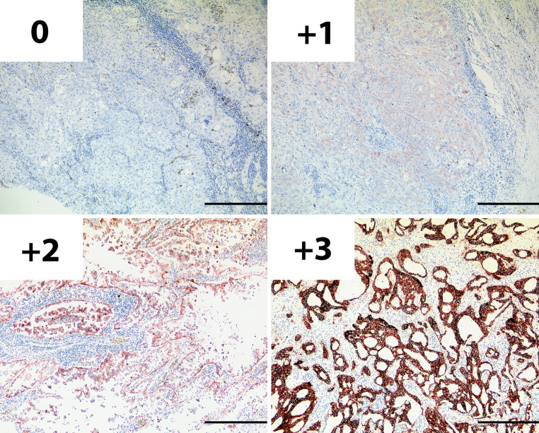
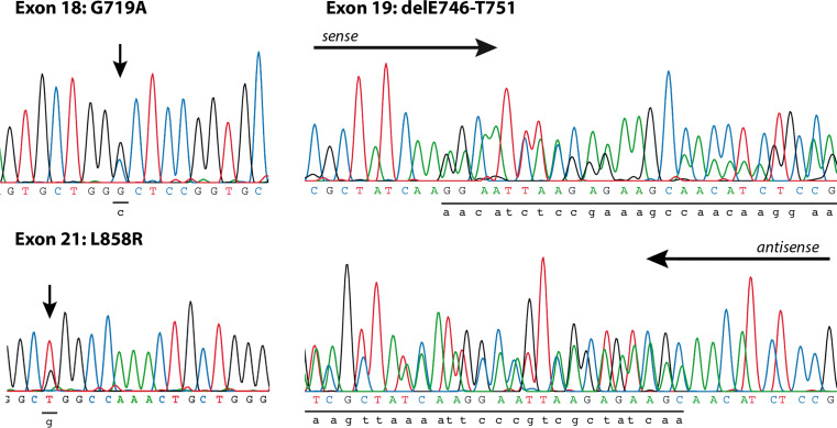
Methods: We have looked for mutations of the EGFR gene in specimens of 67 patients with NSCLC and correlated these with EGFR phosphorylation and the activity of its three main downstream signaling cascades Akt, MAPK and Stat3 by immunohistochemistry.
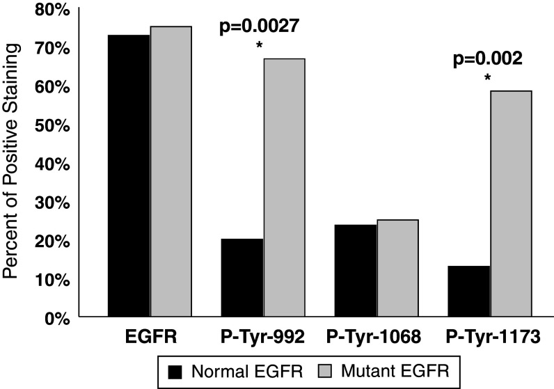
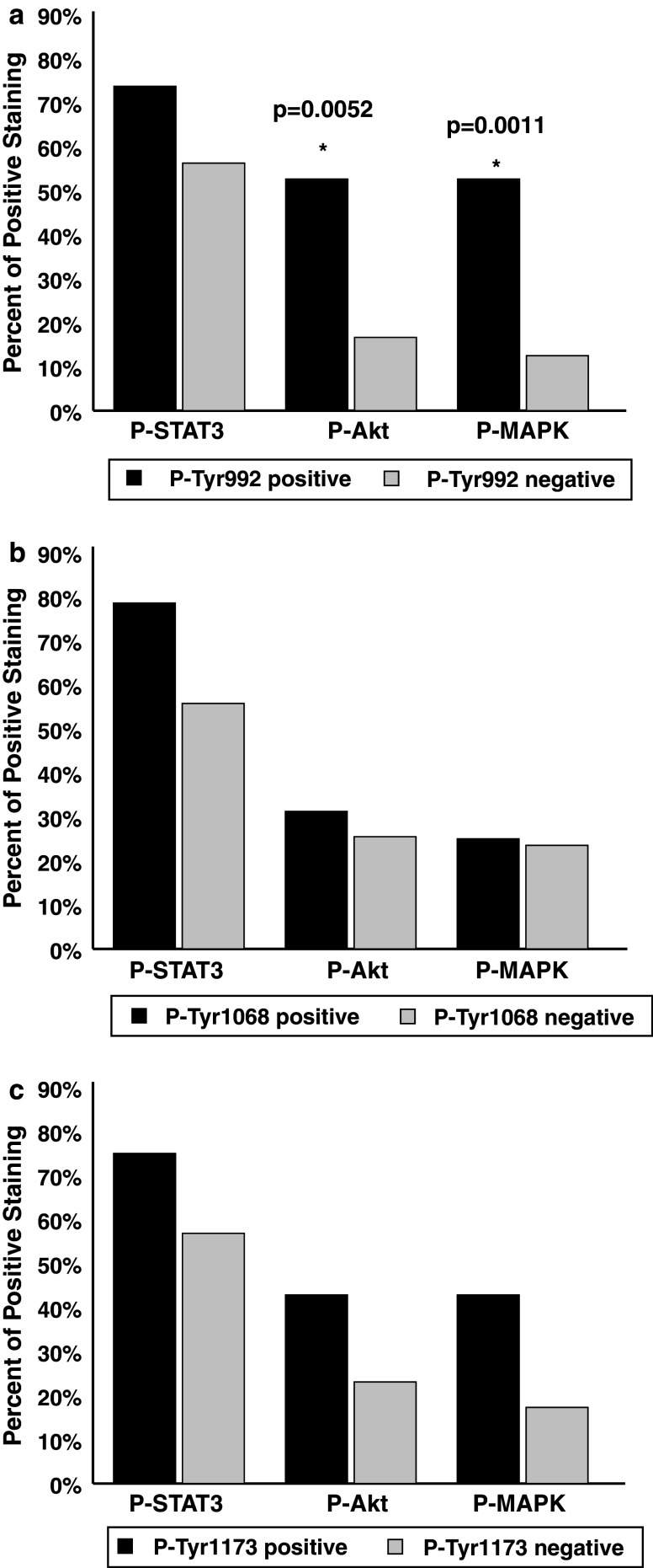
Results: We show that the phosphorylation of tyrosine residues 922 and 1173, but not 1068, are primarily affected by the activating EGFR mutations. Akt activity was significantly higher in patients with EGFR mutations but we found no difference in Stat3 or MAPK phosphorylation. Our results suggest that EGFR mutations not only increase receptor activity, but also alter responses of downstream signaling cascades in human NSCLCs and that these finding differ from results obtained in cell lines.
Figures

References
-
- Alvarez JV, Greulich H, Sellers WR, Meyerson M, Frank DA (2006) Signal transducer and activator of transcription 3 is required for the oncogenic effects of non-small-cell lung cancer-associated mutations of the epidermal growth factor receptor. Cancer Res 66:3162–3168. doi:10.1158/0008-5472.CAN-05-3757 - PubMed
-
- Atkins D, Reiffen KA, Tegtmeier CL, Winther H, Bonato MS, Storkel S (2004) Immunohistochemical detection of EGFR in paraffin-embedded tumor tissues: variation in staining intensity due to choice of fixative and storage time of tissue sections. J Histochem Cytochem 52:893–901. doi:10.1369/jhc.3A6195.2004 - PubMed
-
- Bishnupuri KS, Luo Q, Murmu N, Houchen CW, Anant S, Dieckgraefe BK (2006) Reg IV activates the epidermal growth factor receptor/Akt/AP-1 signaling pathway in colon adenocarcinomas. Gastroenterology 130:137–149. doi:10.1053/j.gastro.2005.10.001 - PubMed
-
- Brambilla E, Travis WD, Colby TV, Corrin B, Shimosato Y (2001) The new World Health Organization classification of lung tumours. Eur Respir J 18:1059–1068. doi:10.1183/09031936.01.00275301 - PubMed
Publication types
MeSH terms
Substances
LinkOut - more resources
Full Text Sources
Medical
Molecular Biology Databases
Research Materials
Miscellaneous
