. 2008 Jan;56(1):19-26.
doi: 10.1007/s10616-007-9099-7. Epub 2007 Oct 16.

Piezotolerance of the cytoskeletal structure in cultured deep-sea fish cells using DNA transfection and protein introduction techniques

Affiliations

Piezotolerance of the cytoskeletal structure in cultured deep-sea fish cells using DNA transfection and protein introduction techniques

Sumihiro Koyama et al. Cytotechnology. 2008 Jan.

Abstract

We used DNA transfection and protein introduction techniques to investigate the pressure tolerance of cytoskeletal structures in pectoral fin cells derived from the deep-sea fish Simenchelys parasiticus (habitat depth, 366-2,630 m). The deep-sea fish cells have G418 resistance. The cell number increased until day 6 of cultivation and all cells had died by day 35 when cultured in 35-mm Petri dishes in medium containing G418. Enhanced yellow fluorescent protein-tagged human beta-actin (EYFP-actin) was stably expressed by 1 in 100,000 deep-sea fish cells. Because almost none of the EYFP-actin was incorporated into actin filaments of the cells, we replaced the relatively large EYFP tag with a chemical fluorescent compound and succeeded in incorporating fluorescently labeled rabbit actins into the deep-sea fish actin filaments. Most of the filament structure in the cells with rabbit actin inserted underwent depolymerization when subjected to pressure of 100 MPa for 20 min, in contrast to control cells. There were no differences in the tubulin filament structure between control cells and deep-sea fish cells with fluorescein-labeled bovine tubulin inserted after the application of pressure ranging from 40 to 100 MPa for 20 min.


Figures

Fig. 1
Fig. 1
Schematic illustration of the pEYFP-actin transfected deep-sea fish cell. In this report, we don’t confirm introduction of the plasmid DNA into chromosomal DNA
Fig. 2
Fig. 2
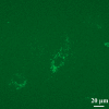
Intracellular distribution of EYFP-actin in deep-sea fish cells 24 h after transfection. Transfection efficiency of plasmid DNA into the cells was 96% when they continually grew with passage at the logarithmic growth phase. When the cells reached 100% confluence, transfection efficiency decreased to 76% even if passaged after trypsinization
Fig. 3
Fig. 3
Inhibitory effects of G418 on deep-sea fish cell growth. G418 4 mg/mL (circles) and G418 1 mg/mL (squares) in salt-enriched L-15 medium containing 20% FBS and 1% antibiotics. Values are the mean ± S.D. of eight independent experiments (*p < 0.05 compared with day 0, Student’s t-test). The deep-sea fish cell density was examined in random 1-mm2 areas
Fig. 4
Fig. 4
EYFP-actin expression in deep-sea fish cells. Red and green fluorescence indicate actin filaments and EYFP-actin, respectively, in the deep-sea fish cells. (a) Stable expression of EYFP-actin in the cells. Stable EYFP-actin expression was seen in cells treated with 4 mg/mL of G418 for 14 days and then further cultured in medium containing 1 mg/mL of G418. Approximately 1 in 100,000 deep-sea fish cells stably expressed EYFP-actin. (b) EYFP-actin inserted into actin filaments in the cells. Approximately 1 in 30,000 cells exhibiting transient expression incorporated EYFP-actin into the actin filaments.
Fig. 5
Fig. 5
Piezotolerance of actin filaments in deep-sea fish cells. Red and green fluorescence indicates Alexa Fluor 594-conjugated rabbit muscle actin and deep-sea fish actin filaments, respectively, in deep-sea fish cells. (a) Rabbit actin was inserted into deep-sea fish cells under atmospheric conditions and (b) after being subjected to pressure of 100 MPa for 20 min. (c) Deep-sea fish cells after being subjected to pressure of 100 MPa for 20 min

Similar articles

Cited by

References

    1. Alberts B, Johnson A, Lewis J, Raff M, Roberts K, Walter P (eds) (2002) Molecular biology of the cell, 4th edn. Garland Science, New York
    1. {'text': '', 'ref_index': 1, 'ids': [{'type': 'PubMed', 'value': '6193810', 'is_inner': True, 'url': 'https://pubmed.ncbi.nlm.nih.gov/6193810/'}]}
    2. Bar-Num S, Shneyour Y, Beckmann J (1983) G-418, an elongation inhibitor of 80S ribosomes. Biochim Biophys Acta 741:123–127 - PubMed
    1. {'text': '', 'ref_index': 1, 'ids': [{'type': 'DOI', 'value': '10.1002/cm.970100305', 'is_inner': False, 'url': 'https://doi.org/10.1002/cm.970100305'}, {'type': 'PubMed', 'value': '3052872', 'is_inner': True, 'url': 'https://pubmed.ncbi.nlm.nih.gov/3052872/'}]}
    2. Bourns B, Franklin S, Cassimeris L, Salmon ED (1988) High hydrostatic pressure effects in vivo: changes in cell morphology, microtubule assembly, and actin organization. Cell Motil Cytoskeleton 10:380–390 - PubMed
    1. {'text': '', 'ref_index': 1, 'ids': [{'type': 'DOI', 'value': '10.1006/excr.1996.0278', 'is_inner': False, 'url': 'https://doi.org/10.1006/excr.1996.0278'}, {'type': 'PubMed', 'value': '8831567', 'is_inner': True, 'url': 'https://pubmed.ncbi.nlm.nih.gov/8831567/'}]}
    2. Crenshaw HC, Allen JA, Skeen V, Harris A, Salmon ED (1996) Hydrostatic pressure has different effects on the assembly of tubulin, actin, myosin II, vinculin, talin, vimentin, and cytokeratin in mammalian tissue cells. Exp Cell Res 227:285–297 - PubMed
    1. {'text': '', 'ref_index': 1, 'ids': [{'type': 'PubMed', 'value': '7001938', 'is_inner': True, 'url': 'https://pubmed.ncbi.nlm.nih.gov/7001938/'}]}
    2. Davies J, Jimenez A (1980) A new selective agent for eukaryotic cloning vectors. Am J Trop Med Hyg 29:1089–1092 - PubMed