. 2008 Jan;56(1):41-8.
doi: 10.1007/s10616-007-9102-3. Epub 2007 Oct 16.

A practical approach for intracellular protein delivery

Affiliations

A practical approach for intracellular protein delivery

Claire O Weill et al. Cytotechnology. 2008 Jan.

Abstract

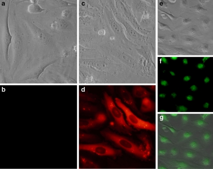
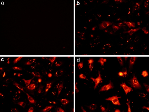
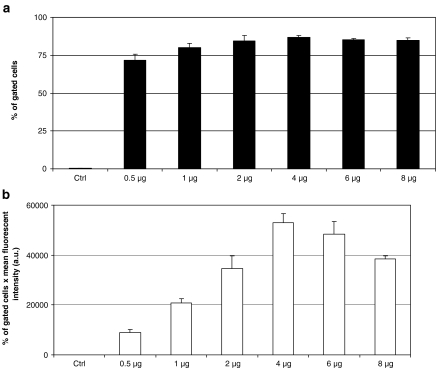
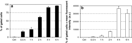
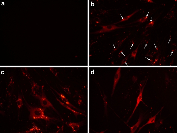
Protein delivery represents a powerful tool for experiments in live cells including studies of protein-protein interactions, protein interference with blocking antibodies, intracellular trafficking and protein or peptide biological functions. Most available reagents dedicated to the protein delivery allow efficient crossing of the plasma membrane. Nevertheless, the major disadvantage for these reagents is a weak release of the delivered protein into the cytoplasm. In this publication we demonstrate efficient protein delivery with a non-peptide based reagent, in human epithelial carcinoma HeLa cells and primary human skin fibroblasts. Using a fluorescent protein in combination with fluorescence microscopy and fluorescence-assisted cell sorting analysis, we show that the delivered protein is indeed released effectively in the cytoplasm, as expected for a dedicated carrier. Furthermore, we present a step-by-step method to optimize conditions for successful intracellular protein delivery.


Figures

Fig. 1
Fig. 1
The intracellular delivery of fluorescent proteins (R-PE and Histone-H1) mediated by a non-peptide based reagent. Two μg of R-PE was diluted in 20 mM Hepes buffer and added with HeLa cells in a 24-well. The plate was incubated for 16 h at 37 °C and cells were analysed by phase-contrast (a) and fluorescence microscopy (b) An amount of 2 μg of R-PE was delivered into HeLa cells after formation of complexes with 4 μL of delivery reagent. Intracellular protein delivery was analyzed after 16 h incubation at 37 °C by phase-contrast (c) and fluorescence microscopy (d) An amount of 4 μg of Histone-H1-AF®488 was delivered into HeLa cells after formation of complexes with 3 μl of delivery reagent. Intracellular protein delivery was analyzed 20 h later. Superimposition (g) of images obtained by phase-contrast (e) and fluorescence microscopy (f) was obtained using Adobe Photoshop software
Fig. 2
Fig. 2
Efficient intracellular protein delivery is dependent on the volume of delivery reagent used. An amount of 2 μg of R-PE was delivered into HeLa cells after formation of complexes with increasing volume of delivery reagent (a: 0 μL; b: 1 μL; c: 2 μL and d: 4 μL). The cells were analysed 7 h after addition of the complexes onto the cells
Fig. 3
Fig. 3
The amount of protein greatly influences the delivery efficiency. Cell sorting experiments were carried out onto HeLa cells after intracellular protein delivery. A volume of 4 μL of delivery reagent was added to various amounts of R-PE (1–8 μg) or 1 μg of BSA (negative control, Ctrl). Complexes were added to cells and further incubated for 20 h before FACS analysis. Gates were set to distinguish between autofluorescence of control cells and highly fluorescent cells. Means of percentage of fluorescent cells ± SEM are shown (a), n = 3 per point. Results are also presented as the percentage of gated fluorescent cells per mean intensity of fluorescence (b), n = 3 per point
Fig. 4
Fig. 4
The incubation time of protein/delivery reagent complexes with cells influences the delivery efficiency. Delivery reagent (4 μL) was added to R-PE (1 μg) or BSA (1 μg, Ctrl) to form complexes. Complexes were thereafter added onto HeLa cells and further incubated for the indicated time (30 min to 6 h). FACS analysis was carried out 16 h post-delivery. The experiment was carried out as described in the figure 3
Fig. 5
Fig. 5
Kinetic of intracellular R-PE delivery into human primary fibroblasts. One μg of R-PE diluted in 100 μL Hepes buffer (20 mM, pH 7.4) was incubated with 4 μL of delivery reagent for 15 min at room temperature. The cells were analyzed 4 (b), 8 (c) and 20 h (d) after addition of protein/reagent complexes onto cells. As a control (a), R-PE alone was added onto human primary fibroblasts and analysed 20 h after addition of the protein onto cells

References

    1. Bar-Sagi D, Feramisco JR (1985) Microinjection of the ras oncogene protein into PC12 cells induces morphological differentiation. Cell 42:841–848 - DOI - PubMed
    1. Campbell PL, McCluskey J, Yeo JP, Toh BH (1995) Electroporation of antibodies into mammalian cells. Methods Mol Biol 48:83–92 - PubMed
    1. Cassinelli G, Lanzi C, Petrangolini G, Tortoreto M, Pratesi G, Cuccuru G, Laccabue D, Supino R, Belluco S, Favini E, Poletti A, Zunino F (2006) Inhibition of c-Met and prevention of spontaneous metastatic spreading by the 2-indolinone RPI-1. Mol Cancer Ther 5:2388–2397 - DOI - PubMed
    1. Chuck AS, Clarke MF, Palsson BO (1996) Retroviral infection is limited by Brownian motion. Hum Gene Ther 7:1527–1534 - PubMed
    1. Dalkara D, Zuber G, Behr JP (2004) Intracytoplasmic delivery of anionic proteins. Mol Ther 9:964–969 - DOI - PubMed