Ultrastructure and melatonin 1a receptor distribution in the ovaries of African ostrich chicks
- PMID: 19002857
- PMCID: PMC2553629
- DOI: 10.1007/s10616-008-9147-y
Ultrastructure and melatonin 1a receptor distribution in the ovaries of African ostrich chicks
Abstract
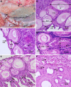
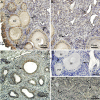
Healthy 90-day-old ostrich chicks were used in the present study. The ultrastructure and melatonin 1a receptor (MT1) distribution in the ovaries of ostrich chicks was observed by transmission electron microscope and light microscope. The results showed that the ostrich chick ovary contained primordial follicles, primary follicles and secondary follicles, but no mature follicles. There are some unique ultrastructural characteristics observed in the secondary follicle, such as the cortical granule, which was located in cytoplasm beside the nucleus and appeared first in the oocyte. The zona radiata appeared in the secondary follicle, and there was an obvious vitelline membrane. There were intraovarian rete, connecting rete, and extraovarian rete in the ovaries of ostrich chicks. This is the first study that provides immunohistochemical evidence for the localization of the melatonin MT1 in the ostrich chick ovary. The germinal epithelium, follicular cell layer of every grade of follicle, cytoplasm of the oocyte and interstitial cells all expressed MT1. The expression of positive immunoreactivity materials was the strongest in the follicular cell layer of the primordial follicle and germinal epithelium, was weaker in the follicular cell layer of the primary follicle and secondary follicle, and was weakest in the oocytes of all grades of follicle. In addition, the extraovarian rete displayed strong positive expression of MT1, while there was no positive expression in the intraovarian rete or connecting rete. The positive expression of MT1 immunoreactivity in the ovary was very strong, implying that the ovary is an important organ for synthesizing MT1.
Figures


References
-
- {'text': '', 'ref_index': 1, 'ids': [{'type': 'PubMed', 'value': '1517707', 'is_inner': True, 'url': 'https://pubmed.ncbi.nlm.nih.gov/1517707/'}]}
- Ayre EA, Yuan H, Pang SF (1992) The identification of 125I-labelled iodomelatonin-binding sites in the testes and ovaries of the chicken (Gallus domesticus). J Endocrinol 133:5–11 - PubMed
-
- {'text': '', 'ref_index': 1, 'ids': [{'type': 'PubMed', 'value': '18044344', 'is_inner': True, 'url': 'https://pubmed.ncbi.nlm.nih.gov/18044344/'}]}
- Boczek-Leszczy KE, Juszczak M (2007) The influence of melatonin on human reproduction. Pol Merkur Lekarski 23:128–130 - PubMed
-
- {'text': '', 'ref_index': 1, 'ids': [{'type': 'PMC', 'value': 'PMC1271596', 'is_inner': False, 'url': 'https://pmc.ncbi.nlm.nih.gov/articles/PMC1271596/'}, {'type': 'PubMed', 'value': '4783416', 'is_inner': True, 'url': 'https://pubmed.ncbi.nlm.nih.gov/4783416/'}]}
- Byskov AG, Lintern-Moore S (1973) Follicle formation in the immature mouse ovary: the role of the rete ovarii. J Anat 116:207–217 - PMC - PubMed
-
- {'text': '', 'ref_index': 1, 'ids': [{'type': 'PMC', 'value': 'PMC1234254', 'is_inner': False, 'url': 'https://pmc.ncbi.nlm.nih.gov/articles/PMC1234254/'}, {'type': 'PubMed', 'value': '838624', 'is_inner': True, 'url': 'https://pubmed.ncbi.nlm.nih.gov/838624/'}]}
- Byskov AG, Skakkebaek NE, Stafanger G, Peters H (1997) Influence of Ovarian surface epithelium and reteovanii on follicle formation. J Anat 123:77–86 - PMC - PubMed
-
- {'text': '', 'ref_index': 1, 'ids': [{'type': 'PubMed', 'value': '2842214', 'is_inner': True, 'url': 'https://pubmed.ncbi.nlm.nih.gov/2842214/'}]}
- Dubocovich ML (1988) Pharmacology and function of melatonin receptors. FASEB J 2:2765–2773 - PubMed
LinkOut - more resources
Full Text Sources

