. 2006 Oct;52(2):71-85.
doi: 10.1007/s10616-006-9032-5. Epub 2006 Nov 30.

Anoikis-resistant MDCK cells carrying susceptibilities to TNF-alpha and verotoxin that are suitable for influenza virus cultivation

Affiliations

Anoikis-resistant MDCK cells carrying susceptibilities to TNF-alpha and verotoxin that are suitable for influenza virus cultivation

Reiko Tsutsumi et al. Cytotechnology. 2006 Oct.

Abstract

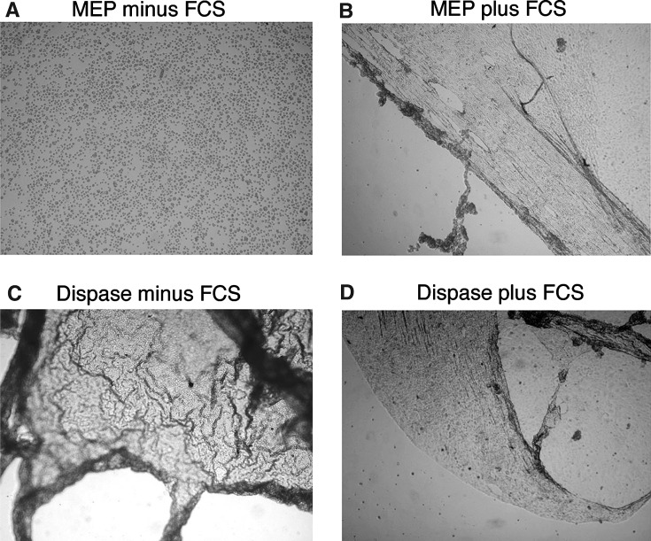
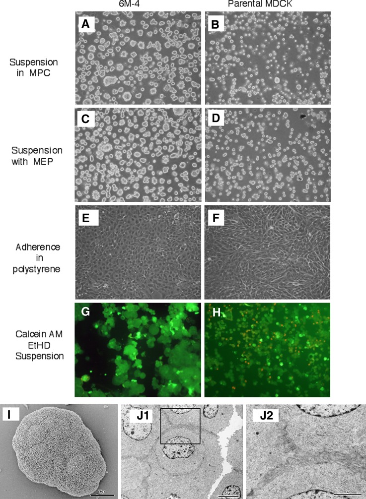
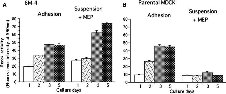
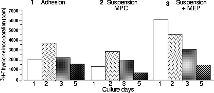
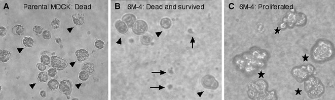
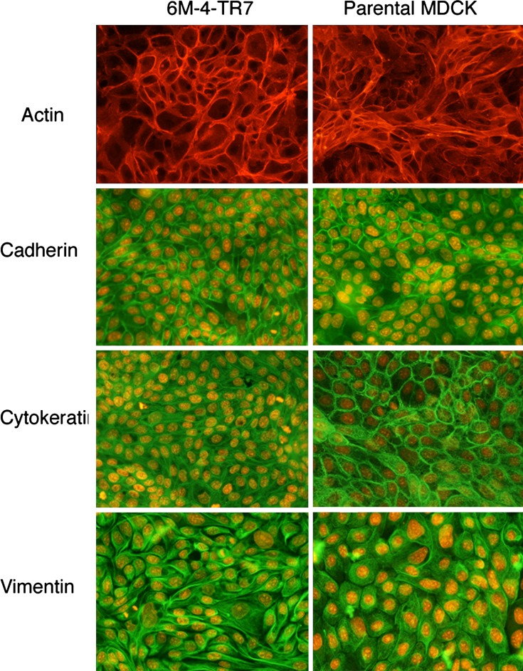
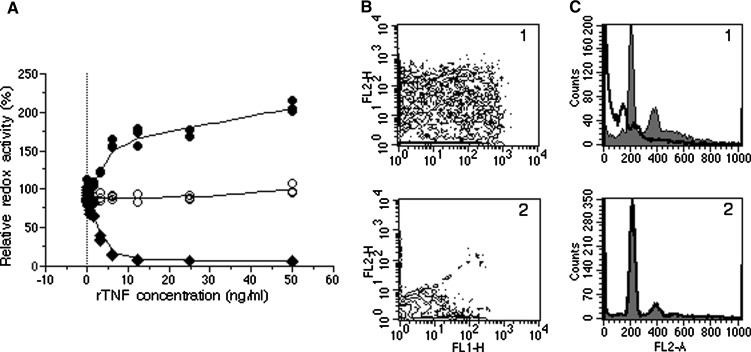
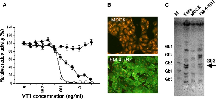
Madin-Darby canine kidney (MDCK) cells were originally anchorage-dependent epithelial cells. Here, we have isolated a novel MDCK-derived cell population, termed 6 M-4, by means of culturing MDCK cells in suspension for nearly 6 months in the presence of Streptomyces griseus metalloendopeptidase (MEP). The isolated cells showed unique proliferation characteristics, which differed from parental MDCK cells. They proliferated adherently on a polystyrene matrix, but proliferated non-adherently both in the presence of MEP and on a non-adhesive matrix coated with poly 2-methacryloyloxyethyl phosphorylcholine (MPC). The 6 M-4 cells consisted of at least two cell types. One type, termed 6 M-4-TR7, would not grow in soft agar and showed a novel phenotype in that the cells were susceptible to both TNF-alpha and verotoxin 1 (VT1). In addition, the isolated adhesion-independent cells sustained epithelial traits of parental MDCK cells. We further show that these MDCK-derivative cells are suitable for influenza virus cultivation. Hemagglutination (HA) titers of influenzaviruses A and B were increased in the suspension culture of 6 M-4-TR7 cells supplemented with the MEP in comparison to adherently growing cells in the presence of trypsin.


Figures

Fig. 1
Fig. 1
MEP and dispase act differently on adherent MDCK cells. MDCK cells grown attached on polystyrene dishes were washed and treated with 50μg/ml MEP in PBS (A), 50 μg/ml MEP in MEM containing 10% FCS (MEM-10)(B), 0.6 U/ml of dispase in PBS (C), and 0.6 U/ml of dispase in MEM containing 10% FCS (D)
Fig. 2
Fig. 2
Viability of 6 M-4 cells cultured adherently and non-adherently.6 M-4 cells (A, C, E and G) and parental MDCK cells (B, D, F and H) were cultured for 7 days in MPC-coated dishes (A, B), in normal polystyrene dishes in the presence (C, D) or absence (E, F) of 50 μg/ml MEP. G and H show 6 M-4 and MDCK cells, respectively, after staining with calcein-AM (live cells) and ethidium homodimer (dead cells). Scanning and transmission electron microscopy of floating 6 M-4 cells are shown (I, J) (J2 is a higher magnification of the rectangular box in J1)
Fig. 3
Fig. 3
Size distribution of 6 M-4 and parental MDCK cells in suspension culture. The confluent monolayer cultures were dispersed with trypsin, re-suspended in the MEM-10 and transferred to MPC-coated flasks. Then the cells were microscopically observed and the maximal diameter was measured after 3 (A) and 7 (B) days of culture. Each size of the aggregates was plotted in order of size, from small (left) to large (right). Marks are for 6 M-4-TR7 (closed circle) and parental MDCK (open circle). Most of the parental MDCK cells were dead on the 3rd (A) and the 7th (B) day of the culture. Many larger aggregates were observed in the 6 M-4 populations at day 3 (A) and at day 7(B)
Fig. 4
Fig. 4
MEP-resistant 6 M-4 cells are anoikis-resistant6 M-4 and parental MDCK cells cultured adherently were detached from the dishes with trypsin and transferred to the non-adherent MPC-coated dishes (A and B) or to normal adherent polystyrene dishes (C and D). After culturing for 8 h, the cells were collected as described in Materials and methods, and subjected to binding reaction for FITC-Annexin V followed by FACS analysis. For control experiments, adherently grown 6 M-4 and MDCK cells were directly stained with FITC-Annexin V immediately after detachment by trypsin, and subjected to FACS analysis (E and F). G and H show FACS analysis of the cells without staining with Annexin V. All these cells were stained with propidium iodide (PI) as well, so that cells exhibiting positive signals for both of Annexin V (FL1) and PI (FL2) are those under apoptosis with inverted cell membranes. The Percentage of the total cells in quadruplet is indicated in the figures (A, B, C and D)
Fig. 5
Fig. 5
Respiratory activities of 6 M-4 cells grown in the presence of MEP. Adherently grown 6 M-4 (A) and parental MDCK (B) cells were detached by trypsin and aliquots (104 cells) were transferred to a 96-well polystyrene plate in the absence (Adhesion) or presence of MEP (Suspension + MEP), as indicated in the figure. After culturing for the indicated time (in days, X-axis), redox activities were assayed using alamarBlueTM (fluorescence intensity of reduced alamarBlue, Y-axis in both A and B) as described in Materials and methods. In suspension condition 6 M-4 cells (A) but not parental MDCK (B) cells increased redox activity during culture in contrast to adhesion condition where 6 M-4 cells and parental MDCK cells showed the same activity
Fig. 6
Fig. 6
DNA synthesis activities of 6 M-4 cells grown under adherent and non-adherent conditions. 6 M-4 cells were cultured in adherent (polystyrene) dishes (left), in non-adherent (MPC-coated) dishes (middle), or in polystyrene dishes in the presence of MEP (right). The cells were prepared and cultured as shown in Figure 5 but incubated with 3H-thymidine for 18 hrs every day. Incorporation of 3H-thymidine into high molecular weight DNA was measured as described in Materials and methods. Greater incorporation of 3H-thymidine into the suspension cells in the presence of MEP (right). Cell proliferation during each day amounted to the increase of respiratory activity during the culture (Fig 5A). Regardless the culture condition is used, 3H-thymidine incorporation par se decreased toward cell confluence
Fig. 7
Fig. 7
Selection of cells survived as a single cell in the soft agar plate. MDCK cells (A) and 6 M-4 cells (B and C) were cultured in soft agar plates for 10 days. Most MDCK cells died and lost their glossy appearances (A, marked by filled triangles). Three types were seen in 6 M-4 cells; dead cells (B, filled triangles), alive as glossy single cells (B, arrows), and proliferating cells (C, asterisks). Cells that survived as a single cell (marked by arrows in B) were recovered and cultured adherently (termed 6 M-4-TR7 cells, see text)
Fig. 8
Fig. 8
Expression of parental MDCK traits in 6 M-4-TR7 cells. 6 M-4-TR7 and parental MDCK cells grown adherently on the LAB-TEK II Chamber Slide were fixed with methanol and stained with FITC-labeled phalloidin (A and B), anti-E-cadherin (C and D), anti-cytokeratin (E and F) and anti-vimentin (G and H) antibodies. Except for phalloidin, FITC-labeled second antibodies were used for the detection of the proteins. Red PI (1 μg/ml) accumulated in the nucleus. Expression of molecules examined showed essentially the same traits in both cells
Fig. 9
Fig. 9
Susceptibility of 6 M-4-TR7 to TNF-α. Susceptibilities to recombinant TNF-α.were assayed by redox activity (A), binding of Annexin V (B) and flow cytometric analysis of apoptotic cell appearance (C). A: Ten thousand of the cells were seeded in 96-well plates in the presence of the indicated concentrations of rmTNF-α. On day 4, dead cells in the medium were removed by aspiration and the resulting adherent cells were assayed for redox activities by incubating with alamarBlue for 4 h. Values were expressed as the percent of the control (without TNF-α). Marks are for 6 M-4TR7 (diamonds), parental MDCK (filled circles), and Vero cells (open circles). B: Binding of Annexin V to 6 M-4-TR7 (1) and MDCK (2) cells adherently grown for 15 h in the presence (10 ng/ml) of TNF-α. Cells were stained with FITC-Annexin V and PI, as described in the Materials and methods. C: FACS analysis of 6 M-4-TR7 (1) and MDCK (2) cells stained with PI after ethanol fixation. The filled histograms represent control cells. The solid lines indicate TNF-α treated cells as above. The typical apoptotic fraction was detected in 6 M-4-TR7 cells
Fig. 10
Fig. 10
Susceptibility, of 6 M-4-TR7 to VT1. A: Redox activities of 6 M-4, MDCK and Vero cells grown in 96-well plates in the presence of VT1. Symbols and growth conditions were the same as those in the legend to Fig. 9, B: Expression of Gb3 (CD77) in the 6 M-4-TR7. Cells were cultured on the LAB-TEK II Chamber Slides as described in the legend to Fig 8, but fixed with 2% paraformaldehyde and stained with anti-Gb3 antibody followed by FITC-labeled second antibody and PI. 6 M-4-TR7 cells were Gb3 positive in contrast to the parental MDCK cells being negative. C: Thin layer chromatogram of cellular glycolipids. The total glycolipids extracted from 5 × 106 cells and separated by TLC as described in the Materials and methods. M: the standard mixture of Gb1, Gb2, Gb3, Gb4 and Gb5. The bands of Gb3 were detected in the lanes of Vero (positive control) and 6 M-4-TR7 but not in the parental MDCK lane

Similar articles

Cited by

References

    1. Aoki M, Sasaki N, Nomura K, Katamoto H, Kubo K, Kodama H, Mukamoto M, Shimada T, Ohashi F. Cytotoxicity induced by recombinant human tumor necrosis factor-alpha dependent on the types of its receptors on canine cells. J Vet Med Sci. 1998;60:889–95. doi: 10.1292/jvms.60.889. - DOI - PubMed
    1. Arthur JM. The MDCK cell line is made up of populations of cells with diverse resistive and transport properties. Tissue Cell. 2000;32:446–50. doi: 10.1054/tice.2000.0135. - DOI - PubMed
    1. Bian J, Wang Y, Smith MR, Kim H, Jacobs C, Jackman J, Kung HF, Colburn NH, Sun Y. Suppression of in vivo tumor growth and induction of suspension cell death by tissue inhibitor of metalloproteinases (TIMP)-3. Carcinogenesis. 1996;17:1805–11. doi: 10.1093/carcin/17.9.1805. - DOI - PubMed
    1. Cheng TL, Symons M, Jou TS. Regulation of anoikis by Cdc42 and Rac1. Exp Cell Res. 2004;295:497–511. doi: 10.1016/j.yexcr.2004.02.002. - DOI - PubMed
    1. Frisch SM, Vuori K, Ruoslahti E, Chan-Hui PY. Control of adhesion-dependent cell survival by focal adhesion kinase. J Cell Biol. 1996;134:793–9. doi: 10.1083/jcb.134.3.793. - DOI - PMC - PubMed