. 2006 Oct;52(2):87-97.
doi: 10.1007/s10616-006-9033-4. Epub 2006 Dec 5.

Optimized and efficient preparation of astrocyte cultures from rat spinal cord

Affiliations

Optimized and efficient preparation of astrocyte cultures from rat spinal cord

Hao Yang et al. Cytotechnology. 2006 Oct.

Abstract

Astrocytes constitute a major class of glial cells in the CNS, and play crucial roles in physiological functioning, performance and maintenance of the CNS, as well as promotion of neuronal migration and maturation. Astrocytes have also been directly and indirectly implicated in the pathophysiology of various trauma occurrences, development of neurodegenerative diseases and nerve regeneration. To further understand mechanisms by which astrocytes elicit these effects, the first critical step in the study of astrocytes is the preparation of purified astrocytes cultures. Here we describe a simple and convenient procedure for producing rat primary astrocyte cultures of high purity, viability and proliferation. For astrocyte culture, we have optimized the isolation procedures and cultivation conditions including coating substrates, enzyme digestion, seeding density and composition of the culture medium. Using immunofluorescent antibodies against GFAP and OX-42 in combination of Hoechst 33342 fluorescent staining, we found that the purity of the astrocyte cultures was >99%. Astrocytes had high viability as measured by 3-(4, 5-dimethyl-2-yl)-2, 5-diphenyl-2H-tetrazolium bromide (MTT) assay. In addition, flow cytometric analysis was used to measure and observe variations in the cell cycle after 1-2 passages and proliferation of astrocytes was detected with a high percentage of cells stand in S+G(2)/M phase. Therefore, the method described here is ideal for experiments, which require highly pure astrocyte cultures.


Figures

Fig. 1
Fig. 1
Phase-contrast micrographs of primarily dissociated cells from spinal cord under different conditions. (a) Two days in CM followed by a switch to MCM for additional two days. (b) CM for four days, astrocyte-like cells with polygonal irregular shape could be seen in field (arrow). (c) Cells in CM over 12 days, contaminating cells such as fibroblasts (arrowheads) and oligodendrocytes (arrow) are marked. (d) Highly enriched population of astrocyte after MCM over 12 days. Bar = 200 μm
Fig. 2
Fig. 2
(a) Effect of MCM on percentage of GFAP positive cells primarily cultured from rat spinal cord for various durations. In each group, the value represents mean ± SD obtained from three replicates in six wells. ** P < 0.01 for MCM vs. CM at the same time point. (b) Effect of MCM on percentage of GFAP positive cells after subcultured with MCM for various durations. In each group, the value represents mean ± SD obtained from three replicates in six wells. **Compared with CM, P < 0.01 namely, fed with MCM vs with CM at same time point. (c) Percentage of OX-42 (a marker of microglia) positive cells in CM for different time. (d) Effect of MCM on percentage of OX-42 positive cells after sub-cultured with MCM for different time. Cells were cultured in MCM for 15 days, then sub-cultured with MCM or CM for different time. In each group, the value represents mean ± SD obtained from repeated test for three times in six wells
Fig. 3
Fig. 3
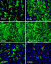
Photomicrographs of dual staining for GFAP immunofluorescence (green) and OX-42 (green) with Hoechst 33342 (blue). (a) Fed with CM for four days. Cytoplasm (green, arrow) and non-astrocytes (blue, arrowhead). (b) CM cultures at 12 days. (c), (d): MCM or CM over 12 days, respectively, after having been precultured in MCM for 12 days. Bar = 200 μm. (e), (f) Fed with CM or MCM over 12 days. The positive double staining for OX-42 and Hoechst 33342 were microglial cells (arrow). Non-microglial cells only labeled in nucleus (arrowheads). Bar = 250 μm
Fig. 4
Fig. 4
Effect of MCM on proliferation and viability of astrocytes subcultured with MCM for different culture durations. ** P < 0.01 μm, * P < 0.05 Fed with MCM vs. with CM at same time point
Fig. 5
Fig. 5
Photomicrographs of formation of cell color-dense formazan from MTT assay. (a) Fed with CM for 10 days. (b) Fed with MCM for 10 days. Bar = 200 μm

Similar articles

Cited by

References

    1. Allan S. The neurovascular unit and the key role of astrocytes in the regulation of cerebral blood flow. Cerebrovasc Dis. 2005;21:137–138. doi: 10.1159/000090447. - DOI - PubMed
    1. Aronica E, Catania MV, Geurts J, Yankaya J, Troost D. Immunohistochemical localization of group I and II metabotropic glutamate receptors in control and amyotrophic lateral sclerosis human spinal cord: upregulation in reactive astrocytes. Neurosci. 2001;105:509–520. doi: 10.1016/S0306-4522(01)00181-6. - DOI - PubMed
    1. Aronica E, Gorter JA, Ijlst-Keizers H, Rozemuller AJ, Yankaya B, Leonstra Troost D. Expression and functional role of mGluR3 and mGluR5 in human astrocytes and glioma cells: opposite regulation of glutamate transporter proteins. Eur J Neurosci. 2003;17:1–13. doi: 10.1046/j.1460-9568.2003.02657.x. - DOI - PubMed
    1. Avola R, Condorelli DF, Surrentino S, Turpeenoja L, Costa A, Giuffrida Stella AM. Effect of epidermal growth factor and insulin on DNA, RNA, and cytoskeletal protein labeing in parmary rat astrocyglial cell cultures. J Neurosci Res. 1988;19:230–238. doi: 10.1002/jnr.490190208. - DOI - PubMed
    1. Avola R, Reale S, Costa A, Insirello L, Spina-Purrello V, Giuffrida-Stella AM. Effects of bFGF and IGF-I on polyadenylated RNA and non-histone chromosomal protein labeling in cultured astrocytes. J Neurochem. 1993;61:200–210. doi: 10.1111/j.1471-4159.1993.tb03556.x. - DOI - PubMed