. 2006 Oct;52(2):125-37.
doi: 10.1007/s10616-007-9043-x. Epub 2007 Feb 2.

Isolation of progenitor cells from cord blood using adhesion matrices

Affiliations

Isolation of progenitor cells from cord blood using adhesion matrices

Sarah Maurice et al. Cytotechnology. 2006 Oct.

Erratum in

Abstract

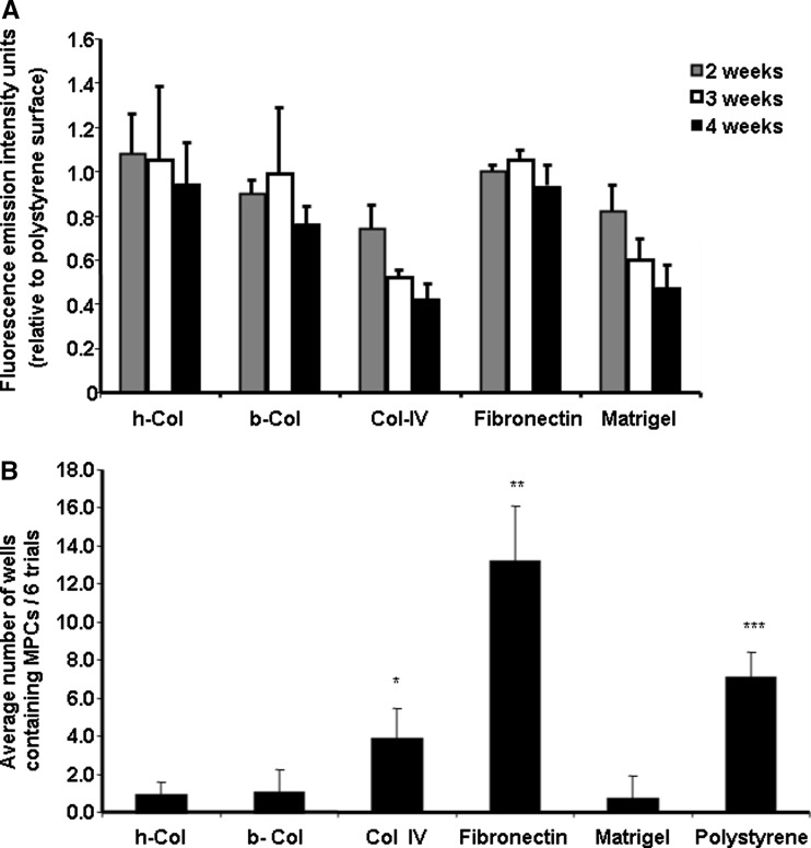
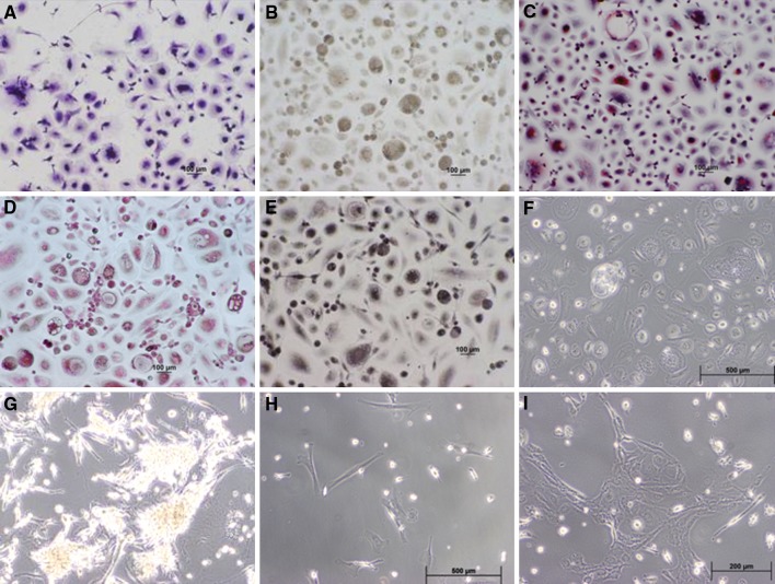
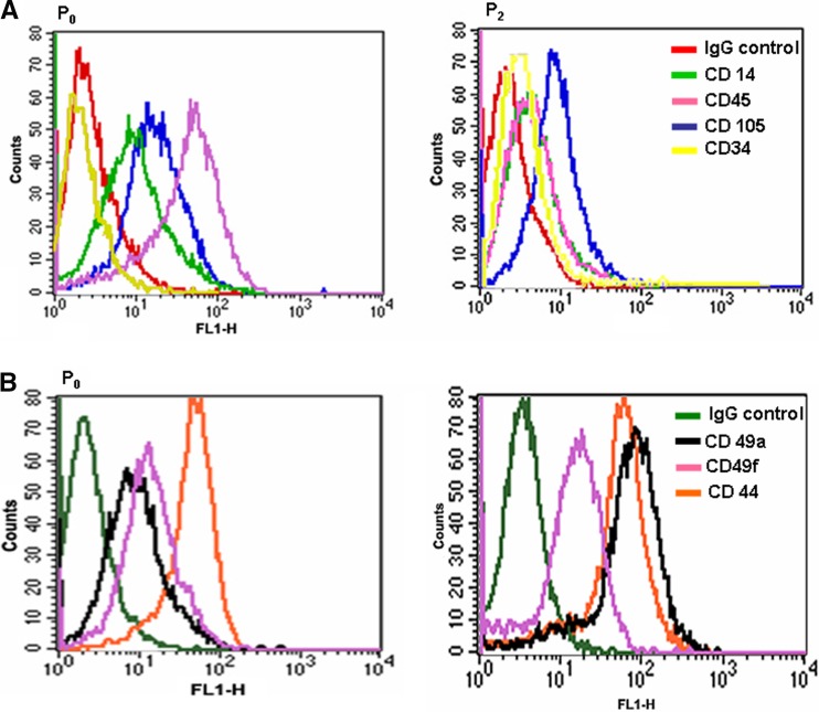
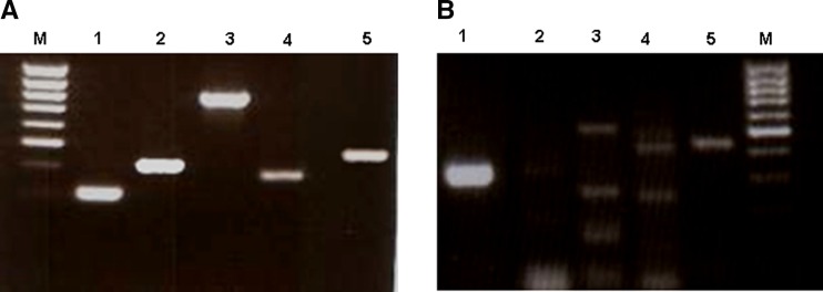
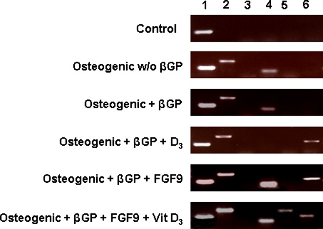
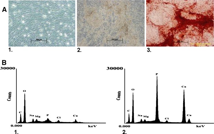
The aim of this study was to develop optimal conditions for selective adhesion and isolation of mesenchymal progenitor cells (PCs) from cord blood and to determine their potential for osteogenic differentiation. Mononuclear cells (MNCs) were isolated by Ficoll-Paque gradient and plated onto 48-well culture plates precoated with: human or bovine collagen type I, human collagen type IV, fibronectin or matrigel. Cultures were incubated in alphaMEM containing fetal calf serum. Viability of the adherent cells was determined by alamarBlue(R) assay after 2, 3, and 4 weeks. After 4 weeks in culture, cells were typsinized and replated. Primary cultures were analyzed by histochemistry and third passage cells by FACS. Isolated fibroblast-like cells were cultured in the presence of osteogenic factors and differentiation determined by Alizarin Red S staining, RT-PCR and electron dispersive spectroscopy (EDS). MNCs adhered to all types of matrices with the greatest adhesion rates on fibronectin. These cells were CD45(+), CD105(+), CD14(+), CD49a(+), CD49f(+), CD44(+) and CD34(-). The highest incidence of PCs was observed on fibronectin and polystyrene. Passages were CD45(-), CD14(-), CD34(-) and weakly CD105(+). Primary cultures expressed endothelial/macrophage RNA markers whether cultured on fibronectin or polystyrene and these markers decreased upon passage. The best osteogenic differentiation was observed in MPCs cultured in osteogenic medium containing Vit D(3) and FGF9. These cells expressed the bone-related mRNA, collagen type I, core binding factor I (Cbfa I), osteocalcin and osteopontin. EDS of deposits produced by these cells demonstrated a calcium/phosphate ratio parallel to hydroxyapatite. It was concluded that fibronectin increased adhesion rates and isolation potential of cord blood mesenchymal progenitor cells.


Figures

Fig. 1
Fig. 1
(a) Relative percentage of cord blood cells which adhered to coated plates. Mononuclear cord blood cells were isolated by Ficoll-Paque gradient and seeded onto 48-well polystyrene culture plates or the same plates pre coated with either h-Col I, b-Col I, human Col IV, fibronectin or matrigel. The presence of live adherent cells was determined 2, 3 and 4 weeks post seeding employing the alamarBlue™ assay and results normalized relative to the level of cell adhesion on uncoated polystyrene culture wells. Redox levels were determined fluorometrically at excitation 530 nm and emission 590 nm with an internal gain of 10. Results are noted as fluorescence emission intensity units and represent the average numbers from six separate experiments with 18 replicates/experiment ± standard deviation. (b) Percentage of PC isolations on different adhesion proteins. Adherent cells were trypsinized from P0 cultures and replated onto matrix coated plates. The number of wells in which PCs developed were counted and displayed on a bar graph. Each bar represents the average number of wells in which PCs developed in all six trials ± standard deviation. Significant differences in PC isolation were observed between protein coatings with fibronectin > polystyrene > collagen type IV > human collagen type I (< 0.05, CI 95)
Fig. 2
Fig. 2
Morphology and histochemistry of P0 adherent cells. Cord blood cells which adhered to polystyrene surfaces for 2 weeks were stained with (a) Giemsa; (b) Alkaline phosphatase; (c) Tartrate-resistant acid phosphatase; (d) Napthol AS-D chloroacetate; (e) α-napthyl acetate esterase; (f) P0 culture on polystyrene (unstained); (g) Morphology of clusters which developed on matrigel coated plates; (h) P1 culture; (i) P2 culture. (Magnification 100×) Two major types; elongated thin cells carrying a funnel-like cytoplasmic extension containing a small but distinct nucleus and larger cells containing cytoplasm surrounding a central nucleus were observed. The rounded population was weakly alkaline phosphates positive, TRAP reactive as well as NCAE and NAE positive. The fibroblast-like elongated cells were mostly alkaline and TRAP negative and weakly NAE and NCAE positive
Fig. 3
Fig. 3
FACS analysis of P0 and P2 cultures employing markers (a) CDs 14, 105, 45, 34; (b) 49a, 49f, and 44. FACS analysis determined that the majority of P0 cells were of the macrophage/monocyte lineage while P3 cultures demonstrated typical mesenchymal markers
Fig. 4
Fig. 4
RT-PCR of cord blood cells cultured on polystyrene plates. 5a P0 cord blood culture after 2 weeks adhesion: left to right: M 100 bp marker (top to bottom 1,031, 900, 800, 700, 600, 500, 400, 300, 200 bp); 1 GAPDH; 2 VEGFR1; 3 VEGFR2; 4 TIE2; 5 eNOS. 5b P1 cord blood culture (PCs) after 2 weeks in culture: 1 GAPDH; 2 VEGFR1; 3 VEGRF2; 4 TIE2; 5 eNOS; M marker (top to bottom 1,031, 900, 800, 700, 600, 500, 400, 300,200 bp). Primary cord blood cultures expressed RNA sequences for VEGFR1, VEGFR2, TIE2 and E-nos (a) while only eNOS was weakly observed in the P1 passage of the same cells (b)
Fig. 5
Fig. 5
RT-PCR of PCs (P2) cultured for 18 days in standard medium and in medium designed for osteogenic differentiation. 1 GAPDH; 2 collagen type I; 3 BSP; 4 CBFAα1; 5 osteocalcin; 6 osteopontin. (w/o without; βGP β-glycerophosphate)
Fig. 6
Fig. 6
PCs were cultured in standard medium or osteogenic medium containing both FGF9 and Vit D3 for a period of 21 days. The presence of calcium deposits was determined by Alizarin Red S and Electron Dispersive Spectroscopy. (a1) PCs cultured in standard medium; (a2) PCs cultured in osteogenic medium containing FGF9 and Vit D3 demonstrating calcium deposits; (a3) Alizarin Red staining culture observed in (a2). Red staining indicates presence of calcium deposits. (b) EDS analysis of culture (a1) and (a2). (b1) EDS of control culture (a1) with very minimal presence of calcium and phosphate; (b2) EDS of osteogenic culture (a2) with strong a signal for phosphate and calcium demonstrating a ratio similar to hydroxyapatite

Similar articles

Cited by

References

    1. Arinzeh TL. Mesenchymal stem cells for bone repair: preclinical studies and potential orthopedic applications. Foot Ankle Clin. 2005;10(4):651–665. doi: 10.1016/j.fcl.2005.06.004. - DOI - PubMed
    1. Bellows CG, Heersche JN, Aubin JE. Inorganic phosphate added exogenously or released from beta-glycerophosphate initiates mineralization of osteoid nodules in vitro. Bone Miner. 1992;17(1):15–29. doi: 10.1016/0169-6009(92)90707-K. - DOI - PubMed
    1. Bhagavati S, Xu W. Isolation and enrichment of skeletal muscle progenitor cells from mouse bone marrow. Biochem Biophys Res Commun. 2004;318(1):119–124. doi: 10.1016/j.bbrc.2004.03.192. - DOI - PubMed
    1. Bieback K, Kern S, Kluter H, et al. Critical parameters for the isolation of mesenchymal stem cells from umbilical cord blood. Stem Cells. 2004;22(4):625–634. doi: 10.1634/stemcells.22-4-625. - DOI - PubMed
    1. Bruder SP, Fink DJ, Caplan AI. Mesenchymal stem cells in bone development, bone repair, and skeletal regeneration therapy. J Cell Biochem. 1994;56(3):283–294. doi: 10.1002/jcb.240560303. - DOI - PMC - PubMed