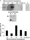
Expression of the hepatitis B virus surface antigen in Drosophila S2 cells
- PMID: 19003172
- PMCID: PMC2553638
- DOI: 10.1007/s10616-008-9154-z
Expression of the hepatitis B virus surface antigen in Drosophila S2 cells
Abstract
Drosophila melanogaster S2 cells were transfected with a plasmid vector (pAcHBsAgHy) containing the S gene, coding for the hepatitis B virus surface antigen (HBsAg), under control of the constitutive drosophila actin promoter (pAc), and the hygromycin B (Hy) selection gene. The vector was introduced into Schneider 2 (S2) Drosophila cells by DNA transfection and a cell population (S2AcHBsAgHy) was selected by its resistance to hygromycin B. The pAcHBsAgHy vector integrated in transfected S2 cell genome and approximately 1,000 copies per cell were found in a higher HBsAg producer cell subpopulation. The HBsAg production varied in different subpopulations, but did not when a given subpopulation was cultivated in different culture flasks. Higher HBsAg expression was found in S2AcHBsAgHy cells cultivated in Insect Xpress medium (13.5 mug/1E7 cells) and SFX medium (7 mug/1E7 cells) in comparison to SF900II medium (0.6 mug/1E7 cells). An increase of HBsAg was observed in culture maintained under hygromycin selection pressure. Data presented in the paper show that S2AcHBsAgHy cells produce efficiently the HBsAg which is mainly found in the cell supernatant, suggesting that HBsAg is secreted from the cells. The data also show that our approach using the Drosophila expression system is suitable for the preparation of other viral protein preparation.
Figures


Similar articles
-
High level expression of hepatitis B virus surface antigen in stably transfected Drosophila Schneider-2 cells.J Virol Methods. 1999 May;79(2):191-203. doi: 10.1016/s0166-0934(99)00021-x. J Virol Methods. 1999. PMID: 10381089
-
Expression of the hepatitis B virus surface antigen in mammalian cells using an Epstein-barr-virus-derived vector.Appl Microbiol Biotechnol. 1996 Dec;46(5-6):533-7. doi: 10.1007/s002530050856. Appl Microbiol Biotechnol. 1996. PMID: 9008886
-
Behavior of wild-type and transfected S2 cells cultured in two different media.Appl Biochem Biotechnol. 2011 Jan;163(1):1-13. doi: 10.1007/s12010-010-8918-z. Epub 2010 Dec 15. Appl Biochem Biotechnol. 2011. PMID: 21161610
-
Identification of a pre-S2 mutant in hepatocytes expressing a novel marginal pattern of surface antigen in advanced diseases of chronic hepatitis B virus infection.J Gastroenterol Hepatol. 2000 May;15(5):519-28. doi: 10.1046/j.1440-1746.2000.02187.x. J Gastroenterol Hepatol. 2000. PMID: 10847439 Review.
-
DNA-mediated immunization to the hepatitis B surface antigen. Activation and entrainment of the immune response.Ann N Y Acad Sci. 1995 Nov 27;772:64-76. doi: 10.1111/j.1749-6632.1995.tb44732.x. Ann N Y Acad Sci. 1995. PMID: 8546414 Review.
Cited by
-
Single-cell cloning enables the selection of more productive Drosophila melanogaster S2 cells for recombinant protein expression.Biotechnol Rep (Amst). 2018 Jul 3;19:e00272. doi: 10.1016/j.btre.2018.e00272. eCollection 2018 Sep. Biotechnol Rep (Amst). 2018. PMID: 29998071 Free PMC article.
-
Expression of recombinant Atlantic salmon serum C-type lectin in Drosophila melanogaster Schneider 2 cells.Cytotechnology. 2013 Aug;65(4):513-21. doi: 10.1007/s10616-012-9505-7. Epub 2012 Oct 18. Cytotechnology. 2013. PMID: 23076800 Free PMC article.
-
Human pathogenic bacteria, fungi, and viruses in Drosophila: disease modeling, lessons, and shortcomings.Virulence. 2014 Feb 15;5(2):253-69. doi: 10.4161/viru.27524. Epub 2014 Jan 7. Virulence. 2014. PMID: 24398387 Free PMC article. Review.
References
-
- {'text': '', 'ref_index': 1, 'ids': [{'type': 'DOI', 'value': '10.1093/nar/19.18.5037', 'is_inner': False, 'url': 'https://doi.org/10.1093/nar/19.18.5037'}, {'type': 'PMC', 'value': 'PMC328807', 'is_inner': False, 'url': 'https://pmc.ncbi.nlm.nih.gov/articles/PMC328807/'}, {'type': 'PubMed', 'value': '1656386', 'is_inner': True, 'url': 'https://pubmed.ncbi.nlm.nih.gov/1656386/'}]}
- Angelichio ML, Beck JA, Johansen H, Ivey-Hole M (1991) Comparison of several promoters and polyadenylation signals for use in heterologous gene expression in cultured Drosophila cells. Nucleic Acids Res 19:5037–5043 - PMC - PubMed
-
- {'text': '', 'ref_index': 1, 'ids': [{'type': 'DOI', 'value': '10.1002/biot.200700179', 'is_inner': False, 'url': 'https://doi.org/10.1002/biot.200700179'}, {'type': 'PubMed', 'value': '18064610', 'is_inner': True, 'url': 'https://pubmed.ncbi.nlm.nih.gov/18064610/'}]}
- Astray RM, Augusto E, Yokomizo AY, Pereira CA (2008) Analytical approach for extraction of recombinant membrane viral glycoprotein from stably transfected Drosophila melanogaster cells. Biotechnol J 3(1):98–103 - PubMed
-
- {'text': '', 'ref_index': 1, 'ids': [{'type': 'DOI', 'value': '10.1073/pnas.80.10.3015', 'is_inner': False, 'url': 'https://doi.org/10.1073/pnas.80.10.3015'}, {'type': 'PMC', 'value': 'PMC393964', 'is_inner': False, 'url': 'https://pmc.ncbi.nlm.nih.gov/articles/PMC393964/'}, {'type': 'PubMed', 'value': '6574469', 'is_inner': True, 'url': 'https://pubmed.ncbi.nlm.nih.gov/6574469/'}]}
- Calos MP, Lebkowski JS, Botchan MR (1983) High mutation frequency in DNA transfected into mammalian cells. Proc Natl Acad Sci USA 80(10):3015–3019 - PMC - PubMed
-
- {'text': '', 'ref_index': 1, 'ids': [{'type': 'DOI', 'value': '10.1016/0378-1119(84)90194-X', 'is_inner': False, 'url': 'https://doi.org/10.1016/0378-1119(84)90194-x'}, {'type': 'PubMed', 'value': '6098537', 'is_inner': True, 'url': 'https://pubmed.ncbi.nlm.nih.gov/6098537/'}]}
- Carloni G, Malpièce Y, Michel ML et al (1984) A transformed Vero cell line stably producing the hepatitis B virus surface antigen. Gene 31(1–3):49–57 - PubMed
-
- {'text': '', 'ref_index': 1, 'ids': [{'type': 'PubMed', 'value': '1806025', 'is_inner': True, 'url': 'https://pubmed.ncbi.nlm.nih.gov/1806025/'}]}
- Chen ZH, Shi YA, Ding JC (1991) Semi-continuous microcarrier culture of rCHO cells secreting HBsAg by feeding microcarriers. Chin J Biotechnol 7(2):153–159 - PubMed
LinkOut - more resources
Full Text Sources

