. 2008 Oct;14(5):376-88.
doi: 10.1080/13550280802199898. Epub 2008 Nov 12.

Mechanisms of minocycline-induced suppression of simian immunodeficiency virus encephalitis: inhibition of apoptosis signal-regulating kinase 1

Affiliations

Mechanisms of minocycline-induced suppression of simian immunodeficiency virus encephalitis: inhibition of apoptosis signal-regulating kinase 1

Susan C Follstaedt et al. J Neurovirol. 2008 Oct.

Abstract

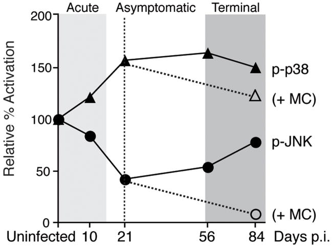
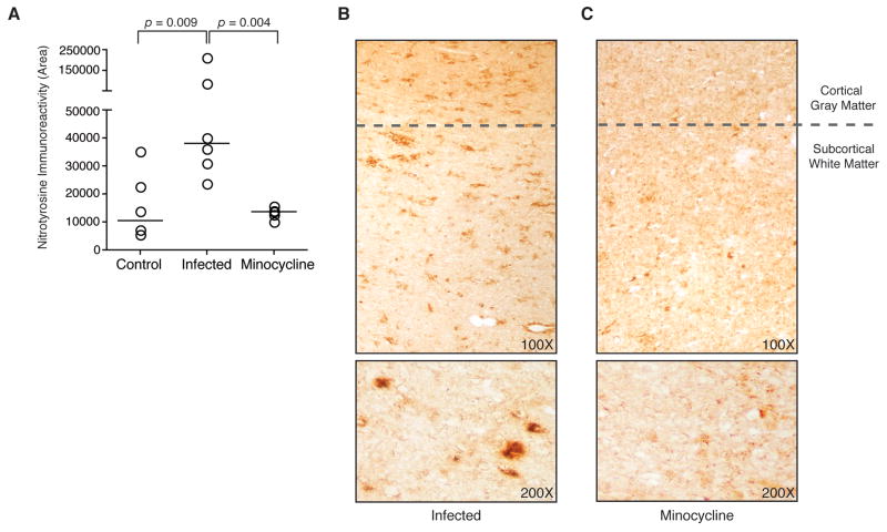
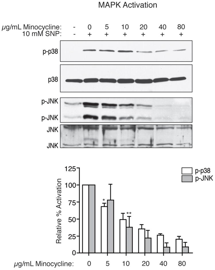
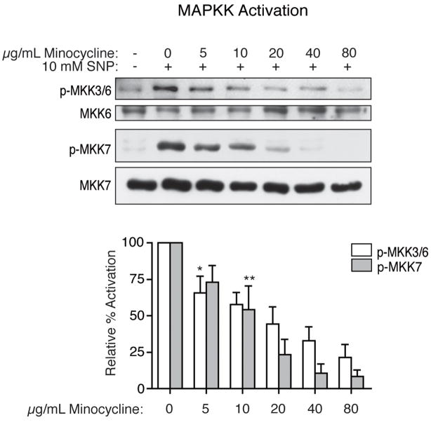
Human immunodeficiency virus (HIV) infection of the central nervous system (CNS) can lead to cognitive dysfunction, even in individuals treated with highly active antiretroviral therapy. Using an established simian immunodeficiency virus (SIV)/macaque model of HIV CNS disease, we previously reported that infection shifts the balance of activation of mitogen-activated protein kinase (MAPK) signaling pathways in the brain, resulting in increased activation of the neurodegenerative MAPKs p38 and JNK. Minocycline treatment of SIV-infected macaques reduced the incidence and severity of SIV encephalitis in this model, and suppressed the activation of p38 in the brain. The purpose of this study was to further examine the effects of minocycline on neurodegenerative MAPK signaling. We first demonstrated that minocycline also decreases JNK activation in the brain and levels of the inflammatory mediator nitric oxide (NO). We next used NO to activate these MAPK pathways in vitro, and demonstrated that minocycline suppresses p38 and c-Jun N-terminal kinase (JNK) activation by reducing intracellular levels, and hence, activation of apoptosis signal-regulating kinase 1 (ASK1), a MAPK kinase capable of selectively activating both pathways. We then demonstrated that ASK1 activation in the brain during SIV infection is suppressed by minocycline. By suppressing p38 and JNK activation pathways, which are important for the production of and responses to inflammatory mediators, minocycline may interrupt the vicious cycle of inflammation that both results from, and promotes, virus replication in SIV and HIV CNS disease.


Figures

Figure 1
Figure 1
Schematic of changes in p38 and JNK activation seen in the brain longitudinally throughout SIV infection, as determined by quantitative immunohistochemistry in subcortical white matter. Solid lines depict mean immunohistochemical data obtained from multiple SIV-infected macaques at each time point and presented as percent activation relative to mean uninfected levels (data adapted from Barber et al, 2004). Dashed lines indicate decreased levels of active phoshpo-p38 (Zink et al, 2005) and phospho-JNK observed terminally at 84 days post-infection (p.i.) in minocycline-treated SIV-infected animals (+MC; treatment initiated at 21 days p.i.).
Figure 2
Figure 2
Quantitative immunohistochemical detection of phospho-JNK in macaque brain sections. (A) Staining for phospho-JNK in subcortical white matter was quantitated in samples from uninfected control animals (n = 4), SIV-infected animals (n = 6), and SIV-infected minocycline-treated animals (n = 5). Each data point represents the mean of 20 repeated measures of phospho-JNK staining in adjacent fields (bars represent group medians). Levels of active phospho-JNK in control animals were not significantly different from levels in SIV-infected untreated animals (p = 0.171; Mann-Whitney test), while SIV-infected minocycline-treated animals had significantly lower levels of phospho-JNK (p = 0.004; Mann-Whitney test). Staining for phospho-JNK with hematoxylin counterstaining to aid visualization (top) is shown in representative brain sections from an SIV-infected untreated animal (B) and an SIV-infected minocycline-treated animal (C). Representative staining present in subcortical white matter, without counterstain, as quantitated for (A) is shown at increased magnification (bottom). Prominent axonal staining is seen in untreated animals, and this is diminished in minocycline-treated animals. Original magnifications 100x and 200x.
Figure 3
Figure 3
Quantitative immunohistochemical detection of nitrotyrosine in macaque brain sections. (A) Staining for nitrotyrosine in subcortical white matter was quantitated in samples from uninfected control animals (n = 6), SIV-infected untreated animals (n = 6), and SIV-infected minocycline-treated animals (n = 5) (bars represent group medians). Samples evaluated are serial sections of the same tissue from each animal stained for phospho-JNK in Figure 2. Each data point represents the mean of 20 repeated measures of nitrotyrosine staining in adjacent fields from one sample. Levels of nitrotyrosine staining were significantly increased from control at terminal infection (p = 0.009; Mann-Whitney test). SIV-infected minocycline-treated animals had significantly lower levels of nitrotyrosine immunopositive staining (p = 0.004; Mann-Whitney test). Staining for nitrotyrosine is shown in representative brain sections (top) from an untreated SIV-infected animal (B) and an SIV-infected minocycline-treated animal (C). Representative staining present in subcortical white matter, as quantitated in (A), is also shown at increased magnification (bottom). Original magnifications 100x and 200x.
Figure 4
Figure 4
Inhibition by minocycline of activation of p38 and JNK MAPKs with SNP treatment in differentiated U937 cells. After four hours of treatment with 10 mM SNP, both p38 and JNK were activated, as indicated by phospho-specific western blot analysis (representative blots shown). Pretreatment for four hours with minocycline led to a dose-dependent reduction in the activation of p38 and JNK by SNP. Western blot analyses for total p38 and JNK expression also are shown. Results are the means of four independent experiments, with error bars illustrating the SEM. Densitometric analysis was performed with Kodak MI software. Values are presented as percent activation relative to activated samples (SNP treated, no minocycline). A one-sample t-test was performed to compare the mean activation level of each treatment group to 100%. Asterisks represent the lowest dose at which a statistically significant decrease in activation was observed compared to SNP stimulation (*p = 0.008, **p = 0.018); all higher doses also resulted in significant decreases.
Figure 5
Figure 5
Inhibition by minocycline of the activation of MKK3/6 and MKK7 in vitro. After four hours of treatment with 10 mM SNP, MKK3/6 and MKK7 were activated, as indicated by phospho-specific western blot analysis (representative blots shown). Pretreatment for four hours with minocycline prior to the addition of SNP led to a decrease in the activation of these MAPKKs. Western blot analyses for total MKK6 and MKK7 expression also are shown. Results are the means of four independent experiments, with error bars representing the SEM. Values illustrated are percent activation relative to activated samples (SNP treated, no minocycline). A one-sample t-test was performed to compare the mean activation level of each treatment group to 100%. Asterisks represent the lowest dose at which a statistically significant decrease in activation was seen compared to SNP stimulation (*p = 0.040, **p = 0.006); all higher doses also resulted in significant decreases.
Figure 6
Figure 6
Effects of minocycline on activation and expression of ASK1 in vitro. (A) ASK1 was activated after three hours of SNP treatment, as shown by an increase in phosphorylation of MBP in in vitro kinase assay. With minocycline pretreatment, decreased phosphorylation of MBP by immunoprecipitated ASK1 was observed (*p = 0.033 for 5 μg/mL, all higher doses also resulted in significant decreases; one sample t-test). A corresponding dose-dependent decrease in the amount of ASK1 immunoprecipitated from minocycline-treated cells was quantitated in metabolic labeling experiments (*p = 0.035 for 5 μg/mL, all higher doses also resulted in significant decreases; one sample t-test) (B) Western blot analysis for ASK1 illustrates a dose-dependent decrease in total ASK1 present in whole cell lysates, normalized to actin expression (*p = 0.050 for 5 μg/mL, all higher doses also resulted in significant decreases; one-sample t-test). (C) RT-PCR for ASK1 normalized to GAPDH showed no decrease in levels of ASK1 transcript produced with minocycline treatment (p > 0.05 for all comparisons; one sample t-test). All results are representative of three or four independent experiments, with error bars representing the SEM. Values shown are percent activation or expression relative to activated samples (SNP treated, no minocycline).
Figure 7
Figure 7
Activation and expression of ASK1 in the macaque brain. (A) Active phospho-Thr845 ASK1 in macaque brain homogenates as observed by western blot analysis after immunoprecipitation of ASK1. The total levels of ASK1 immunoprecipitated for each animal also are shown. Determination of a ratio of phospho-ASK1 to total ASK1 for each animal from densitometric analysis of bands allowed for the comparison of relative levels of ASK1 activation between animals. Activation of ASK1 was observed in SIV-infected untreated animals, and this was significantly decreased by minocycline treatment (p = 0.004; Mann-Whitney test; results are the means of values obtained in duplicate blots). (B) ASK1 expression in white matter brain homogenate. By western blot analysis (performed in duplicate), there was no significant difference in relative ASK1 expression between any of the treatment groups (p > 0.05; Mann-Whitney tests). Using immunohistochemistry, total ASK1 expression was observed clearly in axons (C), as well as in astrocytes (as shown by colocalization with GFAP, D) in the subcortical white matter of all macaques, regardless of group. ASK1 expression also was observed in activated microglia and macrophages when they were detected in sites of inflammation (as shown by colocalization with CD68, E). Original magnification 200x.

Similar articles

Cited by

References

    1. Akterin S, Cowburn RF, Miranda-Vizuete A, Jimenez A, Bogdanovic N, Winblad B, Cedazo-Minguez A. Involvement of glutaredoxin-1 and thioredoxin-1 in beta-amyloid toxicity and Alzheimer’s disease. Cell Death Differ. 2006;13:1454–65. - PubMed
    1. Ances BM, Ellis RJ. Dementia and neurocognitive disorders due to HIV-1 infection. Semin Neurol. 2007;27:86–92. - PubMed
    1. Barber SA, Uhrlaub JL, DeWitt JB, Tarwater PM, Zink MC. Dysregulation of mitogen-activated protein kinase signaling pathways in simian immunodeficiency virus encephalitis. Am J Pathol. 2004;164:355–62. - PMC - PubMed
    1. Borderie D, Hernvann A, Hilliquin P, Lemarchal H, Kahan A, Ekindjian OG. Tetracyclines inhibit nitrosothiol production by cytokine-stimulated osteoarthritic synovial cells. Inflamm Res. 2001;50:409–14. - PubMed
    1. Borsello T, Forloni G. JNK signalling: a possible target to prevent neurodegeneration. Curr Pharm Des. 2007;13:1875–86. - PubMed

Publication types

MeSH terms