

ADAM8 is selectively up-regulated in endothelial cells and is associated with angiogenesis after spinal cord injury in adult mice
- PMID: 19003792
- PMCID: PMC2746684
- DOI: 10.1002/cne.21902
ADAM8 is selectively up-regulated in endothelial cells and is associated with angiogenesis after spinal cord injury in adult mice
Abstract
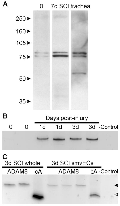
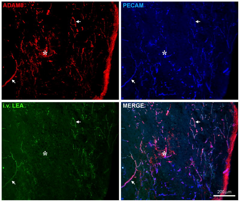
Endothelial cell (EC) loss and subsequent angiogenesis occur over the first week after spinal cord injury (SCI). To identify molecular mechanisms that could be targeted with intravenous (i.v.) treatments, we determined whether transmembrane "a disintegrin and metalloprotease" (ADAM) proteins are expressed in ECs of the injured spinal cord. ADAMs bind to integrins, which are important for EC survival and angiogenesis. Female adult C57Bl/6 mice with a spinal cord contusion had progressively more ADAM8 (CD156) immunostaining in blood vessels and individual ECs between 1 and 28 days following injury. Uninjured spinal cords had little ADAM8 staining. The increase in ADAM8 mRNA and protein was confirmed in spinal cord lysates, and ADAM8 mRNA was present in FACS-enriched ECs. ADAM8 colocalized extensively and exclusively with the EC marker PECAM and also with i.v.-injected lectins. Intravenous isolectin B4 (IB4) labels a subpopulation of blood vessels at and within the injury epicenter 3-7 days after injury, coincident with angiogenesis. Both ADAM8 and the proliferation marker Ki-67 were present in IB4-positive microvessels. ADAM8-positive proliferating cells were seen at the leading end of IB4-positive blood vessels. Angiogenesis was confirmed by BrdU incorporation, binding of i.v.-injected nucleolin antibodies, and MT1-MMP immunostaining in a subset of blood vessels. These data suggest that ADAM8 is vascular selective and plays a role in proliferation and/or migration of ECs during angiogenesis following SCI.
Figures


Similar articles
-
Blockade of the ADAM8-Fra-1 complex attenuates neuroinflammation by suppressing the Map3k4/MAPKs axis after spinal cord injury.Cell Mol Biol Lett. 2024 May 16;29(1):75. doi: 10.1186/s11658-024-00589-3. Cell Mol Biol Lett. 2024. PMID: 38755530 Free PMC article.
-
Griffonia simplicifolia isolectin B4 identifies a specific subpopulation of angiogenic blood vessels following contusive spinal cord injury in the adult mouse.J Comp Neurol. 2008 Mar 1;507(1):1031-52. doi: 10.1002/cne.21570. J Comp Neurol. 2008. PMID: 18092342 Free PMC article.
-
Developmental and injury-induced expression of alpha1beta1 and alpha6beta1 integrins in the rat spinal cord.Brain Res. 2007 Jan 26;1130(1):54-66. doi: 10.1016/j.brainres.2006.10.072. Epub 2006 Dec 11. Brain Res. 2007. PMID: 17161391 Free PMC article.
-
Physiological Insights Into the Role of Pericytes in Spinal Cord Injury.J Cell Physiol. 2025 Jan;240(1):e31500. doi: 10.1002/jcp.31500. J Cell Physiol. 2025. PMID: 39757951 Free PMC article. Review.
-
ADAM8 in invasive cancers: links to tumor progression, metastasis, and chemoresistance.Clin Sci (Lond). 2019 Jan 11;133(1):83-99. doi: 10.1042/CS20180906. Print 2019 Jan 15. Clin Sci (Lond). 2019. PMID: 30635388 Review.
Cited by
-
Matrix metalloproteinase signals following neurotrauma are right on cue.Cell Mol Life Sci. 2019 Aug;76(16):3141-3156. doi: 10.1007/s00018-019-03176-4. Epub 2019 Jun 6. Cell Mol Life Sci. 2019. PMID: 31168660 Free PMC article. Review.
-
Vascular Pathology as a Potential Therapeutic Target in SCI.Transl Stroke Res. 2011 Dec;2(4):556-74. doi: 10.1007/s12975-011-0128-7. Epub 2011 Nov 29. Transl Stroke Res. 2011. PMID: 24323683
-
Gene expression and locomotor recovery in adult rats with spinal cord injury and plasma-synthesized polypyrrole/iodine application combined with a mixed rehabilitation scheme.Front Neurol. 2023 May 23;14:1124245. doi: 10.3389/fneur.2023.1124245. eCollection 2023. Front Neurol. 2023. PMID: 37288064 Free PMC article.
-
Potential prognostic and immunologic significances of ADAM8 in clear cell renal cell carcinoma.Medicine (Baltimore). 2025 Jan 31;104(5):e41375. doi: 10.1097/MD.0000000000041375. Medicine (Baltimore). 2025. PMID: 39889162 Free PMC article.
-
ADAM8: a new therapeutic target for asthma.Expert Opin Ther Targets. 2009 May;13(5):523-40. doi: 10.1517/14728220902889788. Expert Opin Ther Targets. 2009. PMID: 19397475 Free PMC article. Review.
References
-
- Amour A, Knight CG, English WR, Webster A, Slocombe PM, Knauper V, Docherty AJ, Becherer JD, Blobel CP, Murphy G. The enzymatic activity of ADAM8 and ADAM9 is not regulated by TIMPs. FEBS Lett. 2002;524:154–158. - PubMed
-
- Baker KL, Daniels SB, Lennington JB, Lardaro T, Czap A, Notti RQ, Cooper O, Isacson O, Frasca S, Jr, Conover JC. Neuroblast protuberances in the subventricular zone of the regenerative MRL/MpJ mouse. J Comp Neurol. 2006;498:747–761. - PubMed
-
- Bell JH, Herrera AH, Li Y, Walcheck B. Role of ADAM17 in the ectodomain shedding of TNF-alpha and its receptors by neutrophils and macrophages. J Leukoc Biol. 2007;82:173–176. - PubMed
Publication types
MeSH terms
Substances
Grants and funding
LinkOut - more resources
Full Text Sources
Other Literature Sources
Medical
Molecular Biology Databases
