

Neuroprotective effects of testosterone on the morphology and function of somatic motoneurons following the death of neighboring motoneurons
- PMID: 19003970
- PMCID: PMC2592503
- DOI: 10.1002/cne.21885
Neuroprotective effects of testosterone on the morphology and function of somatic motoneurons following the death of neighboring motoneurons
Abstract
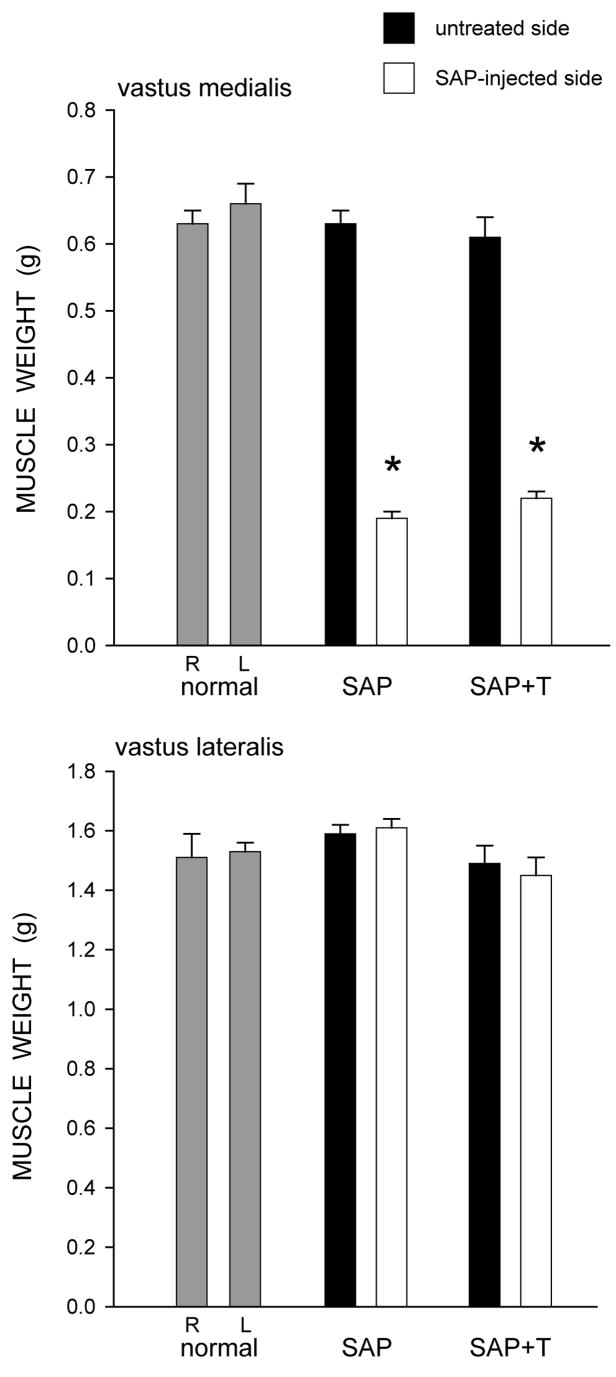
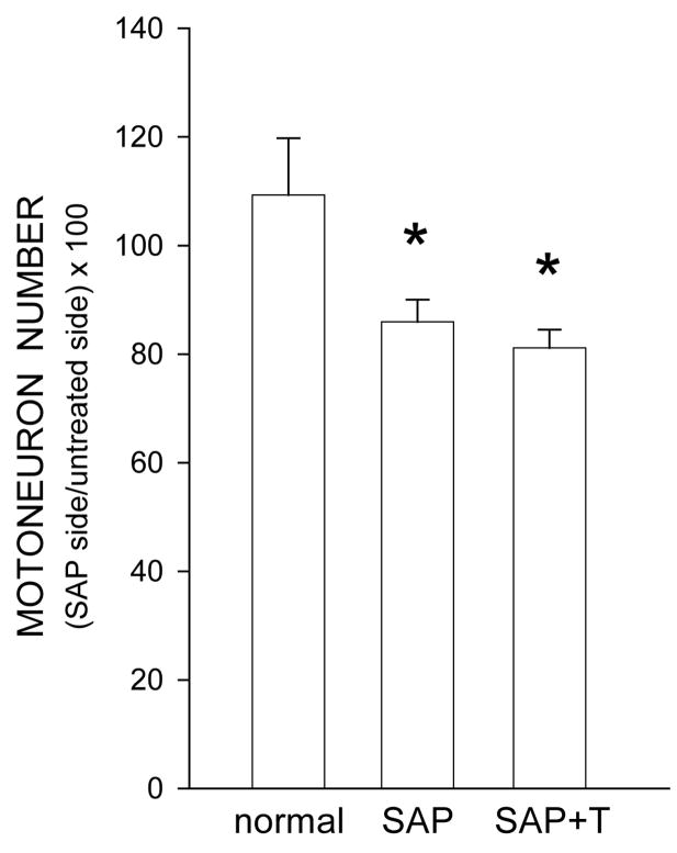
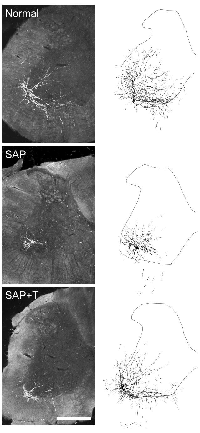
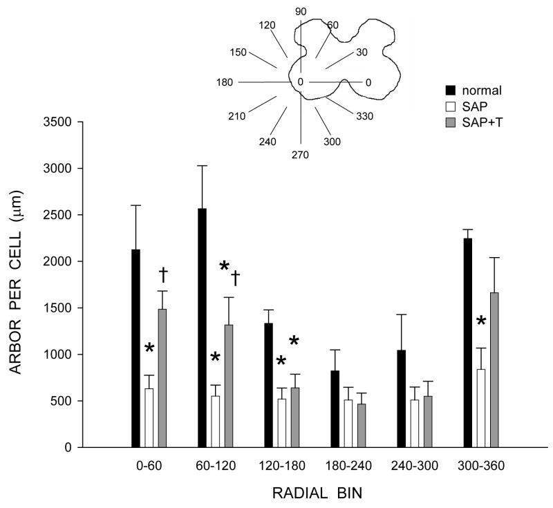
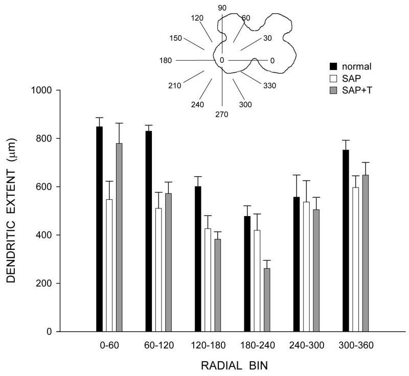
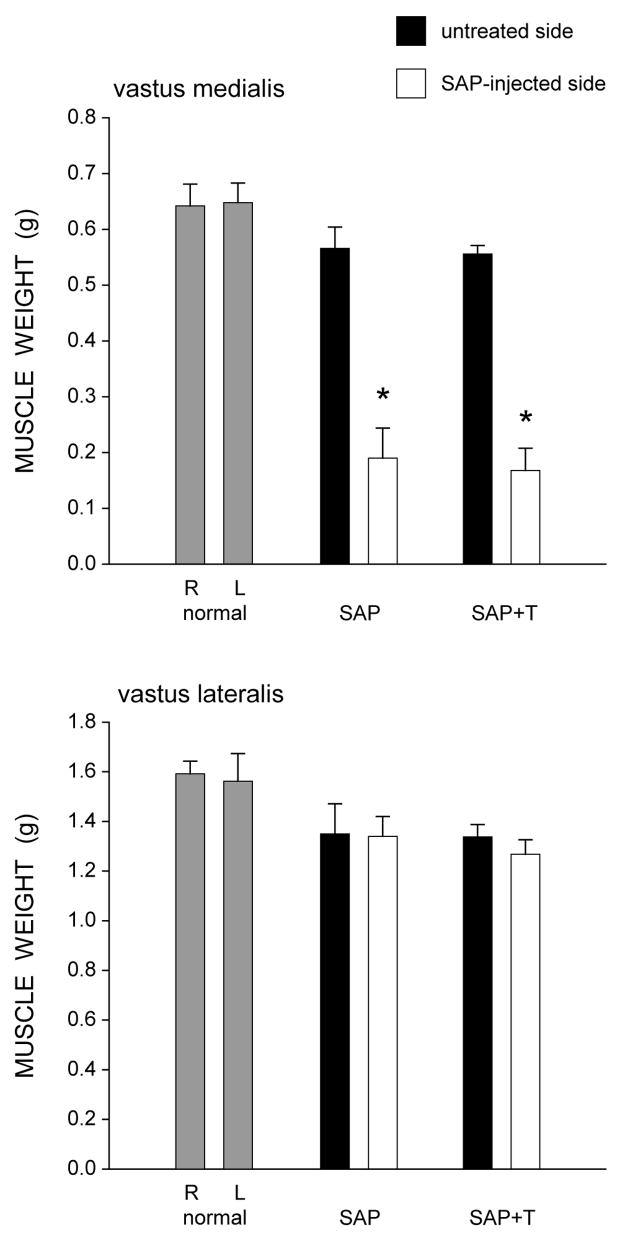
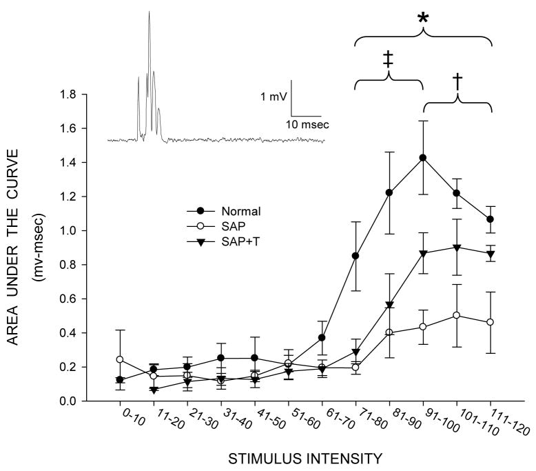
Motoneuron loss is a significant medical problem, capable of causing severe movement disorders or even death. We have previously shown that partial depletion of motoneurons from sexually dimorphic, highly androgen-sensitive spinal motor populations induces dendritic atrophy in remaining motoneurons, and this atrophy is attenuated by treatment with testosterone. To test whether testosterone has similar effects in more typical motoneurons, we examined potential neuroprotective effects in motoneurons innervating muscles of the quadriceps. Motoneurons innervating the vastus medialis muscle were selectively killed by intramuscular injection of cholera toxin-conjugated saporin. Simultaneously, some saporin-injected rats were given implants containing testosterone or left untreated. Four weeks later, motoneurons innervating the ipsilateral vastus lateralis muscle were labeled with cholera toxin-conjugated horseradish peroxidase, and dendritic arbors were reconstructed in three dimensions. Compared with intact normal males, partial motoneuron depletion resulted in decreased dendritic length in remaining quadriceps motoneurons, and this atrophy was attenuated by testosterone treatment. To examine the functional consequences of the induced dendritic atrophy, and its attenuation with testosterone treatment, the activation of remaining quadriceps motoneurons was assessed using peripheral nerve recording. Partial motoneuron depletion resulted in decreased amplitudes of motor nerve activity, and these changes were attenuated by treatment with testosterone, providing a functional correlate to the neuroprotective effects of testosterone treatment on quadriceps motoneuron morphology. Together these findings suggest that testosterone has neuroprotective effects on morphology and function in both highly androgen-sensitive as well as more typical motoneuron populations, further supporting a role for testosterone as a neurotherapeutic agent in the injured nervous system.
Figures

Similar articles
-
Neuroprotective actions of androgens on motoneurons.Front Neuroendocrinol. 2009 Jul;30(2):130-41. doi: 10.1016/j.yfrne.2009.04.005. Epub 2009 Apr 23. Front Neuroendocrinol. 2009. PMID: 19393684 Free PMC article. Review.
-
Neuroprotective effects of testosterone on dendritic morphology following partial motoneuron depletion: efficacy in female rats.Neurosci Lett. 2009 Nov 13;465(2):123-7. doi: 10.1016/j.neulet.2009.09.007. Epub 2009 Sep 6. Neurosci Lett. 2009. PMID: 19735695 Free PMC article.
-
Neuroprotective effects of testosterone metabolites and dependency on receptor action on the morphology of somatic motoneurons following the death of neighboring motoneurons.Dev Neurobiol. 2017 Jun;77(6):691-707. doi: 10.1002/dneu.22445. Epub 2016 Oct 3. Dev Neurobiol. 2017. PMID: 27569375 Free PMC article.
-
Neuroprotective Effects on the Morphology of Somatic Motoneurons Following the Death of Neighboring Motoneurons: A Role for Microglia?Dev Neurobiol. 2019 Feb;79(2):131-154. doi: 10.1002/dneu.22652. Epub 2019 Jan 7. Dev Neurobiol. 2019. PMID: 30430756 Free PMC article.
-
Neuroprotective effects of gonadal steroids on regenerating peripheral motoneurons.Brain Res Brain Res Rev. 2001 Nov;37(1-3):372-82. doi: 10.1016/s0165-0173(01)00107-2. Brain Res Brain Res Rev. 2001. PMID: 11744101 Review.
Cited by
-
Mutant androgen receptor induces neurite loss and senescence independently of ARE binding in a neuronal model of SBMA.Proc Natl Acad Sci U S A. 2024 Jul 16;121(29):e2321408121. doi: 10.1073/pnas.2321408121. Epub 2024 Jul 8. Proc Natl Acad Sci U S A. 2024. PMID: 38976730 Free PMC article.
-
Androgen regulates brain-derived neurotrophic factor in spinal motoneurons and their target musculature.Endocrinology. 2010 Jan;151(1):253-61. doi: 10.1210/en.2009-1036. Epub 2009 Oct 30. Endocrinology. 2010. PMID: 19880806 Free PMC article.
-
Protective Effects of Estradiol and Dihydrotestosterone following Spinal Cord Injury.J Neurotrauma. 2018 Mar 15;35(6):825-841. doi: 10.1089/neu.2017.5329. Epub 2018 Jan 11. J Neurotrauma. 2018. PMID: 29132243 Free PMC article.
-
Neuroprotective actions of androgens on motoneurons.Front Neuroendocrinol. 2009 Jul;30(2):130-41. doi: 10.1016/j.yfrne.2009.04.005. Epub 2009 Apr 23. Front Neuroendocrinol. 2009. PMID: 19393684 Free PMC article. Review.
-
Neuroprotective effects of testosterone on dendritic morphology following partial motoneuron depletion: efficacy in female rats.Neurosci Lett. 2009 Nov 13;465(2):123-7. doi: 10.1016/j.neulet.2009.09.007. Epub 2009 Sep 6. Neurosci Lett. 2009. PMID: 19735695 Free PMC article.
References
-
- Ahlbom E, Prins GS, Ceccatelli S. Testosterone protects cerebellar granule cells from oxidative stress-induced cell death through a receptor mediated mechanism. Brain Res. 2001;892:255–262. - PubMed
-
- Al-Majed AA, Brushart TM, Gordon T. Electrical stimulation accelerates and increases expression of BDNF and trkB rnRNA in regenerating rat femoral motoneurons. Eur J Neurosci. 2000;12:4381–4390. - PubMed
-
- Basso DM, Beattie M, Bresnahan JC. A sensitive and reliable locomotor rating scale for open field testing in rats. J Neurotrauma. 1995;12:1–21. - PubMed
-
- Bisby MA, Tetzlaff W. Changes in cytoskeletal protein synthesis following axon injury and during regeneration. Mol Neurobiol. 1992;6:107–123. - PubMed
-
- Brännström T, Havton L, Kellerth J-O. Changes in size and dendritic arborization patterns of adult cat spinal a-motoneurons following permanent axotomy. J Comp Neurol. 1992;318:439–451. - PubMed
Publication types
MeSH terms
Substances
Grants and funding
LinkOut - more resources
Full Text Sources
