. 2008 Nov 18;105(46):18012-7.
doi: 10.1073/pnas.0808417105. Epub 2008 Nov 12.

Epigenetic regulation of neural cell differentiation plasticity in the adult mammalian brain

Affiliations

Epigenetic regulation of neural cell differentiation plasticity in the adult mammalian brain

Jun Kohyama et al. Proc Natl Acad Sci U S A. .

Abstract

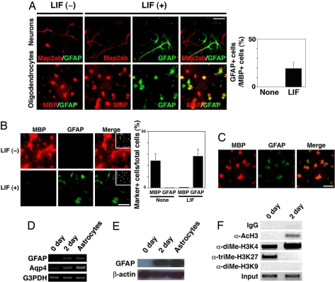
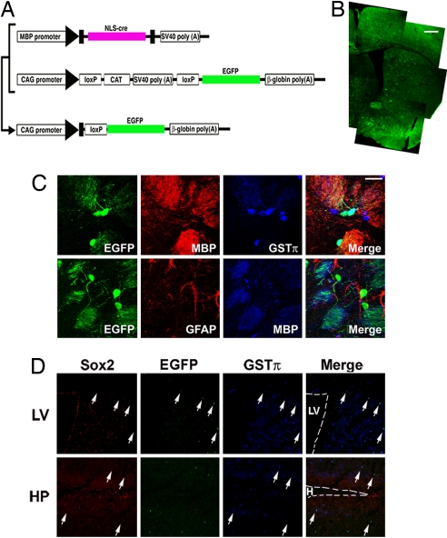
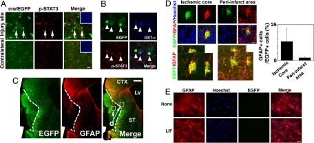
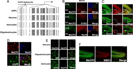
Neural stem/progenitor cells (NSCs/NPCs) give rise to neurons, astrocytes, and oligodendrocytes. It has become apparent that intracellular epigenetic modification including DNA methylation, in concert with extracellular cues such as cytokine signaling, is deeply involved in fate specification of NSCs/NPCs by defining cell-type specific gene expression. However, it is still unclear how differentiated neural cells retain their specific attributes by repressing cellular properties characteristic of other lineages. In previous work we have shown that methyl-CpG binding protein transcriptional repressors (MBDs), which are expressed predominantly in neurons in the central nervous system, inhibit astrocyte-specific gene expression by binding to highly methylated regions of their target genes. Here we report that oligodendrocytes, which do not express MBDs, can transdifferentiate into astrocytes both in vitro (cytokine stimulation) and in vivo (ischemic injury) through the activation of the JAK/STAT signaling pathway. These findings suggest that differentiation plasticity in neural cells is regulated by cell-intrinsic epigenetic mechanisms in collaboration with ambient cell-extrinsic cues.


Conflict of interest statement

The authors declare no conflict of interest.

Figures

Fig. 1.
Fig. 1.
Differentiation plasticity of oligodendrocytes. (A) AHP-derived neurons and oligodendrocytes were cultured for 2 days with or without LIF (50 ng/ml). Cells were then stained with sets of antibodies for Map2ab (Upper, red)/GFAP (Upper, green) and MBP (Bottom, red)/GFAP (Lower, green). (Scale bar: 50 μm.) The percentage of GFAP-positive cells in MBP-positive cells was quantified (Right). Data are mean ± SD. (B) Oligodendrocytes were cultured with or without LIF (50 ng/ml) for 4 days and subsequently stained for MBP (red) and GFAP (green). Insets: Hoechst nuclear staining of each field. (Scale bar: 50 μm.) Percentages of MBP- and GFAP-positive cells in total cells were quantified (Right). Data are mean ± SD. (C) GFAP-positive astrocytes appeared with LIF treatment for 2 days even when the cells were growth-arrested by aphidicolin (10 μg/ml). MBP (red), GFAP (green). (Scale bar: 50 μm.) (D) AHP-derived oligodendrocytes (0 day), oligodendrocytes incubated with LIF (50 ng/ml) for 2 days (2 day), and AHP-derived astrocytes were analyzed by RT-PCR using specific sets of primers against gfap, Aqp4 and G3PDH. (E) AHP-derived oligodendrocytes (0 day), oligodendrocytes incubated with LIF (50 ng/ml) for 2 days (2 day), and AHP-derived astrocytes were analyzed by Western blot using an antibody against GFAP. A band corresponding to the molecular weight of GFAP protein (∼50 kDa) was detected. (F) Differentiated oligodendrocytes (0 day) were incubated with LIF (50 ng/ml) for 2 days and then subjected to ChIP assay using control IgG, anti-diMe-H3K4, -diMe-H3K9, -triMe-H3K27 and -AcH3 antibodies. Co-immunoprecipitated gfap gene fragment (−18bp to + 513bp) was amplified by PCR with a specific set of primers. Nucleotide positions are those of GenBank accession number Z48978.
Fig. 2.
Fig. 2.
Oligodendrocyte-specific labeling with EGFP. (A) Schematic of transgenic constructs used to trace the lineage of oligodendrocytes. In double-transgenic mice harboring the upper two transgenes, Cre recombinase is expressed in MBP-expressing oligodendrocytes and excises the floxed CAT-SV40 poly(A) fragment, resulting in constitutive expression of EGFP under the control of the ubiquitous CAG promoter. (B) Distribution of EGFP-positive cells in the adult brain. (Scale bar: 500 mm.) (C) EGFP expression was confined to MBP- and GST-π-positive mature oligodendrocytes in the adult brain under normal conditions (Upper). Expression of EGFP and GFAP was mutually exclusive (Lower). (Scale bar: 20 μm.) (D) Expression of EGFP in neurogenic regions of the adult brain. Arrows indicate representative cells expressing EGFP and the oligodendrocyte marker GST-π. Sox2 expression was not observed in the EGFP-positive cells. LV, lateral ventricle; HP, hippocampus; H, hilus. (Scale bar: 100 mm.)
Fig. 3.
Fig. 3.
STAT activation and O-A conversion after ischemic injury. (A) Activation of the JAK/STAT pathway in EGFP-positive cells was evaluated by immunostaining using an antibody against phospo-STAT3 (p-STAT3) in the injured and contralateral sides of the striata 72 h after ischemic injury. Arrows indicate representative cells positive for both EGFP and p-STAT3. (Scale bar: 20 μm.) Insets: Hoechst nuclear staining of each field. (B) p-STAT3 was also detected in GSTπ-expressing EGFP-positive cells 72 h after injury (arrowheads). (Scale bar: 20 μm.) (C) A representative brain section stained with anti-GFP and -GFAP antibodies 2 weeks after MCAO surgery. Scale bar = 500 μm. CTX, cortex; CC, corpus callosum; LV, lateral ventricle; ST, striatum. (D) Higher magnification views of square areas in C. GFAP expression appeared in EGFP-expressing cells in the ischemic core (Left square arrowed in C, Upper Left) and peri-infarct (Right square arrowed in C, Upper Right) areas. Representative cells positive for both GFAP and EGFP are shown; Lower Right image in each group is a superimposition of the other three images. (Scale bar: 20 μm.) Three-dimensional digital images of each area are also shown (ischemic core area, Bottom Left; peri-infarct area, Bottom Right). Percentage of GFAP-positive cells in EGFP-positive cells was quantified for each area (graph). Data are mean ± SD. (E) Astrocytes prepared from adult MBP-Cre/EGFP transgenic mice were left untreated or treated with LIF for 4 days, and then stained for EGFP (green) and GFAP (red). (Scale bar: 50 μm.) EGFP expression was not induced even when the cells were stimulated with LIF.
Fig. 4.
Fig. 4.
MeCP2 is predominantly expressed in neurons and suppresses astrocytic gene expression in oligodendrocytes when ectopically expressed. (A) Schematic representation of gfap locus (Upper). Methylation status of five CpG sites around the STAT3 binding site (black box), which is critical for LIF-induced activation of the gfap promoter, and of 30 CpG sites around the transcription initiation site (arrow) were examined for each cell type by bisulfite sequencing. Open and closed circles indicate unmethylated and methylated CpG sites, respectively. (B) MeCP2 expression (green) was examined in adult mouse brain sections. Neurons, oligodendrocytes, and astrocytes are identified with specific antibodies for NeuN (red, Upper middle), GST-π (red, center), and S100β (red, Lower middle), respectively. Staining of the hippocampal dentate gyrus region is shown. Insets: higher magnification views of square areas in each field. (Scale bar: 50 μm.) (C) MeCP2 is predominantly expressed in neurons. MeCP2 expression (green) was examined in the adult mouse cortex and corpus callosum. Oligodendrocytes, astrocytes, and neurons were identified with specific antibodies for GST-π (red, Upper rows), S100β (red, middle rows), and NeuN (red, Bottom rows), respectively. Insets: Hoechst nuclear images of each field. (Scale bar: 100 μm.) CTX, cortex; CC, corpus callosum. (D) EGFP-positive cells do not express MeCP2. MeCP2 expression (Upper Left) was observed in NeuN-positive (Upper Right) but not in EGFP-positive (Lower Left) cells. (E) MeCP2 (red) is expressed in Map2ab-positive neurons (Top, green), but not in RIP-positive oligodendrocytes (middle, green) or GFAP-positive astrocytes (Bottom, green) generated from AHPs in vitro. (Scale bar: 50 μm.) (F) Redundant expression of MBDs in vivo. Expression of MeCP2 (green, Left) and MBD1 (red, middle), another member of the MBD family, was examined in the adult hippocampal region. These two MBDs showed the same expression pattern (Right). (Scale bar: 100 μm.)
Fig. 5.
Fig. 5.
MeCP2 expression is sufficient for inhibition of astrocyte differentiation of NSCs/NPCs and oligodendrocytes. (A and B) AHPs were infected with recombinant retrovirus engineered to express only EGFP (pMY), or MeCP2 together with EGFP (pMY-MeCP2-IRES-EGFP), cultured with LIF for 4 days and subjected to immunostaining. GFP (A and B, green), S100β (A, red), GFAP (B, red). (Scale bar: 50 μm.) Percentages of S100β- and GFAP-positive cells in EGFP-positive cells were quantified (Upper and Lower graphs, respectively). Data are mean ± SD. (C) AHPs were transfected with MBP-Cre together with CAG-CAT-EGFP and CAG floxed neo, or with CAG-CAT-EGFP and CAG floxed neo-MeCP2, and were allowed to differentiate into oligodendrocytes for 4 days. The cells were then cultured with LIF for an additional 4 days, followed by immunocytochemical staining using antibodies against GFAP and S100β. (Scale bar: 50 μm.) Percentages of GFAP- and S100β-positive cells in EGFP-expressing cells were quantified (Right). Data are mean ± SD.

References

    1. Temple S. The development of neural stem cells. Nature. 2001;414:112–117. - PubMed
    1. Gage FH. Mammalian neural stem cells. Science. 2000;287:1433–1438. - PubMed
    1. Hsieh J, Gage FH. Epigenetic control of neural stem cell fate. Curr Opin Genet Dev. 2004;14:461–469. - PubMed
    1. Bonni A, et al. Regulation of gliogenesis in the central nervous system by the JAK-STAT signaling pathway. Science. 1997;278:477–483. - PubMed
    1. Nakashima K, et al. Synergistic signaling in fetal brain by STAT3-Smad1 complex bridged by p300. Science. 1999;284:479–482. - PubMed

Publication types

Substances

LinkOut - more resources