. 2008 Nov 18;105(46):17736-41.
doi: 10.1073/pnas.0807142105. Epub 2008 Nov 12.

Structure of influenza hemagglutinin in complex with an inhibitor of membrane fusion

Affiliations

Structure of influenza hemagglutinin in complex with an inhibitor of membrane fusion

Rupert J Russell et al. Proc Natl Acad Sci U S A. .

Abstract

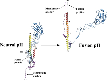
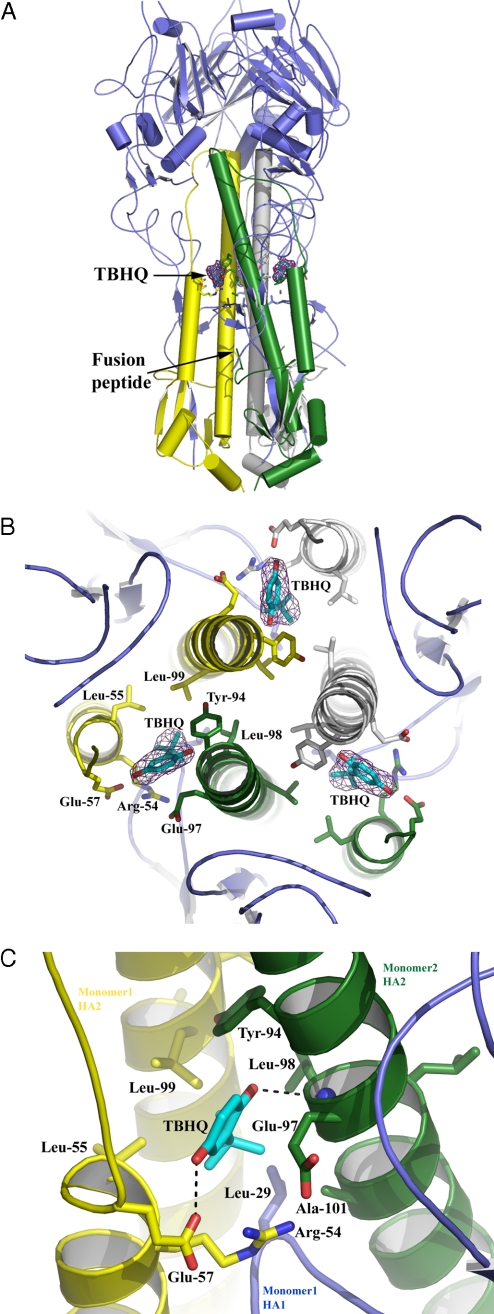
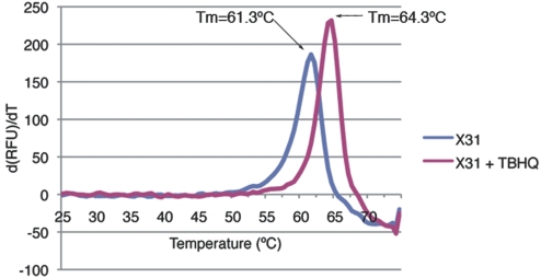
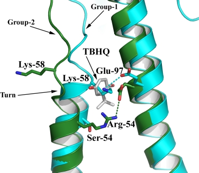
The influenza surface glycoprotein hemagglutinin (HA) is a potential target for antiviral drugs because of its key roles in the initial stages of infection: receptor binding and the fusion of virus and cell membranes. The structure of HA in complex with a known inhibitor of membrane fusion and virus infectivity, tert-butyl hydroquinone (TBHQ), shows that the inhibitor binds in a hydrophobic pocket formed at an interface between HA monomers. Occupation of this site by TBHQ stabilizes the neutral pH structure through intersubunit and intrasubunit interactions that presumably inhibit the conformational rearrangements required for membrane fusion. The nature of the binding site suggests routes for the chemical modification of TBHQ that could lead to the development of more potent inhibitors of membrane fusion and potential anti-influenza drugs.


Conflict of interest statement

The authors declare no conflict of interest.

Figures

Fig. 1.
Fig. 1.
Diagrams of the structure of a subunit of the HA trimer at neutral pH (18) (Left) and after incubation at fusion pH (pH 5.0, 20°) (19) (Right). The disulfide-linked polypeptide chains, HA1 and HA2, are colored blue and multicolored, respectively. Their N and C termini are labeled N1, C1, and N2, C2, respectively. The fusion pH diagram was constructed by combining the HA1 domain structure (39) and the fusion pH HA2 structure (20). Comparison of the diagrams shows that at fusion pH the HA1 membrane distal domains (blue) detrimerize. The fusion peptide at the N terminus of HA2 (gray) is relocated to become the N terminus of a new 100-Å-long α-helix that forms a triple-helical coiled-coil in the fusion pH trimer. Each monomer of the new coiled-coil is composed of the repositioned (turned through 180°) short α-helix (red) from the neutral pH α-helical hairpin (red-orange-yellow); the interhelical extended chain of the hairpin (orange), refolded into an α-helix; and the N-terminal half (yellow) of the central, neutral pH, long α-helix. Two turns (green) of the central, neutral pH long α-helix refold at fusion pH to form a 180° reverse turn at the C terminus of the new coiled-coil. As a consequence the C-terminal half of the neutral pH long α-helix (dark blue) and the C-terminal region of HA2 (purple) are reoriented and refolded in the fusion pH structure, antiparallel to the new α-helix and packed into the grooves of the coiled-coil, positioning the C-terminal membrane anchor at the same end of the fusion pH HA as the relocated fusion peptide (dashed lines indicate regions of structure that are not determined). The numbered residues in HA2, 58, 75, and 106 are in regions that differ in structure between group 1 and group 2 HAs.
Fig. 2.
Fig. 2.
Phylogenetic tree of influenza A HAs. The 2 groups are colored cyan (group 1) and green (group 2), each of which can be further subdivided into 3 clades (H8, H9, and H12; H1, H2, H5, and H6; H11, H13, and H16) and 2 clades (H3, H4, and H14; H7, H10, and H15). The tree was drawn by using the program TreeIllustrator (40).
Fig. 3.
Fig. 3.
The location and composition of the TBHQ binding site. (A) Representation of the H14 HA trimer. The 3 HA1s are colored in blue, and the 3 HA2s are in yellow, green, and gray. The locations of the 3 TBHQ molecules and the fusion peptide are highlighted. (B) View down the 3-fold axis of H14 HA to show the 3 TBHQ binding sites. A 2 FoFc electron density map (contoured at 1σ) is shown for TBHQ. Selected residues are shown as sticks. The same color scheme as in A is used. (C) Close-up view of a single TBHQ binding site. Selected residues are shown as sticks, and potential hydrogen bonds are shown as dotted lines.
Fig. 4.
Fig. 4.
A representative fluorometric trace of the first derivative of the change in RFU against temperature, demonstrating the thermostabilization of X31 H3 HA by TBHQ. Applying the methodology of Mayhood and Windsor (41), a binding affinity of TBHQ for X13 H3 HA of between 5 and 50 μM at 20 °C can be calculated.
Fig. 5.
Fig. 5.
The structural basis for HA group-specific inhibition by TBHQ. Superposition of H14 (green) and H5 (cyan) HAs clearly shows that the additional turn of helix A in the group 1 HAs precludes TBHQ binding. Potential hydrogen bonds are shown as dotted lines.

Comment in

  • How influenza virus is locked out of the cell.
    Modis Y. Modis Y. Proc Natl Acad Sci U S A. 2008 Dec 2;105(48):18647-8. doi: 10.1073/pnas.0810508106. Epub 2008 Nov 24. Proc Natl Acad Sci U S A. 2008. PMID: 19033206 Free PMC article. No abstract available.

Similar articles

Cited by

References

    1. Skehel JJ, Wiley DC. Receptor binding and membrane fusion in virus entry: The influenza hemagglutinin. Annu Rev Biochem. 2000;69:531–569. - PubMed
    1. Pinto LH, Lamb RA. The M2 proton channels of influenza A and B viruses. J Biol Chem. 2006;281:8997–9000. - PubMed
    1. Murphy BR, Webster RG. In: Fields Virology. 3rd Ed. Fields DBN, Knipe M, Howley PM, editors. Philadelphia: Lippincott–Raven; 1996. pp. 1397–1445.
    1. Hay AJ, Wolstenholme AJ, Skehel JJ, Smith MH. The molecular basis of the specific anti-influenza action of amantadine. EMBO J. 1985;4:3021–3024. - PMC - PubMed
    1. Von Itzstein M, et al. Rational design of potent sialidase-based inhibitors of influenza virus replication. Nature. 1993;363:418–423. - PubMed

Publication types

MeSH terms

Substances

LinkOut - more resources