. 2009 Jan 9;284(2):1040-9.
doi: 10.1074/jbc.M806638200. Epub 2008 Nov 12.

Correlation of fragile histidine triad (Fhit) protein structural features with effector interactions and biological functions

Affiliations

Correlation of fragile histidine triad (Fhit) protein structural features with effector interactions and biological functions

Flavia Pichiorri et al. J Biol Chem. .

Abstract

We have previously shown that Fhit tumor suppressor protein interacts with Hsp60 chaperone machinery and ferredoxin reductase (Fdxr) protein. Fhit-effector interactions are associated with a Fhit-dependent increase in Fdxr stability, followed by generation of reactive oxygen species and apoptosis induction under conditions of oxidative stress. To define Fhit structural features that affect interactions, downstream signaling, and biological outcomes, we used cancer cells expressing Fhit mutants with amino acid substitutions that alter enzymatic activity, enzyme substrate binding, or phosphorylation at tyrosine 114. Gastric cancer cell clones stably expressing mutants that do not bind substrate or cannot be phosphorylated showed decreased binding to Hsp60 and Fdxr and reduced mitochondrial localization. Expression of Fhit or mutants that bind interactor proteins results in oxidative damage and accumulation of cells in G(2)/M or sub-G(1) fractions after peroxide treatment; noninteracting mutants are defective in these biological effects. Gastric cancer clones expressing noncomplexing Fhit mutants show reduction of Fhit tumor suppressor activity, confirming that substrate binding, interaction with heat shock proteins, mitochondrial localization, and interaction with Fdxr are important for Fhit tumor suppressor function.


Figures
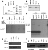
FIGURE 1.
FIGURE 1.
Direct interactions of purified Fhit forms with Hsp60/10 and Fdxr. A, immunoblot of preparations of purified GST-tagged proteins for in vitro interaction studies. Examples of the purified proteins made in bacteria are shown. B–D, interactions between purified Fhit and interactor proteins. GST-tagged or 35S-labeled proteins made by in vitro transcription-translation were prepared and combined in pairs as indicated, followed by radiographic imaging of pulled down or immunoprecipitated partner proteins. E, interaction of purified Fhit and phospho-Fhit forms with effectors. Purified Fhit and phospho-Fhit forms were combined with labeled interactors; IP was with anti-Fhit. The upper panel shows the radiographic image of co-IP of Hsp60 and the lower panel co-IP of Fdxr with the Fhit forms. F, anti-Fhit immunoprecipitates phospho-Fhit monomers. This panel shows the immunoblotted Fhit proteins precipitated for each of the IP experiments shown in E; the anti-Fhit serum precipitates and detects phosphorylated and unphosphorylated forms of Fhit.
FIGURE 2.
FIGURE 2.
In vivo effect of abrogation of the Fhit-Fdxr interaction. Human colon cancer cells, HCT116 FDXR+/+/+, and FDXR+/-/- (1 × 10⁶) cells were injected into the flanks of nude mice subcutaneously, and siFHIT or nonspecific sequence (siNSR) oligonucleotides with Lipofectamine were injected into the tumors on days 7 and 14. Mice were sacrificed on day 21 and tumor volumes calculated. A and B, FDXR+/-/- tumors treated with siFHIT were significantly smaller than tumors of other groups (scale bar, 10 mm). A, time course of tumor growth in all groups. B, tumors were excised on day 21, and portions of each tumor were taken for fixation, processing, and immunohistochemical analyses. C, tissue sections of siFHIT-treated tumors were stained for Fhit and Fdxr expression; representative results are shown; staining for apoptotic cells (by ISOL method) is also shown on the right. Expression of Fhit was low in both si-FHIT-treated tissues (panels a and d); expression of Fdxr was low in the tissue from FDXR+/-/- tumor (panel b) and high in tissue from FDXR+/+/+ tumor (panel e). Many apoptotic cells with black-brown nuclei were observed in tissue from FDXR+/-/- tumor (panel c) but not in tissue from FDXR+/+/+ tumor (panel f) panels a, b, d, and e, ×200; panels c and f, ×400. Similarly stained tissue sections of si-NSR treated tumors are shown below the si-FHIT-treated sections.
FIGURE 3.
FIGURE 3.
Localization and co-immunoprecipitation of mutant Fhit proteins in stable expressors. A, mitochondrial and cytosol fractions of MKN74 transfectants were isolated using mitochondria/cytosol fractionation kit (BioVision), and fractionated lysates were separated on a polyacrylamide gel and probed with anti-Fhit serum (left panel). Western blot images were analyzed with GS800 calibrated densitometer (Bio-Rad) and Quantity One software system to determine the ratio of Fhit or mutant Fhit protein in mitochondria versus cytosol (right panel). Note that the Y114D residue adds a negative charge to the protein, effecting slower migration on the gel, whereas the Y114F and Y114A mutants migrate faster than WT Fhit. Note also that the Y114D mutant was made to mimic a phospho-Fhit and is apparently much less stable than WT, in accord with the report that phospho-Fhit is targeted for degradation (15). B, MKN74/FHIT and FHIT mutant transfectants (WT, Y114A, Y114D, Y114F, H96N, and L25W) were cultured, and 106 cells of each were collected. Cells were lysed; DSP cross-linker was added, and immunoprecipitation with Fdxr or Hsp60 antiserum was carried out; proteins were separated on polyacrylamide gel and probed with anti-Fhit serum. Western blot images were analyzed with GS800 calibrated densitometer (Bio-Rad) and Quantity One software system.
FIGURE 4.
FIGURE 4.
FhitY114 mutants bind poorly to Fhit effector proteins. A, H1299 cells were infected with AdFHIT or AdFHIT mutants at a multiplicity of infection of 5. At 48 h after infection cells were lysed for immunoprecipitation experiments. In the left panel, the immunoblot shows the level of Fhit or mutant Fhit protein expressed in infected cells, normalized to the level of Vinculin. The lysates, in amounts that would give equivalent amounts of Fhit protein, were then used in IP experiments, as shown in the middle and right panels; upper nonspecific bands in these panels include rabbit IgG. Numbers below each lane indicate the relative amounts of Fhit or Fhit mutant protein co-precipitated with Hsp60 or Fdxr, respectively. B, similar experiment was performed using Y114F, Y114A, and Y114D mutants; the left panel shows the input level of Hsp60 and Fhit after infection of H1299 cells. The right panel shows the IP with Hsp60 and Fhit antisera. Western blot images were analyzed with GS800 calibrated densitometer (Bio-Rad) and Quantity One software system.
FIGURE 5.
FIGURE 5.
H2O2 induction of 8-OHdG in DNA of Fhit and Fhit mutant stable and induced expressors. A, stable MKN74 Fhit and mutant expressors were treated with 0.5 mm H2O2, and DNA damage in the nuclear genomes was assessed by immunofluorescent detection of 8-OHdG residues in the DNA of the respective cell lines. The red nuclear stain detects 8-OHdG and the blue stain is 4′,6-diamidino-2-phenylindole nuclear DNA staining (insets). Negative controls for all cells showed no staining (data not shown). B, bar graph ± S.D. shows the relative frequency of 8-OHdG detection in MKN74 WT and mutant expressors. The double asterisk on Y114A and H96D denotes a p value >0.05. C, immunoblot analysis of Fdxr, Fhit, and glyceraldehyde-3-phosphate dehydrogenase in H1299 cells expressing Fhit WT, Y114A, and Y114D proteins, showing the Fdxr level after cycloheximide (CHX) chase (30 μg/ml) for 6–24 h. Densitometry based on glyceraldehyde-3-phosphate dehydrogenase levels shows enhanced stability of Fdxr in presence of WT Fhit.
FIGURE 6.
FIGURE 6.
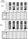
Cell cycle kinetics of H2O2-treated MKN74/Fhit and Fhit mutant expressors. MKN74 stable Fhit or mutant-expressing cells were treated with 0.25 mm H2O2 and incubated for 24 (top panels) or 48 h (bottom panels). Cells were collected, washed with PBS, and resuspended in cold 70% ethanol. For analysis, cells were spun down, washed in PBS, and suspended in 0.1 mg/ml propidium iodide/Triton X-100 staining solution (0.1% Triton X-100, 0.2 μg/ml DNase-free RNase A) for 30 min at room temperature and analyzed by flow cytometry.
FIGURE 7.
FIGURE 7.
Tumorigenicity of MKN74/Fhit and Fhit mutant expressors in nude mice. 5 × 10⁶ WT or mutant (Y114A, Y114D, Y114F, H96N, and L25W) stable expressing MKN74 cells were injected subcutaneously into the right flanks of four nude mice for each cell line. Tumor sizes were measured over a 2-week period and volumes of tumors calculated and graphed with standard deviations as shown. Significant differences in pairwise comparisons of tumor sizes of different MKN74/Fhit and mutant expressors were determined by Student's t test. The tumors from MKN74/Fhit WT cells were significantly smaller than tumors of all mutant expressors (values ranging from p = 0.008 for Y114A to p = 0.04 for Y114D and L25W) except H96N (p = 0.2). The MKN74/H96N tumors were significantly smaller than Y114A (p = 0.01) and Y114F (p = 0.02) tumors. There were no other significant differences.

Similar articles

Cited by

References

    1. Huebner, K., and Croce, C. M. (2001) Nat. Rev. Cancer 1 214-221 - PubMed
    1. Huebner, K., and Croce, C. M. (2003) Br. J. Cancer 88 1501-1506 - PMC - PubMed
    1. Barnes, L. D., Garrison, P. N., Siprashvili, Z., Guranowski, A., Robinson, A. K., Ingram, S. W., Croce, C. M., Ohta, M., and Huebner, K. (1996) Biochemistry 35 11529-11535 - PubMed
    1. Ji, L., Fang, B., Yen, N., Fong, K., Minna, J. D., and Roth, J. A. (1999) Cancer Res. 59 3333-3339 - PubMed
    1. Ishii, H., Dumon, K. R., Vecchione, A., Trapasso, F., Mimori, K., Alder, H., Mori, M., Sozzi, G., Baffa, R., Huebner, K., and Croce, C. M. (2001) Cancer Res. 61 1578-1584 - PubMed

Publication types

MeSH terms

Substances