. 2009 Jan 2;284(1):486-494.
doi: 10.1074/jbc.M806468200. Epub 2008 Nov 11.

The roles of individual domains of RNase R in substrate binding and exoribonuclease activity. The nuclease domain is sufficient for digestion of structured RNA

Affiliations

The roles of individual domains of RNase R in substrate binding and exoribonuclease activity. The nuclease domain is sufficient for digestion of structured RNA

Helen A Vincent et al. J Biol Chem. .

Abstract

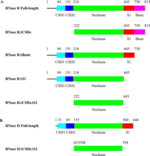
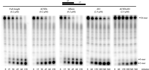
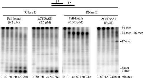
RNase R and RNase II are the two representatives from the RNR family of processive, 3' to 5' exoribonucleases in Escherichia coli. Although RNase II is specific for single-stranded RNA, RNase R readily degrades through structured RNA. Furthermore, RNase R appears to be the only known 3' to 5' exoribonuclease that is able to degrade through double-stranded RNA without the aid of a helicase activity. Consequently, its functional domains and mechanism of action are of great interest. Using a series of truncated RNase R proteins we show that the cold-shock and S1 domains contribute to substrate binding. The cold-shock domains appear to play a role in substrate recruitment, whereas the S1 domain is most likely required to position substrates for efficient catalysis. Most importantly, the nuclease domain alone, devoid of the cold-shock and S1 domains, is sufficient for RNase R to bind and degrade structured RNAs. Moreover, this is a unique property of the nuclease domain of RNase R because this domain in RNase II stalls as it approaches a duplex. We also show that the nuclease domain of RNase R binds RNA more tightly than the nuclease domain of RNase II. This tighter binding may help to explain the difference in catalytic properties between RNase R and RNase II.


Figures

FIGURE 1.
FIGURE 1.
Linear domain organization of RNase R and RNase II proteins. The CSDs are colored in cyan and blue for CSD1 and CSD2, respectively, the nuclease domains are in green, the S1 domains are red, and the low complexity, highly basic region, found in RNase R only, is in magenta. A, RNase R. RNase R full-length is the full-length wild-type RNase R protein. RNase RΔCSDs lacks both CSD1 and CSD2. RNase RΔBasic is missing the low complexity, highly basic region. RNase RΔS1 is missing both the S1 domain and the low complexity, highly basic region. RNase RΔCSDsΔS1 consists of the nuclease domain alone. B, RNase II. RNase II full-length is the full-length wild-type RNase II protein. RNase IIΔCSDsΔS1 contains the nuclease domain alone.
FIGURE 2.
FIGURE 2.
Exoribonuclease activity of truncated RNase R proteins on a substrate containing a duplex. Assays were carried out as described under “Experimental Procedures” with 10 μm ds17-A17 substrate and the indicated enzyme concentrations. Aliquots were taken at the indicated times and analyzed by denaturing PAGE.
FIGURE 3.
FIGURE 3.
Comparison of RNase II and RNase R full-length proteins and nuclease domain-truncated proteins on a substrate containing a duplex. Assays were carried out as described under “Experimental Procedures” with 10 μm ds17-A17 substrate and the indicated enzyme concentrations. Aliquots were taken at the indicated times and analyzed by denaturing PAGE. The origin of the band at the approximate position for an 8-mer that first appears at the 30-min time point with RNase RΔCSDsΔS1 is unknown. However, it is not observed reproducibly.

Similar articles

Cited by

References

    1. Deutscher, M. P. (2006) Nucleic Acids Res. 34 659-666 - PMC - PubMed
    1. Donovan, W. P., and Kushner, S. R. (1986) Proc. Natl. Acad. Sci. U. S. A. 83 120-124 - PMC - PubMed
    1. Mohanty, B. K., and Kushner, S. R. (2003) Mol. Microbiol. 50 645-658 - PubMed
    1. Khemici, V., and Carpousis, A. J. (2004) Mol. Microbiol. 51 777-790 - PubMed
    1. Cheng, Z. F., and Deutscher, M. P. (2005) Mol. Cell 17 313-318 - PubMed

Publication types

MeSH terms

LinkOut - more resources