. 2009 Jan 9;284(2):1177-89.
doi: 10.1074/jbc.M805144200. Epub 2008 Nov 11.

Biomolecular characterization of CD44-fibrin(ogen) binding: distinct molecular requirements mediate binding of standard and variant isoforms of CD44 to immobilized fibrin(ogen)

Affiliations

Biomolecular characterization of CD44-fibrin(ogen) binding: distinct molecular requirements mediate binding of standard and variant isoforms of CD44 to immobilized fibrin(ogen)

Christina S Alves et al. J Biol Chem. .

Abstract

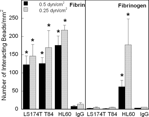
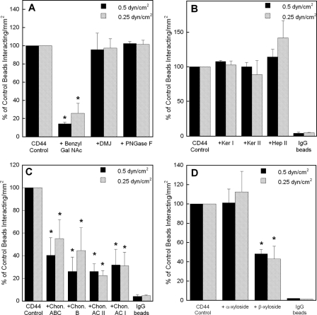
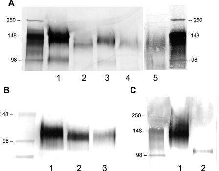
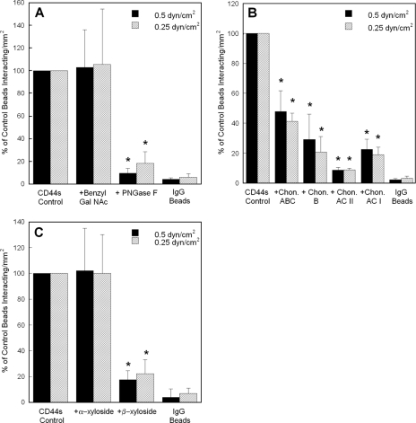
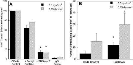
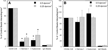
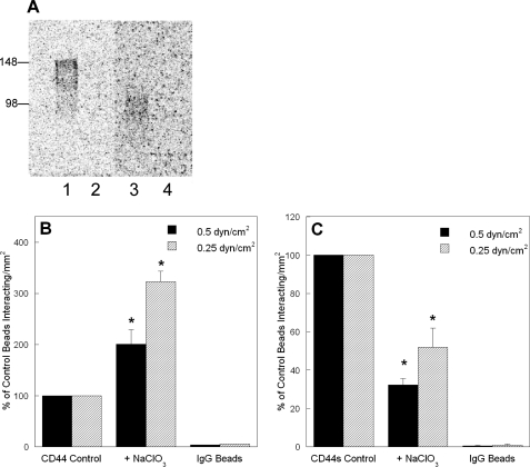
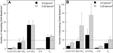
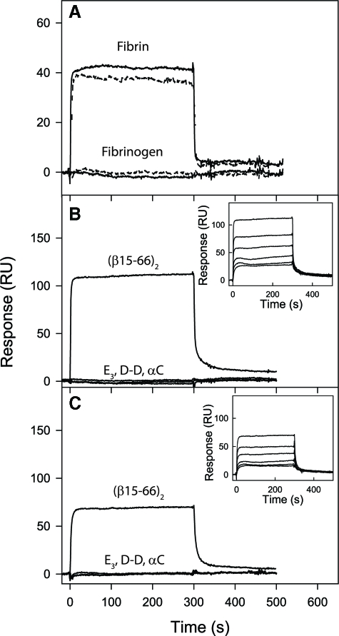
CD44 and fibrin(ogen) play critical roles in the hematogenous dissemination of tumor cells, including colon carcinomas. We recently reported that CD44 is the primary fibrin, but not fibrinogen, receptor on LS174T colon carcinomas. However, the biochemical nature of this interaction and the roles of CD44 standard (CD44s) versus CD44 variant (CD44v) isoforms in fibrin(ogen) recognition have yet to be delineated. Microspheres, coated with CD44 immunopurified from LS174T or T84 colon carcinoma cells, which express primarily CD44v, effectively bind to immobilized fibrin, but not fibrinogen, in shear flow. In contrast, CD44s from HL-60 cells binds to both immobilized fibrin and fibrinogen under flow. Use of highly specific enzymes and metabolic inhibitors reveals that LS174T CD44 binding to fibrin is dependent on O-glycosylation of CD44, whereas CD44s-fibrin(ogen) interaction has an absolute requirement for N-, but not O-, linked glycans. The presence of chondroitin and dermatan sulfate on CD44 standard and variant isoforms facilitates fibrin recognition. Use of the anti-CD44 function-blocking monoclonal antibody Hermes-1 nearly abolishes binding of LS174T CD44 to fibrin, although it has no effect on CD44s-fibrin(ogen) interaction. The CD44-binding site is localized within the N-terminal portion of the fibrin beta chains, including amino acid residues (beta15-66). Surface plasmon resonance experiments revealed high affinity binding of immobilized CD44 with solubilized fibrin but not fibrinogen. Collectively, these data suggest that immobilization of fibrinogen exposes a cryptic site that mediates binding to CD44s but not CD44v. Our findings may provide a rational basis for designing novel therapeutic strategies to combat metastasis.


Figures

FIGURE 1.
FIGURE 1.
CD44 from colon carcinoma cells and HL60 CD44s effectively bind to fibrin under flow, but only CD44s binds to immobilized fibrinogen. Immunoprecipitated CD44 from LS174T or T84 colon carcinoma whole cell lysates or immunopurified CD44s from HL60 human myeloid cell whole cell lysate was adsorbed onto 10-μm microspheres. The CD44-coated microspheres were then perfused over 1.0 mg/ml immobilized fibrin or fibrinogen at the specified wall shear stresses in a parallel plate flow chamber. The number of beads interacting with fibrin or fibrinogen was quantified using video microscopy. Data represent the mean ± S.E. of n = 3-4 experiments. *, p < 0.05 with respect to IgG control-coated microspheres.
FIGURE 2.
FIGURE 2.
A, effect of O-versus N-glycosylation on the adhesion of LS174T CD44-coated microspheres to immobilized fibrin under flow. CD44-coated microspheres were generated using CD44 immunopurified from LS174T cells pretreated with either 2 mm benzyl-GalNAc or 1 mm DMJ, or from LS174T whole cell lysate treated with 8 units/ml PNGase F. B, contribution of keratan and heparan sulfates to the adhesion of LS174T CD44-coated microspheres to immobilized fibrin under flow. CD44-coated microspheres were generated using CD44 immunoprecipitated from LS174T cells, which was subsequently treated with either 70 milliunits/ml keratanase (Ker) I, 5 milliunits/ml keratanase II, or 0.5 unit/ml heparinase (Hep) II for 24 h at 37 °C prior to their use in flow-based adhesion assays. C, effect of chondroitin and dermatan sulfates on the adhesion of LS174T CD44-coated microspheres to immobilized fibrin under flow. CD44-coated microspheres were generated using CD44 immunopurified from LS174T cells, which was subsequently treated with either 1 unit/ml chondroitinase (Chon) ABC, B, AC II, or AC I for 60 min at 37 °C. D, effect of chondroitin and dermatan sulfate chain coupling to the CD44 core protein on CD44-fibrin binding. CD44-coated microspheres were generated using CD44 immunoprecipitated from LS174T cells, which was previously treated with either 1 mm p-nitrophenyl α-d-xylopyranoside or p-nitrophenyl β-xylopyranoside for 48 h at 37 °C. In all experiments (A-D), CD44-coated microspheres (2 × 10⁶/ml) were perfused over 1 mg/ml fibrin for 5 min at the specified wall shear stresses. Data are reported as percent of untreated control beads that interacted with immobilized fibrin and represent the mean ± S.E. of n = 3-4 experiments. *, p < 0.05 with respect to untreated control microspheres.
FIGURE 3.
FIGURE 3.
A, Western blots of LS174T whole cell lysates using cells pretreated with highly specific glycoconjugate biosynthesis inhibitors. CD44 was immunoprecipitated from untreated control LS174T cells (lane 1) and subjected to SDS-PAGE followed by Western blotting with the anti-CD44 mAb 2C5. Lysate was also immunoprecipitated from LS174T colon carcinoma cells cultured for 48 h in medium containing 2 mm benzyl-GalNAc (to inhibit O-linked glycosylation; lane 2) or 1 mm DMJ (to disrupt N-linked processing; lane 3). The efficacy of the DMJ treatment was verified by incubating CD44 from DMJ-treated cells with 0.12 unit/ml endoglycosidase H for 3 h at 37 °C, which cleaves high mannose and hybrid but not complex oligosaccharides from glycoproteins (lane 4). Alternatively, LS174T whole cell lysate was treated with 8 units/ml PNGase F for 48 h at 37 °C prior to immunoprecipitation (to cleave N-linked glycans from the glycoprotein core, lane 5). B, Western blots of HL60 cell lysates using cells pretreated with highly specific glycoconjugate biosynthesis inhibitors. CD44s was immunoprecipitated from untreated control HL60 cells (lane 1) and subjected to SDS-PAGE followed by Western blotting with the anti-CD44 mAb 156-3C11. Lysate was also immunopurified from HL60 cells cultured for 48 h in medium containing 2 mm benzyl-GalNAc (to inhibit O-linked glycosylation; lane 2). Alternatively, HL60 whole cell lysate was treated with 8 units/ml PNGase F for 48 h at 37 °C prior to immunoprecipitation (to remove N-linked glycans from the glycoprotein core; lane 3). C, detection of HECA-452 reactive epitopes on LS174T CD44 and HL60 CD44s proteins. CD44 immunoprecipitated from LS174T colon carcinoma cells using the anti-CD44 mAb 2C5 (lane 1) and CD44s immunopurified from HL60 cells using the anti-CD44 mAb 515 (lane 2) were separated by SDS-PAGE followed by Western blotting with the HECA-452 mAb.
FIGURE 4.
FIGURE 4.
A, effect of O-versus N-glycosylation on the adhesion of HL60 CD44s-coated microspheres to immobilized fibrin under flow. CD44s-coated microspheres were generated using CD44s immunoprecipitated from HL60 cells pretreated with 2 mm benzyl-GalNAc or from HL60 whole cell lysate treated with 8 units/ml PNGase F. B, effect of chondroitin and dermatan sulfates on the adhesion of HL60 CD44s-coated microspheres to immobilized fibrin under flow. CD44s-coated microspheres were generated using CD44s immunopurified from HL60 cells, which was subsequently treated with 1 unit/ml chondroitinase (Chon) ABC, B, AC II, or AC I for 60 min at 37 °C. C, effect of chondroitin and dermatan sulfate chain coupling to the CD44s core protein on CD44s-fibrin binding. CD44s-coated microspheres were generated using CD44s immunoprecipitated from HL60 cells treated with either 1 mm p-nitrophenyl α-d-xylopyranoside or p-nitrophenyl β-xylopyranoside for 48 h at 37 °C. In all experiments (A-C), CD44s-coated microspheres (2 × 10⁶/ml) were perfused over 1 mg/ml fibrin for 5 min at the specified wall shear stresses. Data are reported as percent of untreated control beads that interacted with immobilized fibrin and represent the mean ± S.E. of n = 3-4 experiments. *, p < 0.05 with respect to untreated control microspheres.
FIGURE 5.
FIGURE 5.
A, effect of O- and N-glycosylation on the adhesion of HL60 CD44s-coated microspheres to immobilized fibrinogen under flow. CD44s-coated microspheres were generated using CD44s immunoprecipitated from HL60 cells pretreated with 2 mm benzyl-GalNAc or from HL60 whole cell lysate treated with 8 units/ml PNGase F. CD44s-coated microspheres (2 × 10⁶/ml) were perfused over 1 mg/ml fibrinogen for 5 min at the specified wall shear stresses. Data are reported as percent of untreated control beads that interacted with immobilized fibrinogen, and represent the mean ± S.E. of n = 3 experiments. *, p < 0.05 with respect to untreated control microspheres. B, impact of cleavage of HECA-452 reactive epitopes from CD44 variant isoforms on the adhesion of LS174T CD44-coated microspheres to immobilized fibrinogen under flow. CD44-coated microspheres were treated with 0.1 unit/ml sialidase for 90 min at 37 °C. CD44-coated microspheres (2 × 10⁶/ml) were perfused over 1 mg/ml fibrinogen for 5 min at the specified wall shear stresses. Data are reported as the number of interacting microspheres per mm2, and represent the mean ± S.E. of n = 3 experiments. *, p < 0.05; §, p < 0.1 with respect to untreated control microspheres.
FIGURE 6.
FIGURE 6.
The anti-CD44 mAb Hermes-1 and soluble hyaluronan inhibit the adhesion of CD44-, but not CD44s-, coated microspheres to immobilized fibrin under shear. A, CD44-coated microspheres, generated using CD44 immunopurified from LS174T colon carcinoma cells, were incubated with 20 μg/ml of the anti-CD44 mAb Hermes-1 or 1 mg/ml soluble hyaluronic acid for 1 h at 37 °C. CD44-coated microspheres were subsequently washed once in DPBS and perfused at a concentration of 2 × 10⁶ per ml over 1 mg/ml fibrin for 5 min at a wall shear stress of 0.5 or 0.25 dyne/cm2. Data are reported as percent of untreated control (Cntl) beads that interacted with immobilized fibrin, and represent the mean ± S.E. of n = 5 experiments. *, p < 0.05 with respect to untreated control microspheres. B, CD44s-coated microspheres, generated using CD44s immunoprecipitated from HL60 cells, were incubated with 20 μg/ml of the anti-CD44 mAb Hermes-1 or 1 mg/ml soluble hyaluronic acid for 1 h at 37 °C, and perfused at a concentration of 2 × 10⁶/ml over 1 mg/ml fibrin for 5 min at the specified wall shear stresses. Data are reported as percent of untreated control beads that interacted with immobilized fibrin and represent the mean ± S.E. of n = 3 experiments. As a negative control, IgG-coated microspheres were perfused over immobilized fibrin at prescribed wall shear stresses.
FIGURE 7.
FIGURE 7.
Effect of sulfation on the adhesion of CD44-coated microspheres to immobilized fibrin under flow. A, effectiveness of sodium chlorate treatment in inhibiting sulfation of CD44. CD44 immunoprecipitated from untreated (control) LS174T colon carcinoma cells (lane 1) or LS174T cells treated with 60 mm sodium chlorate (lane 2) both cultured in the presence of 20 μCi/ml Na35SO4 for 48 h was subjected to SDS-PAGE. CD44s immunoprecipitated from untreated (control) HL60 cells (lane 3) or HL60 cells treated with 20 mm sodium chlorate (lane 4) both cultured in the presence of 20 μCi/ml Na35SO4 for 48 h was subjected to SDS-PAGE. B, removal of sulfate groups increases LS174T CD44 binding to immobilized fibrin. CD44-coated microspheres were generated using CD44 immunopurified from LS174T colon carcinoma cells cultured in the presence or absence of 60 mm sodium chlorate for 48 h. C, inhibition of sulfation diminishes HL60 CD44s-fibrin binding under flow. CD44s-coated microspheres were generated using CD44s immunopurified from HL60 cells cultured in the presence or absence of 20 mm sodium chlorate for 48 h. In all experiments, CD44-coated microspheres (2 × 10⁶/ml) were perfused over 1 mg/ml fibrin for 5 min at the specified wall shear stresses. Data are reported as percent of untreated control beads that interacted with immobilized fibrin and represent the mean ± S.E. of n = 3 experiments. *, p < 0.05 with respect to untreated control microspheres.
FIGURE 8.
FIGURE 8.
Analysis of CD44- versus CD44s-coated microsphere binding to immobilized fibrin(ogen) fragments under flow. A, LS174T CD44 effectively binds to the (β15-66)2 fibrin fragment. CD44-coated microspheres, generated using CD44 immunopurified from LS174T colon carcinoma cells, were perfused for 5 min over fibrin(ogen) fragments (100 μg/ml) immobilized onto polystyrene dishes at prescribed wall shear stresses. B, HL60 CD44s binds to the (β15-66)2 and (Bβ1-66)2 fibrin(ogen) fragments. CD44s-coated microspheres, generated using CD44s immunopurified from HL60 human myeloid cells, were perfused for 5 min over fibrin(ogen) fragments (100 μg/ml) immobilized onto polystyrene dishes at the specified wall shear stresses. A and B, data are reported as the number of interacting beads per mm2 and represent the mean ± S.E. of n = 3 experiments. The dashed lines in each graph represent base-line microsphere binding.
FIGURE 9.
FIGURE 9.
Analysis of binding of soluble fibrin, fibrinogen, and their fragments to the immobilized LS174T CD44 and HL60 CD44s receptors by surface plasmon resonance. A, fibrinogen or fibrin solubilized with GPRP, both at 1 μm, were added to immobilized LS174T CD44 (solid curves) or HL60 CD44s (dashed curves), and their association/dissociation was monitored in real time while registering the resonance signal (response). The E3, D-D, and recombinant (β15-66)2 and αC fragments, all at 1 μm, were added to immobilized LS174T CD44 (B) or HL60 CD44s (C), and their association/dissociation was monitored in real time while registering the resonance signal (response). The curves for E3, D-D, and αC in B and C essentially coincide. The insets show dose-dependent binding of the (β15-66)2 fragment added at 25, 50, 100, 250, 500, and 1000 nm to LS174T CD44 (B) or HL60 CD44s (C). The determined Kd values are listed in Table 2. Note that GPRP-solubilized fibrin was monomeric at 1 μm but formed polymers at higher concentration precluding its dose-dependent binding study.

Similar articles

Cited by

References

    1. Ponta, H., Sherman, L., and Herrlich, P. A. (2003) Nat. Rev. Mol. Cell Biol. 4 33-45 - PubMed
    1. Martin, T. A., Harrison, G., Mansel, R. E., and Jiang, W. G. (2003) Crit. Rev. Oncol. Hematol. 46 165-186 - PubMed
    1. Harada, N., Mizoi, T., Kinouchi, M., Hoshi, K., Ishii, S., Shiiba, K., Sasaki, I., and Matsuno, S. (2001) Int. J. Cancer 91 67-75 - PubMed
    1. Hofmann, M., Rudy, W., Zoller, M., Tolg, C., Ponta, H., Herrlich, P., and Gunthert, U. (1991) Cancer Res. 51 5292-5297 - PubMed
    1. Wielenga, V. J., Heider, K. H., Offerhaus, G. J., Adolf, G. R., van den Berg, F. M., Ponta, H., Herrlich, P., and Pals, S. T. (1993) Cancer Res. 53 4754-4756 - PubMed

Publication types