. 2008 Dec;135(24):4037-4047.
doi: 10.1242/dev.028308. Epub 2008 Nov 12.

Dual requirement for Pax6 in retinal progenitor cells

Affiliations

Dual requirement for Pax6 in retinal progenitor cells

Varda Oron-Karni et al. Development. 2008 Dec.

Abstract

Throughout the developing central nervous system, pre-patterning of the ventricular zone into discrete neural progenitor domains is one of the predominant strategies used to produce neuronal diversity in a spatially coordinated manner. In the retina, neurogenesis proceeds in an intricate chronological and spatial sequence, yet it remains unclear whether retinal progenitor cells (RPCs) display intrinsic heterogeneity at any given time point. Here, we performed a detailed study of RPC fate upon temporally and spatially confined inactivation of Pax6. Timed genetic removal of Pax6 appeared to unmask a cryptic divergence of RPCs into qualitatively divergent progenitor pools. In the more peripheral RPCs under normal circumstances, Pax6 seemed to prevent premature activation of a photoreceptor-differentiation pathway by suppressing expression of the transcription factor Crx. More centrally, Pax6 contributed to the execution of the comprehensive potential of RPCs: Pax6 ablation resulted in the exclusive generation of amacrine interneurons. Together, these data suggest an intricate dual role for Pax6 in retinal neurogenesis, while pointing to the cryptic divergence of RPCs into distinct progenitor pools.


Figures

Fig. 1
Fig. 1. Differential response to Pax6 loss in subpopulations of Pax6lacZ/lacZ OV progenitors
(A-L) Crx expression was characterized by fluorescent in situ hybridization (A-D,I-L, green), the distribution of VC1.1 epitope (E-H, green K,L; red) and Pax6 (A-H, red) was monitored by antibody labeling, in the control Pax6+/+ (A-H) and Pax6lacZ/lacZ (I-L) embryos. The insets in A,E,I,C,G,K are enlarged in B,F,J,D,H,L. inl, inner nuclear layer; le, lens; nr, neuroretina; oc, optic cup; os, optic stalk; prp, prospective photoreceptor layer; rpe, retinal pigmented epithelium. Scale bars: in A, 50 μm for A,E,I,C,G,K; in B, 10 μm for B,F,J,D,H,L.
Fig. 2
Fig. 2. Pax6 plays a unique role in each of two spatially distinct subsets of RPCs in the Pax6flox/flox;α-Cre OC
The expression of Pax6, VC1.1 (A-H; green and red, respectively), Crx (I-P), BrdU (Q-V) and syntaxin (W,X red) were characterized on adjacent sections by antibody labeling (A-H,Q-X) or in situ hybridization (I-P) in control (Pax6flox/flox) and mutant (Pax6flox/flox;α-Cre) retinas in the course of eye development. In the Pax6flox/flox;α-Cre OC, Pax6 was eliminated from the peripheral regions (B,D,F,H; the Pax6-deficient domain is flanked with arrowheads). Two spatially distinct populations of Pax6-deficient RPCs were identified (diagram): the Pax6 cells that are located in the OC periphery upregulate Crx, whereas the Pax6 cells located towards the central OC do not upregulate Crx. The border between the two Pax6-deficient cell types is indicated with an arrow and a broken line, and their margins are marked with arrowheads (labeled as region 1 or region 2, respectively). (Y) The percentage of the Crx-expressing domain (region 1, white bars) and the Crx domain (region 2, gray bars) relative to the total Pax6-deficient area was calculated for E12, E14, E16 OCs (n=4 eyes for all embryonic stages). (Z) Significant reduction in the percentage of BrdU+ cells was detected for both Pax6-deficient regions at all stages of development (**P<0.005 and *P<0.05 by Student's t-test; n=4 eyes for all Pax6flox/flox;α-Cre retinas and three eyes for controls). The reduction in the proliferation index was significantly more extensive in region 1 than in region 2 at E14 (P<0.01 by Student's t-test). inl, inner nuclear layer; gcl, ganglion cell layer; le, lens; nr, neuroretina; prp, prospective photoreceptor layer; rpe, retinal pigmented epithelium. Scale bar: 100 μm.
Fig. 3
Fig. 3. Altered expression profile of bHLH transcription factors in the two regions of Pax6 mutant RPCs
(A-J) At E15, the expression in control (A-E) and Pax6flox/flox;α-Cre (F-J) of Crx (A,F), Atoh4 (B,G), Atoh3 (C,H), Neurod1 (D,I) and Atoh7 (E,J) was characterized on adjacent sections by fluorescent in situ hybridization (green). On the same sections, Pax6 expression was determined by indirect immunofluorescence analysis (red). In the control, Crx was detected in the prospective photoreceptor layer (A), while in the Pax6flox/flox;α-Cre, Crx was upregulated in the peripheral region of the Pax6-deficient OC (F, surrounded by broken lines labeled 1), but was downregulated in the mutated RPCs that are located more centrally (F, surrounded by broken lines labeled 2). inl, inner nuclear layer; le, lens; nbl, neuroblast layer; nnp, non-neuronal progenitors; rpe, retinal pigmented epithelium. Asterisk indicates cells that escaped the recombination. Scale bar: 75 μm.
Fig. 4
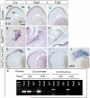
Fig. 4. Pax6 Crx+ RPCs do not complete the photoreceptor-specification program
(A-I) The expression pattern of factors involved in photoreceptor differentiation; Crx (A,D,G), Otx2 (B,E,H) and Trβ2 (C,F,I) in control (A-C), Pax6lacZ/lacZ (D-F) and Pax6flox/flox;a-Cre;Z/AP (G-I) E15 eyes. (J) The region of Pax6 inactivation was determined by detection of human alkaline phosphatase (hAP) expressed from the Z/AP reporter (J is adjacent to G-I). (K) Chromatin immunoprecipitation (ChIP) was conducted on chromatin from E13 eyes with Pax6 or rabbit IgG (IgG). PCR amplification was carried out with specific primers for detection of the Crx promoter. The Crx 3′ UTR sequence was amplified as a control that does not bind Pax6 in vivo. The same pairs of primers were used for amplification of the chromatin samples prior to immunoprecipitation (input lane). When ChIP was conducted on limb tissue, where Pax6 is not expressed, no amplification of Crx promoter sequences was detected. co, cornea; le, lens; oc, optic cup; ov, optic vesicle; prp, photoreceptor layer; rpe, retinal pigmented epithelium. Scale bar: 100 μm.
Fig. 5
Fig. 5. The misexpression of Crx is a cell-autonomous response to Pax6 loss in RPCs
(A-E) Pax6 and Crx were detected on the same sections from control (A,B) and Pax6flox/flox;Chx10-Cre (C-E) E14.5 eyes using indirect immunofluorescence analysis (Pax6, red) or fluorescent in situ hybridization (Crx, green). Only in some of the Pax6-deficient cells was misexpression of Crx detected, whereas other cells were negative for both Pax6 and Crx (white arrowhead in C,D). The Pax6Crx+ cells (D,E) were detected both adjacent to Pax6-expressing cells (white asterisk) or in regions distant from Pax6 expression (red arrowhead). (F-I) At E16.5, the expression of VC1.1 (red) was compared with Pax6 protein (F,H green) or Crx transcripts (G,I green) in control (F,G) and Pax6flox/flox;Chx10-Cre embryos (H,I). (J) The distribution of Pax6Crx+ cells along the central-peripheral regions of the OC was quantified. A significant difference in the proportion of Crx-expressing cells in the Pax6-deficient areas between central and peripheral OC was identified (*P<0.001 by Student's t-test, n=6 eyes). The scheme illustrates the arbitrary division of the OC into central/peripheral regions. co, cornea; le, lens; nnp, non-neuronal progenitors; oc, optic cup; prp, photoreceptor layer; rpe, retinal pigmented epithelium. Scale bar: in A, 200 μm; in B,E, 25 μm; in C, 75 μm; in D,F,G,H,I, 50 μm.
Fig. 6
Fig. 6. A model summarizing Pax6 functions in retinal progenitor cells
(A) In the central retina, close to the differentiation front (arrows), Pax6 is required for the normal expression profile of transcription factors that play a role in the execution of specific retinal lineages but is dispensable for the completion of neurogenesis. (B) In the peripheral RPCs, Pax6 inhibits Crx expression and at the same time is essential for proneural gene expression and completion of the neurogenic program. The involvement of Pax6 in the regulation of Crx and Otx places Pax6 upstream in the transcriptional network that regulates PR specification and differentiation. The requirement for Otx and Crx encompasses the determination and specification of both rod and cone photoreceptors. Later, the terinoids and thyroid hormone (e.g. Trβ2) nuclear receptors function within the cone precursors for the distinction between the M and S cone types (Hennig et al., 2008). nnp, non-neuronal progenitors; nbl, neuroblast layer; prp, photoreceptor precursor layer.

References

    1. Adler R, Canto-Soler MV. Molecular mechanisms of optic vesicle development: complexities, ambiguities and controversies. Dev. Biol. 2007;305:1–13. - PMC - PubMed
    1. Alexiades MR, Cepko CL. Subsets of retinal progenitors display temporally regulated and distinct biases in the fates of their progeny. Development. 1997;124:1119–1131. - PubMed
    1. Applebury ML, Farhangfar F, Glosmann M, Hashimoto K, Kage K, Robbins JT, Shibusawa N, Wondisford FE, Zhang H. Transient expression of thyroid hormone nuclear receptor TRbeta2 sets S opsin patterning during cone photoreceptor genesis. Dev. Dyn. 2007;236:1203–1212. - PubMed
    1. Aramant R, Seiler M, Ehinger B, Bergstrom A, Adolph AR, Turner JE. Neuronal markers in rat retinal grafts. Brain Res. Dev. Brain Res. 1990;53:47–61. - PubMed
    1. Arendt D, Tessmar-Raible K, Snyman H, Dorresteijn AW, Wittbrodt J. Ciliary photoreceptors with a vertebrate-type opsin in an invertebrate brain. Science. 2004;306:869–871. - PubMed

Publication types

MeSH terms