. 2008 Dec;135(24):4025-35.
doi: 10.1242/dev.028209. Epub 2008 Nov 12.

TGF-beta signals regulate axonal development through distinct Smad-independent mechanisms

Affiliations

TGF-beta signals regulate axonal development through distinct Smad-independent mechanisms

Julian Ng. Development. 2008 Dec.

Abstract

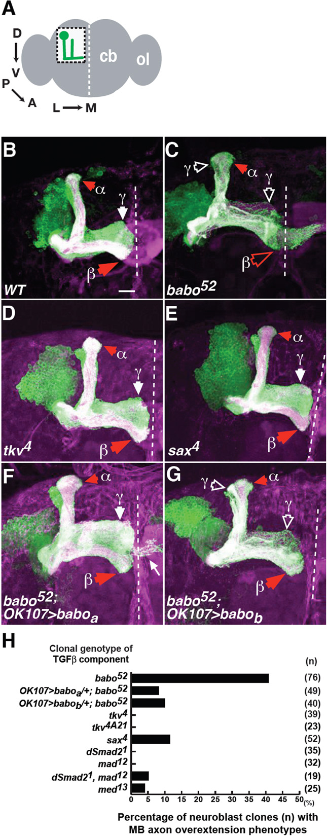
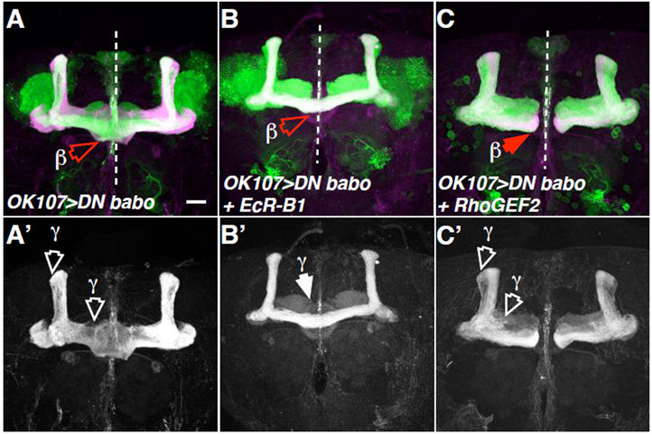
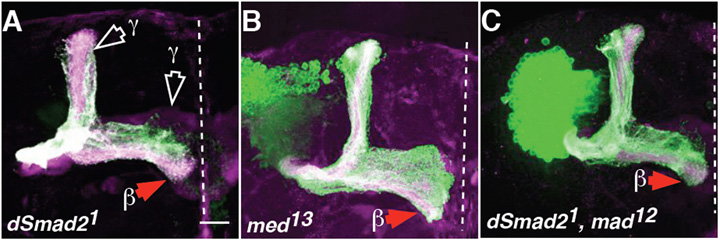
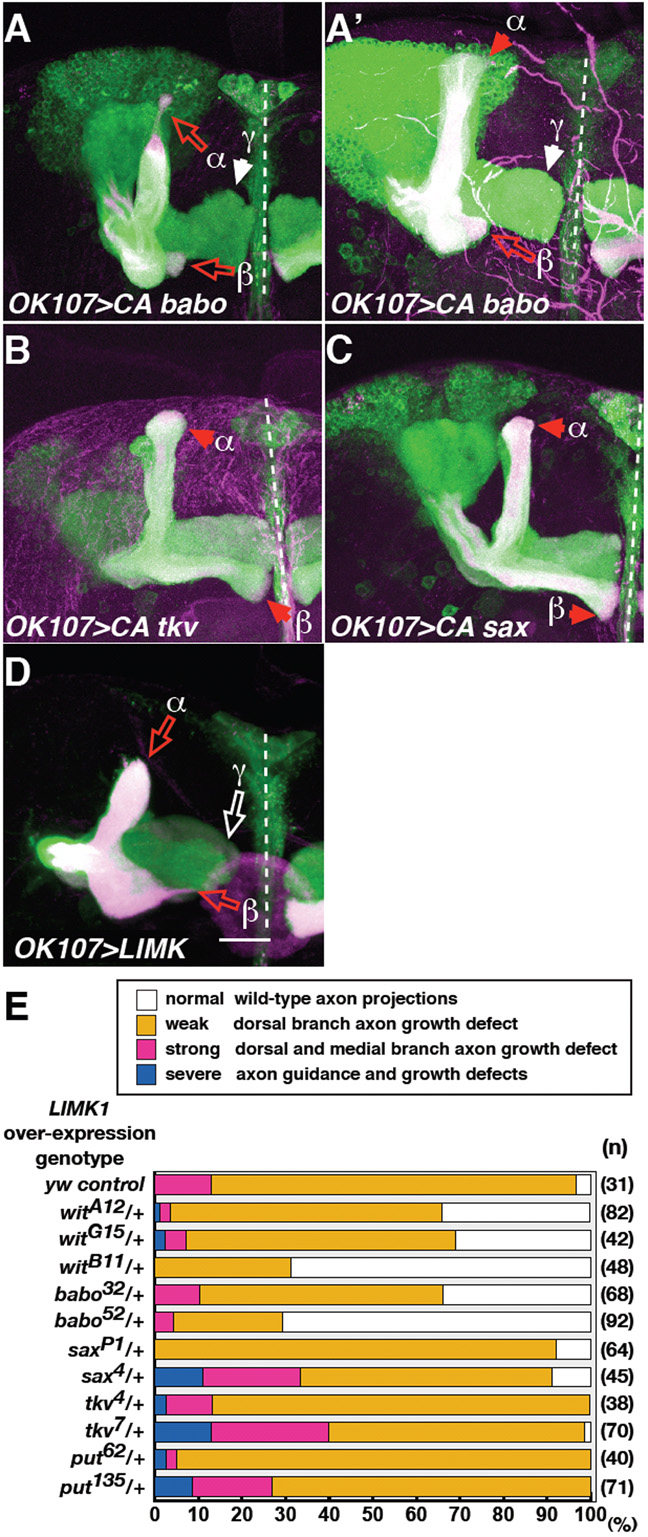
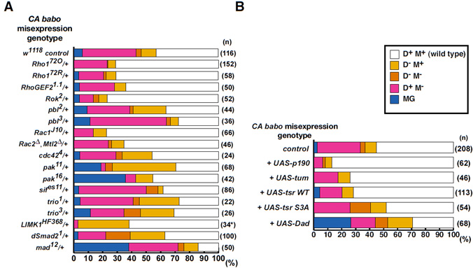
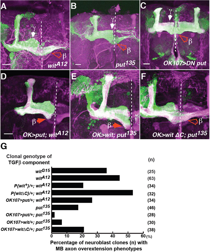
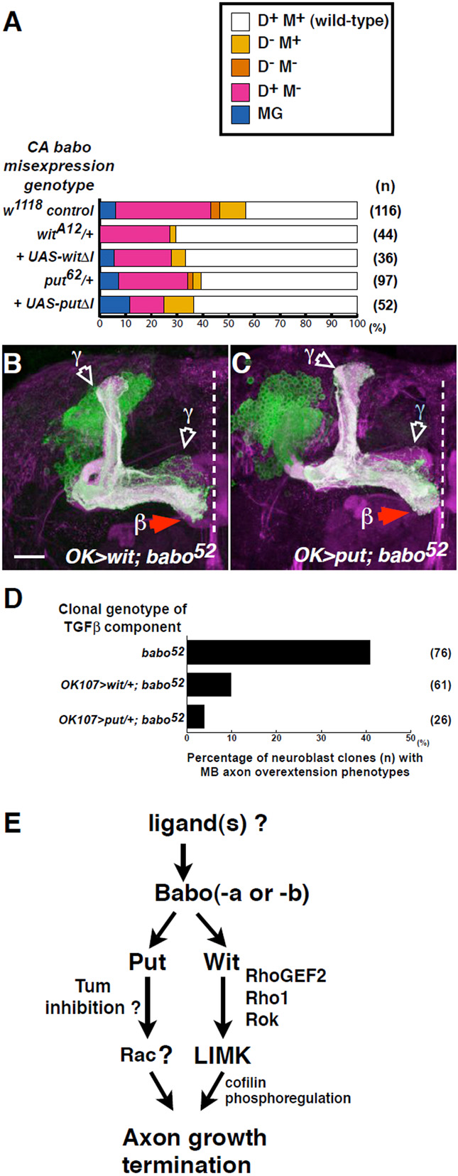
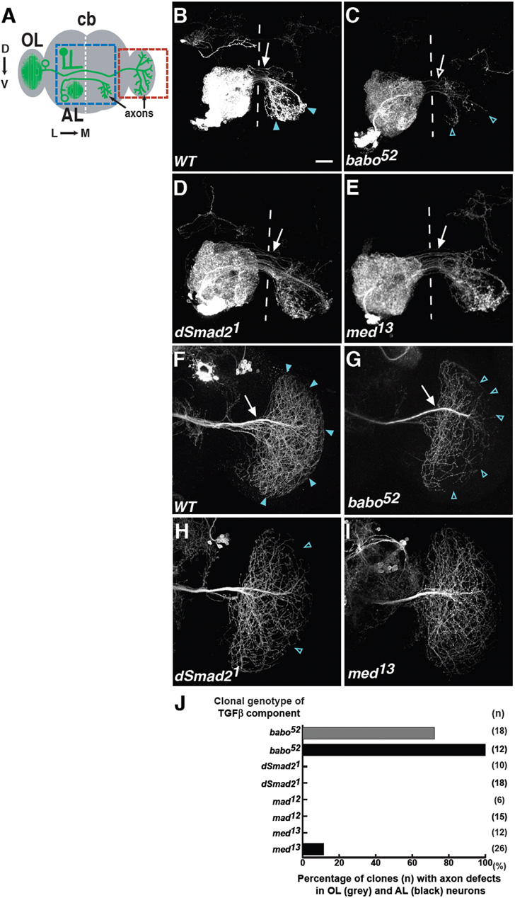
Proper nerve connections form when growing axons terminate at the correct postsynaptic target. Here I show that Transforming growth factor beta (TGFbeta) signals regulate axon growth. In most contexts, TGFbeta signals are tightly linked to Smad transcriptional activity. Although known to exist, how Smad-independent pathways mediate TGFbeta responses in vivo is unclear. In Drosophila mushroom body (MB) neurons, loss of the TGFbeta receptor Baboon (Babo) results in axon overextension. Conversely, misexpression of constitutively active Babo results in premature axon termination. Smad activity is not required for these phenotypes. This study shows that Babo signals require the Rho GTPases Rho1 and Rac, and LIM kinase1 (LIMK1), which regulate the actin cytoskeleton. Contrary to the well-established receptor activation model, in which type 1 receptors act downstream of type 2 receptors, this study shows that the type 2 receptors Wishful thinking (Wit) and Punt act downstream of the Babo type 1 receptor. Wit and Punt regulate axon growth independently, and interchangeably, through LIMK1-dependent and -independent mechanisms. Thus, novel TGFbeta receptor interactions control non-Smad signals and regulate multiple aspects of axonal development in vivo.


Figures

Fig. 1
Fig. 1. Babo inactivation results in axon overextension
Babo regulates axon growth through Babo-a and -b isoforms. (A) Schematic of the adult Drosophila whole brain. The boxed region shows mushroom body (MB) neurons in the left hemisphere of the central brain (cb). Arrows show the MB axon trajectory extending from posterior dorsal cell bodies, projecting anteroventrally and then turning towards the midline. The MB images shown are either from the left hemisphere in this orientation, or of the central brain, showing both hemispheres. Dashed white lines indicate the midline. ol, optic lobe. D, dorsal; V, ventral; P, posterior; A, anterior; L, lateral; M, medial. (B) A wild-type MB neuroblast clone. Typical adult, wild-type clones generated from newly hatched larvae have axonal projections that terminate either in the dorsal anterior cortex or just prior to the midline. Only γ, α, and β projections are indicated. (C-E) Representative images of babo52 (C), tkv4 (D) and sax4 (E) neuroblast clones. Note β lobe overextensions (open red arrowhead) across the midline in babo clones. Open white arrowheads indicate γ axon pruning defects, in this and subsequent figures. (F,G) Representative images of babo52 neuroblast clones expressing either UAS-baboa (F), or UAS-babob (G). Many axons in the UAS-baboa rescue exhibited small protrusions that were not characteristic of any lobe (thin white arrow in F). These represent ectopic projections of a subclass of MB axons induced in the OK107>baboa genetic background. In these and subsequent figures, solid red or white arrowheads indicate normal α and β or γ lobe termination points, as indicated. All images in this and subsequent figures are z-projections of confocal sections. Green, expression of the marker mCD8::GFP on all MB, neuroblast or single-cell MARCM clones (sometimes multiple single cell clones); magenta, Fas2 staining of all MB γ (weakly stained) and αβ (strongly stained) axons (appearing as white when overlapping with mCD8::GFP). Dashed white line, midline. Scale bar: 20 μm. (H) Quantification of axon overextension defects in the indicated genotype. n, number of neuroblast clones examined.
Fig. 2
Fig. 2. Babo regulates axon pruning and axon growth independently
(A-C) Drosophila MB neurons misexpressing DN babo (A), DN babo plus EcR-B1 (B), or DN babo plus RhoGEF2 (C). Additional panels (A’,B’,C’) indicate the corresponding Fas2 (magenta in A, B and C) positive axon projections. Note β lobe overextensions (open red arrowheads) in A and B, but absent in C, and axon pruning phenotypes (open white arrowheads highlight the aberrant γ-dorsal and medial branches), which are visible in A’ and C’, but absent in B’. Cell body sections were removed from C to clearly show MB axons. Scale bar: 20 μm.
Fig. 3
Fig. 3. Babo-regulated axon growth is Smad-independent
(A-C) Drosophila dSmad21 (A), Med13 (B), and double Smad21, mad12 (C) neuroblast clones did not show β lobe overextensions. Scale bar: 20μm.
Fig. 4
Fig. 4. CA babo misexpression resulted in MB axon truncations
babo and wit genetically interact with LIMK1. (A-D) Drosophila MB neurons misexpressing CA babo (A, A’), CA tkv (B), CA sax (C), or LIMK1 (D). Solid red or white arrowheads indicate normal α, β or γ lobe termination points, as indicated. Open red or white arrows indicate axon truncations in α, and β lobes, or in γ lobes, respectively. In D, the cell body section was removed to clearly reveal axon phenotypes. Scale bar: 20 μm. (E) Quantification of axon growth defects in LIMK1-overexpressing neurons (using intermediate expression line F4) in the presence of control (y, w), or one copy of each TGFβ receptor mutant. Phenotypic quantifications were carried out as previously (Ng and Luo, 2004), and briefly summarised in the key. N; number of hemispheres examined.
Fig. 5
Fig. 5. CA babo genetically interacts with the components of the Rho GTPase pathway
(A) Quantification of CA Babo defects in the presence of control (w1118) or one mutant copy of Rho or Smad, as indicated. CA Babo phenotypes were classed according to the loss or truncation of dorsal (D-M+), medial (D+M-), or both lobes (D-M-). Axon fasciculation defects were also observed (classed as misguidance, ‘MG’; see Fig. S1 in the supplementary material). Based on the level of Babo expression (see Materials and methods), misguidance represents the strongest and loss of dorsal lobes represents the weakest phenotypes (MG > D+M- > D-M- > D-M+). The asterisk denotes CA Babo-induced β lobe overextensions upon the loss of one copy of LIMK1. (B) Quantification of CA Babo defects in control (UAS-mCD8::GFP), or with one copy of the indicated transgene. n, number of hemispheres examined.
Fig. 6
Fig. 6. type 2 receptors Wit and Punt regulate axon growth and can function interchangeably
(A,B) witA12 (A), put135 (B) neuroblast clones show β lobe overextensions (open red arrowheads). (C) DN put expressing neurons show similar overextensions. (D-F) wit clones expressing UAS-put (D), or put clones expressing either UAS-wit (E), or UAS-witΔC (F). (G) Quantification of these defects. n, number of neuroblast clones examined. Scale bars: 20 μm
Fig. 7
Fig. 7. Wit and Punt act downstream of Babo
(A) Quantification of CA Babo defects in control (w1118), or with one mutant copy of wit, put, UAS-witΔI or UAS-putΔI, as indicated. n, number of hemispheres examined. (B,C) babo null clones expressing either UAS-wit (B), or UAS-put (C). Wit or Punt expression suppresses the babo axon overextension but not the axon pruning phenotype. Scale bar: 20 μm D) Quantification of babo axon growth phenotypes in the presence of one copy of UAS-wit, or UAS-put, as indicated. (E) A model of Babo-regulated axon growth derived from data in this study.
Figure 8
Figure 8. Babo regulates extension and targeting of AL and OL axons independently of Smads
(A) Schematic of the adult Drosophila brain. Shown from the left hemisphere, antennal lobe (AL) contralateral projection neurons elaborate dendrites (green) ipsilaterally to one AL but project axons contralaterally to the opposite AL. The blue boxed region shows the location of all represented AL images. Also, from the left hemisphere, optic lobe (OL) contralateral projection neurons elaborate dendrites (green) ipsilaterally to one OL, but project axons contralaterally to the opposite OL. The red boxed region shows the orientation of all representative OL axons. Open green circles indicate cell bodies. (B-I) Wild-type (B,F), babo52 (C,G), Smad21 (D,H), and Med13 (E,I) AL (B-E) and OL (F-I) contralateral projecting neurons. White arrows indicate wild-type number of axons that reach the target zone. Open white arrows indicate axon extension defects. Blue arrowheads indicate wild-type axon termination zones. Open blue arrowheads indicate targeting defects (‘gaps’). Scale bar: 20 μm. See also Fig. S5 in the supplementary material. (J) Quantification of these OL (grey bars) and AL (black bars) phenotypes. n, number of clones analysed.

Similar articles

Cited by

References

    1. Adams MD, Celniker SE, Holt RA, Evans CA, Gocayne JD, Amanatides PG, Scherer SE, Li PW, Hoskins RA, Galle RF, et al. The genome sequence of Drosophila melanogaster. Science. 2000;287:2185–95. - PubMed
    1. Augsburger A, Schuchardt A, Hoskins S, Dodd J, Butler S. BMPs as mediators of roof plate repulsion of commissural neurons. Neuron. 1999;24:127–41. - PubMed
    1. Bamburg JR. Proteins of the ADF/cofilin family: essential regulators of actin dynamics. Annu Rev Cell Dev Biol. 1999;15:185–230. - PubMed
    1. Bashaw GJ, Hu H, Nobes CD, Goodman CS. A novel Dbl family RhoGEF promotes Rho-dependent axon attraction to the central nervous system midline in Drosophila and overcomes Robo repulsion. J Cell Biol. 2001;155:1117–22. - PMC - PubMed
    1. Bhowmick NA, Ghiassi M, Bakin A, Aakre M, Lundquist CA, Engel ME, Arteaga CL, Moses HL. Transforming growth factor-beta1 mediates epithelial to mesenchymal transdifferentiation through a RhoA-dependent mechanism. Mol Biol Cell. 2001;12:27–36. - PMC - PubMed

Publication types

MeSH terms

LinkOut - more resources