. 2008 Dec;135(24):4101-11.
doi: 10.1242/dev.027367. Epub 2008 Nov 12.

Canonical Wnt signaling is required for the maintenance of dorsal retinal identity

Affiliations

Canonical Wnt signaling is required for the maintenance of dorsal retinal identity

Eric S Veien et al. Development. 2008 Dec.

Abstract

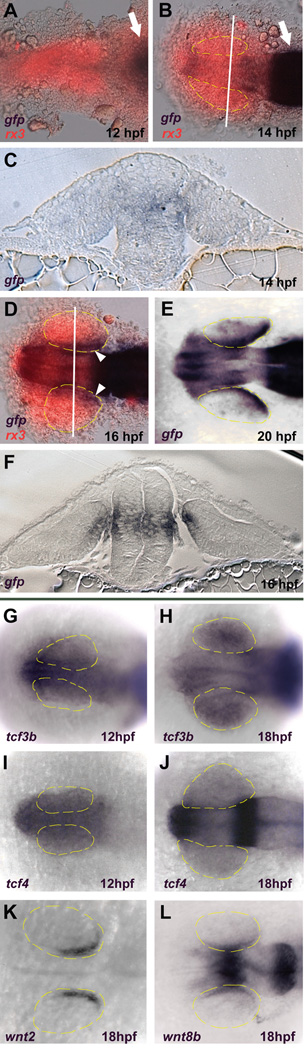
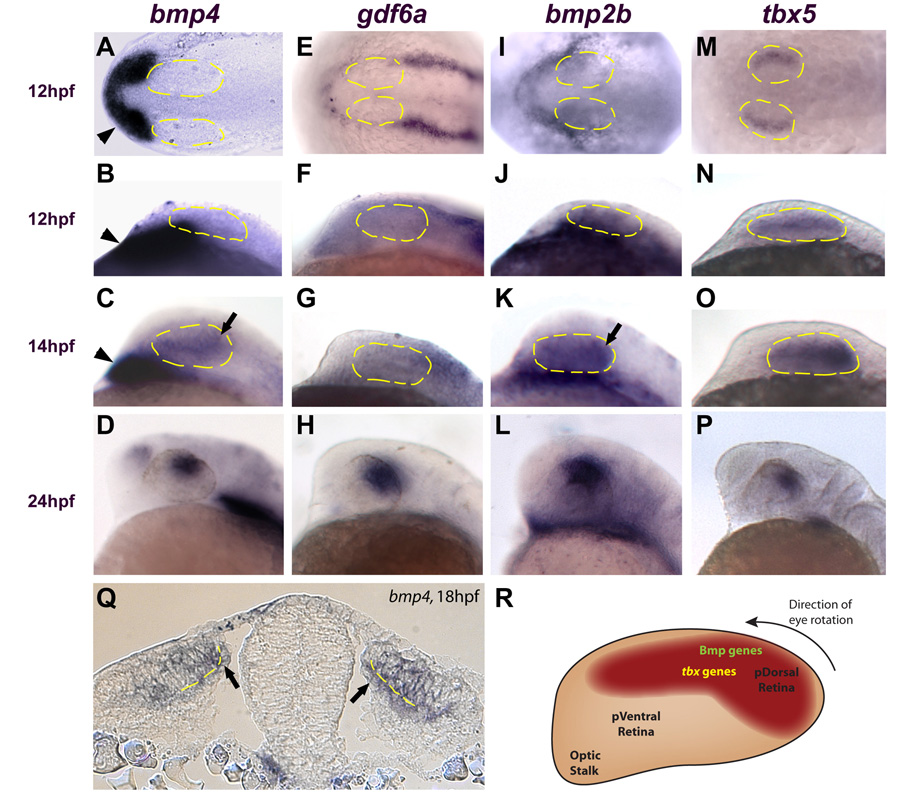
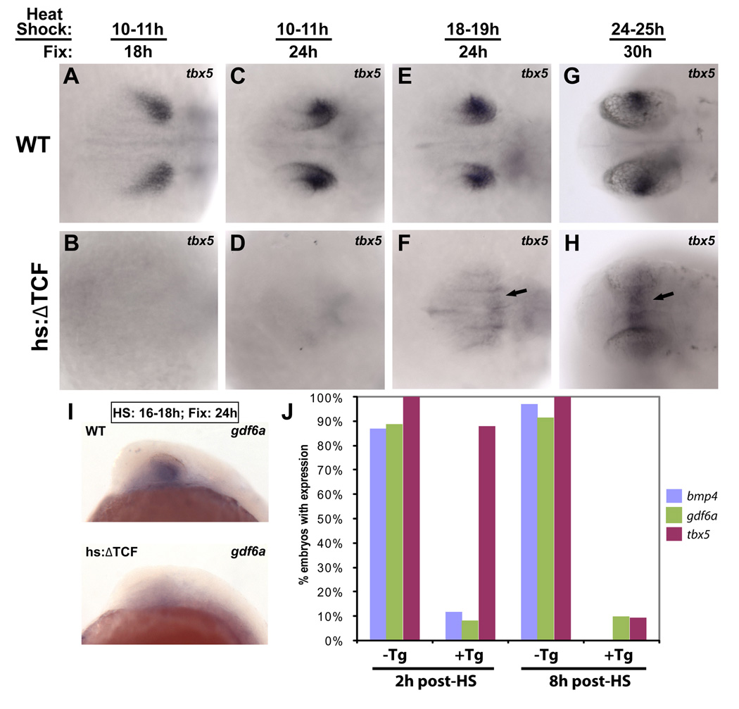
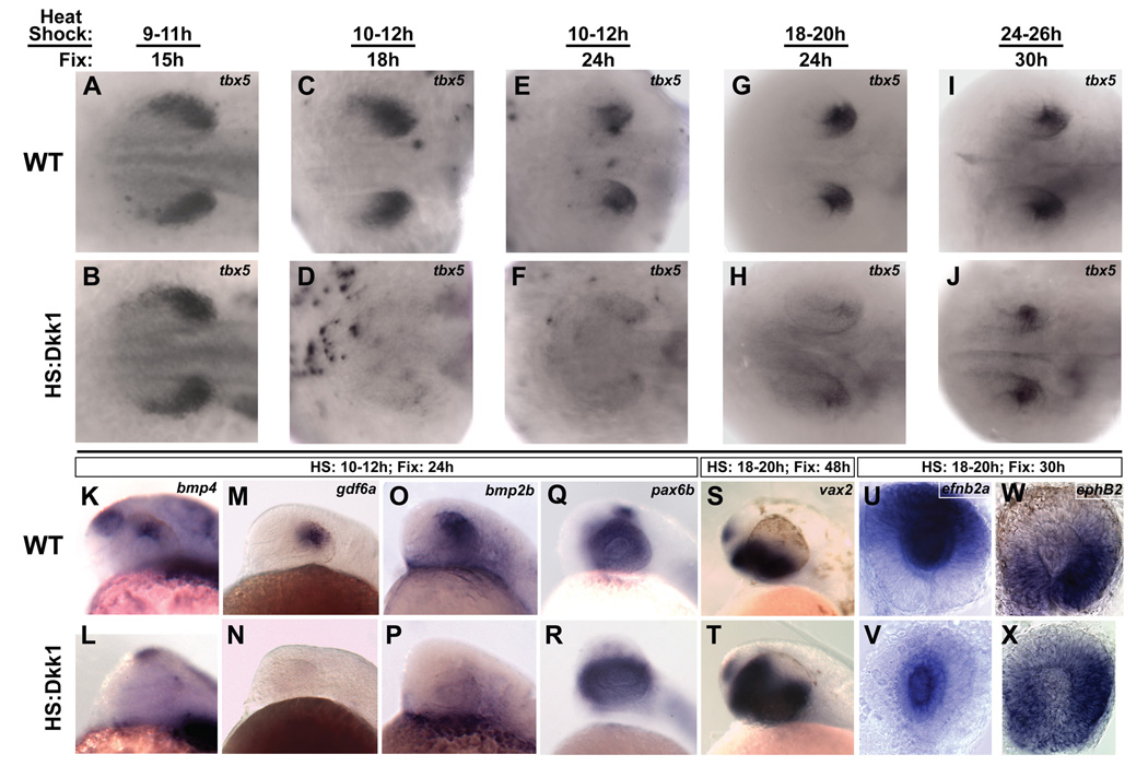
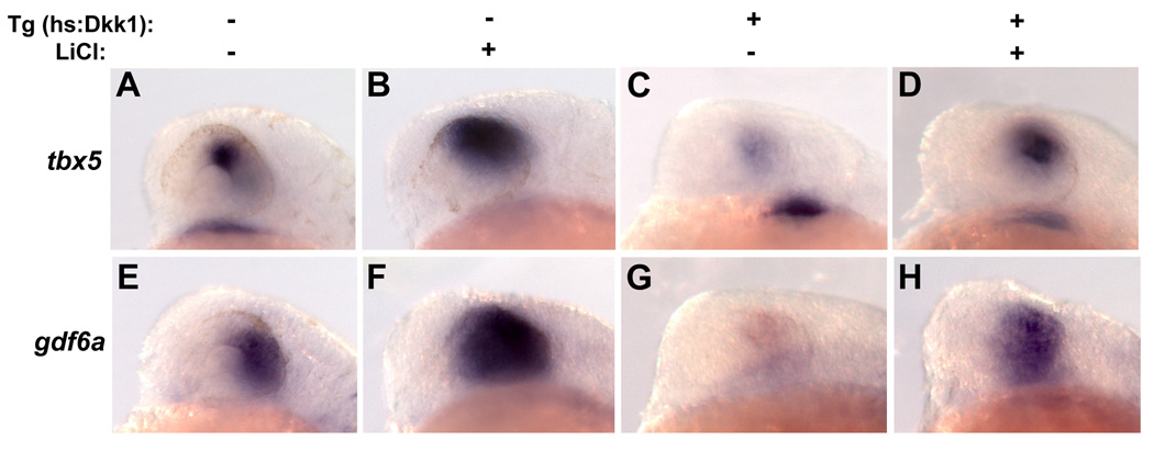
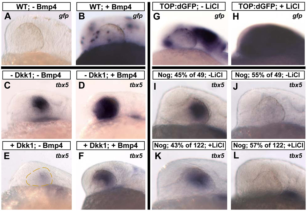
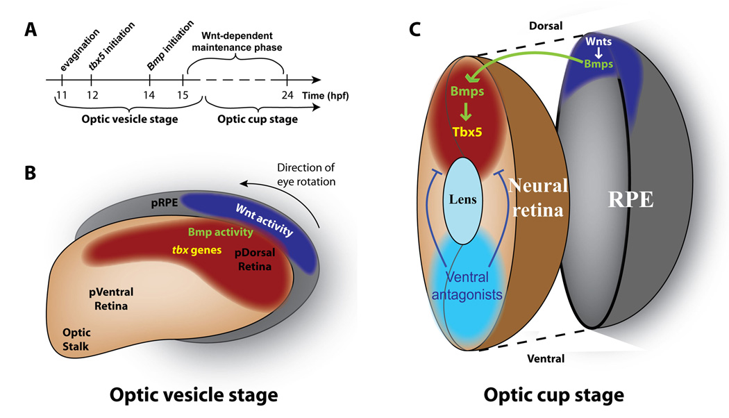
Accurate retinotectal axon pathfinding depends upon the correct establishment of dorsal-ventral retinal polarity. We show that dorsal retinal gene expression is regulated by Wnt signaling in the dorsal retinal pigment epithelium (RPE). We find that a Wnt reporter transgene and Wnt pathway components are expressed in the dorsal RPE beginning at 14-16 hours post-fertilization. In the absence of Wnt signaling, tbx5 and Bmp genes initiate normal dorsal retinal expression but are not maintained. The expression of these genes is rescued by the downstream activation of Wnt signaling, and tbx5 is rescued by Bmp signaling. Furthermore, activation of Wnt signaling cannot rescue tbx5 in the absence of Bmp signaling, suggesting that Wnt signaling maintains dorsal retinal gene expression by regulating Bmp signaling. We present a model in which dorsal RPE-derived Wnt activity maintains the expression of Bmp ligands in the dorsal retina, thus coordinating the patterning of these two ocular tissues.


Figures

Figure 1
Figure 1. Wnt signaling becomes active in the dorso-posterior retinal pigmented epithelium (RPE) between 14–16 hpf
A–F: Expression of the TOP:dGFP Wnt reporter which was detected using in situ hybridization for gfp (blue). In A, B, and D, the embryos were also probed for rx3 expression which marks the eye field (red). A, B: Dorsal views, anterior left. Active Wnt signaling does not extend rostrally past the midbrain-hindbrain boundary (arrows) at 12 hpf and approaches but does not enter the eye field at 14 hpf. C, F: Coronal sections through caudal midbrain/posterior optic vesicles, dorsal up. The lines in B and D indicate the planes of section in C and F, respectively. Active Wnt signaling is seen in the dorso-posterior RPE at 16 hpf, but not at 14 hpf. D, E: Dorsal views, anterior left. Active Wnt signaling is clearly present in the dorso-posterior eye field at 16 and 20 hpf. G–L: Dorsal views, anterior left. G–J: Expression of tcf3b and tcf4 is present in the early eye-field during optic vesicle evagination (12 hpf) and throughout the eye at 18 hpf. K: The Wnt ligand wnt2 is expressed in the dorsal RPE at 18 hpf. L: Expression of wnt8b in the midbrain and RPE at 18 hpf.
Figure 2
Figure 2. Multiple bmp genes and tbx5 are expressed in the retina before canonical Wnt activity
A, E I, M: Dorsal views, anterior left. B–D, F–H, J–L, N–P: Lateral views, dorsal up, anterior left. A–D: bmp4 is expressed in the prechordal mesoderm at 12 and 14 hpf (arrowheads in A–C) but is not expressed in the optic vesicle until 14 hpf (arrow in C). At 24 hpf, bmp4 expression is restricted to the dorsal retina (D). E–L: gdf6a and bmp2b are not expressed in the optic vesicle at 12 hpf (expression of these genes is restricted to the surface ectoderm). Expression of bmp2b is present in the retina at 14 hpf (arrow in K), but gdf6a does not appear in the optic vesicle until 16 hpf (not shown). Both genes are expressed in the dorsal retina at 24 hpf (H, L). M–P: tbx5 expression begins in the optic vesicle at 12 hpf and becomes progressively restricted to the dorsal retina by 24 hpf. Q: Transverse section through the midbrain at 18 hpf. bmp4 is expressed in the presumptive dorsal neural retina and RPE (arrows) Broken yellow lines indicate the interface between the neural retina and RPE. R: Diagram of zebrafish retina at approximately 14 hpf, showing expression domains of bmp genes and tbx5 at this timepoint. At approximately 22 hpf, the entire eye rotates 90° in the direction indicated. Anterior left, dorsal up.
Figure 3
Figure 3. Expression of dorsal retinal genes is lost following repression of Wnt signaling
The Tg(hsp70l:Tcf3-GFP)w26 transgenic zebrafish line, which expresses a dominant-repressor form of Tcf3 (ΔTcf-GFP) upon heat-shock, was used for these experiments. Dorsal views, anterior left. A–H: Embryos were heat-shocked and fixed at the indicated times, and sorted for GFP expression. Repression of Wnt targets led to the downregulation of tbx5 in the dorsal retina at every timepoint. tbx5 expression was upregulated in the dorsal diencephalon at later timepoints (arrows in F, H). I: Expression of gdf6a was also eliminated in embryos expressing ΔTcf at 18 hpf and fixed at 24 hpf. J: To determine the times at which bmp4, gdf6a, and tbx5 are lost in the dorsal retina following repression of Wnt targets, embryos were heat-shocked at 16 hpf and fixed 2 and 8 hours later. bmp4 and gdf6a expression were strongly reduced at the 2 hour timepoint, while tbx5 was still expressed. By 8 hours, expression of all three genes was lost.
Figure 4
Figure 4. Wnt signaling is required for the maintenance of dorsal retinal identity
The Tg(hsp70l:dkk1-GFP)w32 transgenic zebrafish line, which expresses the secreted Wnt pathway inhibitor Dkk1 upon heat-shock, was used for these experiments. A–J: Dorsal views, anterior left. Embryos were heat-shocked and fixed at the indicated times, and sorted for GFP expression. A, B: Embryos fixed just before Wnt signaling becomes active in dorsal RPE express tbx5 normally, showing that tbx5 expression initiates properly in the absence of Wnt signaling. C–J: Inhibition of Wnt signaling caused a strong downregulation of tbx5 at the early timepoints, with a weaker effect at the last timepoint. This demonstrates a requirement for Wnt signaling in the maintenance of tbx5. K–T: Lateral views, dorsal up, anterior left. K–P: Expression of the Bmp ligands bmp4, gdf6a, and bmp2b are lost from the dorsal retina following Wnt inhibition, suggesting loss of dorsal character. Q–T: pax6b is expressed normally and vax2 expands dorsally, suggesting a ventralized retina. U–X: Whole eyes, dorsal up. Expression of ephrinB2a is downregulated in the dorsal retina, but maintained in the lens, and ephB2 expands dorsally following Wnt inhibition.
Figure 5
Figure 5. Activation of Wnt signaling rescues loss of dorsal eye markers in Dkk1-expressing embryos
Dkk1-expressing embryos were treated with the Wnt pathway activator LiCl (150 mM) from 11–14 hpf, heat-shocked at 12 hpf, and fixed at 24 hpf. Expression of tbx5 (A–D) and gdf6a (E–H) were analyzed by in situ hybridization. LiCl led to an expansion of tbx5 and gdf6a expression in embryos not expressing Dkk1 (B, F), and a rescue of tbx5 and gdf6a in embryos expressing Dkk1 (D, H). Lateral views, dorsal up, anterior left.
Figure 6
Figure 6. Bmp signaling is downstream of Wnt signaling in the maintenance of dorsal retinal markers
A–F: Bmp4 can rescue dorsal retinal markers in the absence of Wnt signaling. hs:Dkk1 and control WT embryos at the one-cell stage were injected with a construct that expresses Bmp4 upon heat-shock (pDestTol2pA2;hsp70l:bmp4-IRES-GFP), heat-shocked at 12 hpf, and fixed at 24 hpf. A, B: To illustrate transgene expression following heat-shock, in situ hybridization was performed for gfp. Widespread clonal expression was observed in the retinas in 85% of embryos. C–F: Expression of Bmp4 caused a clear expansion of tbx5 in embryos not expressing Dkk1 (D) and rescue of tbx5 in embryos expressing Dkk1 (F). G–L: Activation of Wnt signaling does not rescue dorsal markers in the absence of Bmp signaling. Embryos heterozygous for the Tg(hsp70l:nog3)fr14 transgene, which express the Bmp pathway inhibitor Noggin upon heat-shock, were outcrossed to TL strain fish and placed in 200 mM LiCl at 18 hpf. A 2 hour heat-shock was performed at 18 hpf, and embryos were fixed at 24 hpf. To illustrate Wnt pathway activation, TOP:dGFP embryos were similarly treated with LiCl from 18–24 hpf and gfp detected by in situ hybridization (G, H). For hs:Noggin embryos untreated with LiCl, 55% of 49 embryos lost expression of tbx5 (J), and for embryos treated with LiCl, 57% of 122 embryos lost expression of tbx5 (L), showing that activation of Wnt signaling cannot rescue tbx5 in the absence of Bmp signaling. Lateral views, dorsal up, anterior left.
Figure 7
Figure 7. Model for Wnt-dependent maintenance of dorsal identity
A: Timeline of dorsal identity establishment. Optic vesicles evaginate from the anterior neural tube at 11 hpf. Soon thereafter, at 12 hpf, the first dorsally restricted marker, tbx5, begins to be expressed. Expression of Bmp ligands within the dorsal retina and RPE begins at 14 hpf, and a phase of Wnt-dependent dorsal identity maintenance begins between 14–16 hpf. B: Diagram of the optic vesicle at approximately 14 hpf. Wnt signaling becomes active in the dorso-posterior presumptive RPE at this point, Bmp signaling is active in the presumptive dorsal retina and presumptive RPE, and tbx5 is expressed in the presumptive dorsal retina. At about 22 hpf, the entire eye undergoes an approximately 90° rotation so that the posterior eye assumes its final dorsal position. C: Model of Wnt-mediated maintenance of dorsal retinal identity. A Wnt signaling center in the dorsal RPE maintains bmp expression in the dorsal RPE and retina. Bmp signaling then maintains tbx5 expression in the dorsal retina. Simultaneously, inhibitors such as Bmp antagonists and transcription factors like Vax2 act to limit the extent of dorsal identity. Abbreviations: pRPE: presumptive retinal pigmented epithelium; pDorsal Retina: presumptive dorsal retina; p Ventral Retina: presumptive ventral retina.

Similar articles

Cited by

References

    1. Arce L, Yokoyama NN, Waterman ML. Diversity of LEF/TCF action in development and disease. Oncogene. 2006;25:7492–7504. - PubMed
    1. Asai-Coakwell M, French CR, Berry KM, Ye M, Koss R, Somerville M, Mueller R, van Heyningen V, Waskiewicz AJ, Lehmann OJ. GDF6, a novel locus for a spectrum of ocular developmental anomalies. Am J Hum Genet. 2007;80:306–315. - PMC - PubMed
    1. Barbieri AM, Broccoli V, Bovolenta P, Alfano G, Marchitiello A, Mocchetti C, Crippa L, Bulfone A, Marigo V, Ballabio A, et al. Vax2 inactivation in mouse determines alteration of the eye dorsal-ventral axis, misrouting of the optic fibres and eye coloboma. Development. 2002;129:805–813. - PubMed
    1. Behesti H, Holt JK, Sowden JC. The level of BMP4 signaling is critical for the regulation of distinct T-box gene expression domains and growth along the dorso-ventral axis of the optic cup. BMC Dev Biol. 2006;6:62. - PMC - PubMed
    1. Bonner J, Gribble SL, Veien ES, Nikolaus OB, Weidinger G, Dorsky RI. Proliferation and patterning are mediated independently in the dorsal spinal cord downstream of canonical Wnt signaling. Dev Biol. 2008;313:398–407. - PMC - PubMed

Publication types

MeSH terms

Substances