. 2009 Feb;83(3):1368-78.
doi: 10.1128/JVI.01263-08. Epub 2008 Nov 12.

Pre-P is a secreted glycoprotein encoded as an N-terminal extension of the duck hepatitis B virus polymerase gene

Affiliations

Pre-P is a secreted glycoprotein encoded as an N-terminal extension of the duck hepatitis B virus polymerase gene

Feng Cao et al. J Virol. 2009 Feb.

Abstract

The duck hepatitis B virus (DHBV) pregenomic RNA is a bicistronic mRNA encoding the core and polymerase proteins. Thirteen AUGs (C2 to C14) and 10 stop codons (S1 to S10) are located between the C1 AUG for the core protein and the P1 AUG that initiates polymerase translation. We previously found that the translation of the DHBV polymerase is initiated by ribosomal shunting. Here, we assessed the biosynthetic events after shunting. Translation of the polymerase open reading frame was found to initiate at the C13, C14, and P1 AUGs. Initiation at the C13 AUG occurred through ribosomal shunting because translation from this codon was cap dependent but was insensitive to blocking ribosomal scanning internally in the message. C13 and C14 are in frame with P1, and translation from these upstream start codons led to the production of larger isoforms of P. We named these isoforms "pre-P" by analogy to the pre-C and pre-S regions of the core and surface antigen open reading frames. Pre-P was produced in DHBV16 and AusDHBV-infected duck liver and was predicted to exist in 80% of avian hepadnavirus strains. Pre-P was not encapsidated into DHBV core particles, and the viable strain DHBV3 cannot make pre-P, so it is not essential for viral replication. Surprisingly, we found that pre-P is an N-linked glycoprotein that is secreted into the medium of cultured cells. These data indicate that DHBV produces an additional protein that has not been previously reported. Identifying the role of pre-P may improve our understanding of the biology of DHBV infection.


Figures

FIG. 1.
FIG. 1.
DHBV3 pgRNA genomic organization. (Top) DHBV3 pgRNA showing the location of the ORFs, ɛ, the cap, and the poly(A) tail. The pgRNA is 3.3-kb polyadenylated RNA with a terminal redundancy of approximately 270 nt. (Bottom) Enlarged view of the 5′ end of the DHBV3 pgRNA. C1 and P1 are the AUGs for C and P. CO2, C2-C14, and P2 are AUG codons. S1 to S10 are stop codons. The open boxes are small ORFs upstream of P. NsiI and EcoRI are sites for the insertion of the BamHI-SL. The lines below the genomic diagram represent the shunting mechanism employed for the initiation of P translation. Dashed line, scanning ribosomes; thin lines, shunting ribosomes; black arrows, ribosomes translating C or P. The nucleotide positions of key sites examined in this study are shown.
FIG. 2.
FIG. 2.
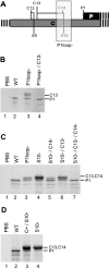
Translation of the P ORF can initiate at the C13, C14, and P1 AUGs. LMH cells were transfected with DHBV C derivatives, and P was detected by Western analysis of lysates on day 1 posttransfection. (A) Mutation sites on the DHBV3 pgRNA. (B) Accumulation of P from the C13, C14, and P1 AUGs in the P1loop background. (C) Accumulation of P from the C13, C14, and P1 AUGs in the S10 background. PBS, pBluescript (empty vector control); WT, wild type (DHBV C). The codons from which the various P isoforms initiate are indicated. (D) Accumulation of pre-P from genomes in which C1 is intact (C+/S10) or ablated (S10).
FIG. 3.
FIG. 3.
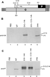
The pre-P region is translated in the S10 background. An HA epitope tag was inserted 21 nt downstream of C13 in frame with P, the expression vectors were transfected into LMH cells, and P expression was measured by Western analysis of cell lysates. (A) Diagram of mutation sites on the DHBV3 pgRNA. (B) Detection of pre-P by anti-HA antibody 3F10. (C) Detection of P and pre-P by anti-P antibody MAb 11. PBS, pBluescript (empty vector control); WT, wild type (DHBV C); HA, HA epitope tag. The codons from which the various P isoforms initiate are indicated.
FIG. 4.
FIG. 4.
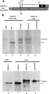
Pre-P is produced during infection of ducks. P was immunoprecipitated from liver lysates from ducks infected with DHBV3, DHBV16, or AusDHBV and detected by Western blotting. (A) Diagram of the DHBV16 and AusDHBV pgRNAs. (B) Detection of pre-P in DHBV-infected duck livers by Western blotting. PBS, pBluescript (empty vector control); WT, wild type (DHBV C).
FIG. 5.
FIG. 5.
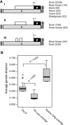
Genomic structure of the pre-P region in avian hepadnaviral sequences. (A) Diagrams showing the structures of the pre-P region (open box) in 57 different avian hepadnavirus sequences in GenBank. Pre-P was predicted to exist in 46 of the 57 isolates. (B) The pre-P region is under selective pressures. The genetic distances among the pre-P terminal protein domains and the spacer domains of P were compared, and the statistical significance was evaluated using one-way analysis of variance with a Games-Howell post hoc test. Pre-P, pre-P sequences from C13 to P1; TP-Core, overlap between the terminal protein domain of P and the C protein; Spacer-SAg, overlap between the spacer domain of P and the S-Ag.
FIG. 6.
FIG. 6.
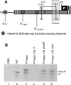
Initiation at C13 occurs by ribosomal shunting. The BamHI-SL was inserted into the 5′ end of the pgRNA coding sequences or at the NsiI and EcoRI sites between C1 and P1 on the P1loop background, the genomic expression plasmids were transfected into LMH cells, and P levels were measured by Western analysis of lysates on day 1 posttransfection. (A) Diagram of BamHI-SL insertion sites on DHBV3 pgRNA. (B) Representative experiment. PBS, pBluescript (empty vector control); WT, wild type (DHBV C).
FIG. 7.
FIG. 7.
Pre-P is not encapsidated. Cell lysates and subviral cores were isolated from transfected LMH cells and liver lysates, and serum-derived virions were isolated from infected and noninfected ducks. Cores were permeabilized and treated with micrococcal nuclease prior to the detection of P by Western blotting. Lanes 4, 5, 10, and 11, DHBV cores; lanes 1 to 3 and 6 to 9, cell lysates. Dpol, P expression vector; CDNA 3.1, empty vector control; WT, wild type (DHBV C); LMH, untransfected LMH cells.
FIG. 8.
FIG. 8.
Pre-P is N-glycosylated. Genomic expression plasmids carrying the DHBV derivatives were transfected into LMH cells. One day posttransfection, cell lysates were harvested and treated or mock treated with Endo H or PNGase F, and P and pre-P were detected by Western blotting. The mobilities of pre-P and P are indicated. (A) Pre-P is N-glycosylated. Odd-numbered lanes, mock treated; even-numbered lanes, Endo H or PNGase F treated, as indicated. (B) Deglycosylated pre-P and P are similar in size. Lanes 1 and 3, mock treated; lane 2, Endo H treated. The mobilities of pre-P initiating from C13, P initiating from P1, and N-terminally truncated P initiating from P2 are indicated. WT, wild type. (C) DHBV16 pre-P is glycosylated. LMH cells were transfected with the indicated genomes (lanes 1 to 4 and 7 to 12), or LMH D2 cells that have been stably transfected with DHBV16 were employed; DHBV16 is in lanes 1 to 6, and DHBV3 is in lanes 7 to 10. The even-numbered lanes were treated with PNGase F, and the odd-numbered lanes were mock treated. The mobility difference between pre-P and deglycosylated pre-P is smaller for DHBV16 than for DHBV3 because DHBV16 pre-P does not appear to be proteolytically trimmed. PBS, pBluescript. (D) Mapping the signal sequence. Odd-numbered lanes, mock treated; even-numbered lanes, Endo H or PNGase F treated, as indicated.
FIG. 9.
FIG. 9.
Pre-P is secreted. DHBV genomic expression plasmids were transfected into LMH cells, or stably transfected LMH D2 cells were employed. Cell lysates were harvested, the medium was collected, and P and pre-P were then immunoprecipitated. The lysates and medium-derived immunocomplexes were treated with Endo H or PNGase F or were mock treated, and P was detected by Western blotting. The mobilities of pre-P and P are indicated. Odd-numbered lanes, mock treated; even-numbered lanes, Endo H or PNGase F treated, as indicated. (A) DHBV3 pre-P is secreted. (B) DHBV16 pre-P is secreted. Pre-P was immunoprecipitated from medium from LMH cells, LMH D2 cells, or LMH cells transfected with the wild-type DHBV16 genome (lanes 2 to 4) and detected by Western analysis. A lysate from cells transfected with wild-type DHBV16 (lane 1) was employed as a mobility control for pre-P.

Similar articles

Cited by

References

    1. Beck, J., H. Bartos, and M. Nassal. 1997. Experimental confirmation of a hepatitis B (HBV) epsilon-like bulge and loop structure in avian HBV RNA encapsidation signals. Virology 227500-504. - PubMed
    1. Bouchard, M. J., and R. J. Schneider. 2004. The enigmatic X gene of hepatitis B virus. J. Virol. 7812725-12734. - PMC - PubMed
    1. Bouchard, M. J., L. H. Wang, and R. J. Schneider. 2001. Calcium signaling by HBx protein in hepatitis B virus DNA replication. Science 2942376-2378. - PubMed
    1. Cao, F., and J. E. Tavis. 2006. Suppression of mRNA accumulation by the duck hepatitis B virus reverse transcriptase. Virology 350475-483. - PubMed
    1. Chang, S. F., H. J. Netter, M. Bruns, R. Schneider, K. Frolich, and H. Will. 1999. A new avian hepadnavirus infecting snow geese (Anser caerulescens) produces a significant fraction of virions containing single-stranded DNA. Virology 26239-54. - PubMed

Publication types

LinkOut - more resources