. 2009 Jan;83(2):969-80.
doi: 10.1128/JVI.01663-08. Epub 2008 Nov 12.

African swine fever virus blocks the host cell antiviral inflammatory response through a direct inhibition of PKC-theta-mediated p300 transactivation

Affiliations

African swine fever virus blocks the host cell antiviral inflammatory response through a direct inhibition of PKC-theta-mediated p300 transactivation

Aitor G Granja et al. J Virol. 2009 Jan.

Abstract

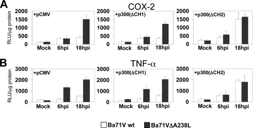
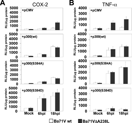
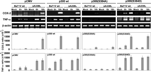
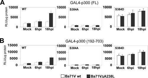
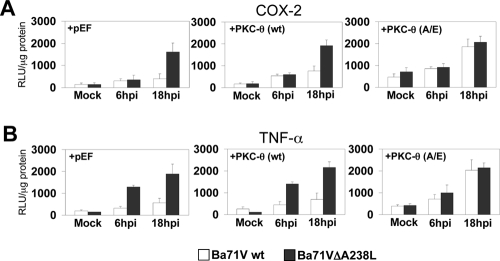
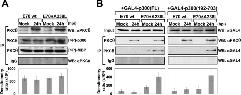
During a viral infection, reprogramming of the host cell gene expression pattern is required to establish an adequate antiviral response. The transcriptional coactivators p300 and CREB binding protein (CBP) play a central role in this regulation by promoting the assembly of transcription enhancer complexes to specific promoters of immune and proinflammatory genes. Here we show that the protein A238L encoded by African swine fever virus counteracts the host cell inflammatory response through the control of p300 transactivation during the viral infection. We demonstrate that A238L inhibits the expression of the inflammatory regulators cyclooxygenase-2 (COX-2) and tumor necrosis factor alpha (TNF-alpha) by preventing the recruitment of p300 to the enhanceosomes formed on their promoters. Furthermore, we report that A238L inhibits p300 activity during the viral infection and that its amino-terminal transactivation domain is essential in the A238L-mediated inhibition of the inflammatory response. Importantly, we found that the residue serine 384 of p300 is required for the viral protein to accomplish its inhibitory function and that ectopically expressed PKC-theta completely reverts this inhibition, thus indicating that this signaling pathway is disrupted by A238L during the viral infection. Furthermore, we show here that A238L does not affect PKC-theta enzymatic activity, but the molecular mechanism of this viral inhibition relies on the lack of interaction between PKC-theta and p300. These findings shed new light on how viruses alter the host cell antiviral gene expression pattern through the blockade of the p300 activity, which represents a new and sophisticated viral mechanism to evade the inflammatory and immune defense responses.


Figures

FIG. 1.
FIG. 1.
p300 protein structure and functional domains. Diagram of the p300 coactivator protein showing its functional domains clustered in two different regulatory regions: the amino-terminal region, containing the CH1 and KIX functional domains and also a bromo domain, and the carboxyl-terminal region, containing the CH2 and CH3 domains, which are part of the HAT catalytic domain. Both regulatory regions can act independently and interact simultaneously with the transcriptional machinery and/or with different transcription factors to build the transcriptional activity mediated by these coactivators. The amino acid (aa) position of each functional domain is also indicated in the scheme.
FIG. 2.
FIG. 2.
Effect of p300 overexpression in the transcriptional activation of COX-2 and TNF-α promoters. (A) Vero cells were transfected with COX-2 (left panel) or TNF-α (right panel) promoter reporter plasmids (250 ng/106 cells), together with pCI-p300wt or p300 HAT deletion mutant (pCI-p300ΔHAT) expression plasmids (1 μg/106 cells). Sixteen hours after transfection, the cells were mock infected (−) or infected (+) with the Vero-adapted isolate Ba71Vwt or Ba71VΔA238L at a MOI of 5 PFU/cell. Whole-cell extracts were prepared at 24 hpi and assayed for luciferase activity. Extracts were normalized to Renilla luciferase activity as described in Materials and Methods. RLU per μg of protein from triplicate transfections (means ± standard deviations) are shown. (B) Jurkat-pcDNA or Jurkat-A238L cells were transiently transfected with COX-2 (left panel) or TNF-α (right panel) promoter reporter plasmids (250 ng/106 cells), together with pCI-p300wt or p300 HAT deletion mutant (pCI-p300ΔHAT) expression plasmids (1 μg/106 cells). Sixteen hours after transfection, the cells were cultured in the absence (−) or presence (+) of 15 ng/ml of PMA plus 1 μM Ion (PMA/Ion) during 4 h and assayed for luciferase activity. Extracts were normalized to Renilla luciferase activity as described in Materials and Methods. Results from triplicate assays are shown in RLU per μg of protein (means ± standard deviations).
FIG. 3.
FIG. 3.
A238L inhibits the transcriptional initiation complex recruitment to the enhancer elements located in COX-2 and TNF-α. Nuclear extracts from Jurkat-pcDNA and Jurkat-A238L cells untreated (−) or treated (+) with 15 ng/ml of PMA plus 1 μM Ion (PMA/Ion) for 4 h were incubated with the indicated biotinylated probes. (A) Distal and proximal NFAT binding sites (d-NFAT and p-NFAT) and distal and proximal NF-κB binding sites (d-NFκB and p-NFκB) from the COX-2 promoter. (B) κ1 and κ2 NF-κB binding sites, the (−157)NFAT binding site, and the composite element CRE/κ3 from the TNF-α promoter. The complexes were pulled down with streptavidin-agarose beads, as described in Materials and Methods. After extensive washing, proteins in the complexes were analyzed by Western blotting using antibodies against p300, RNA Pol II, or TFIIB. Inputs were also included to show the presence of the analyzed proteins in the nuclear protein extracts. A control probe was included to rule out unspecific binding of the analyzed proteins (data not shown).
FIG. 4.
FIG. 4.
A238L inhibits p300 transactivation in ASFV-infected Vero cells. Vero cells were cotransfected with the reporter plasmid GAL4-luc (250 ng/106 cells) and with the following GAL4-fused mutant constructs of p300, at a final concentration of 250 ng/106 cells: panel A, full-length p300 [GAL4-p300(FL)]; panel B, mutant p300 construct encoding amino acids from 192 to 703 [GAL4-p300(192-703)]; and panel C, mutant p300 construct encoding amino acids from 1239 to 2414 [GAL4-p300(1239-2414)]. Sixteen hours after transfection, Vero cells were infected with Ba71Vwt or Ba71VΔA238L at a MOI of 5 PFU/cell. A sample was infected and also activated with 15 ng/ml of PMA, to check the functionality of the constructs assayed. At the indicated poststimulation times, whole-cell extracts were prepared and luciferase activity was assayed. Extracts were normalized to Renilla luciferase activity as described in Materials and Methods. RLU per μg of protein from triplicate transfections (means ± standard deviations) are shown.
FIG. 5.
FIG. 5.
CH1 and KIX domains of p300 are required in the A238L-mediated inhibition of COX-2 and TNF-α gene expression. Vero cells were transiently cotransfected with COX-2 (A) or TNF-α (B) promoter reporter plasmids (250 ng/106 cells) as indicated in the figure, together with pCMV-p300(ΔCH1) or pCMV-p300(ΔCH2) deletion mutant expression plasmids (1 μg/106 cells). Sixteen hours after transfection, Vero cells were infected with Ba71Vwt or Ba71VΔA238L at a MOI of 5 PFU/cell. At the indicated postinfection times, whole-cell extracts were prepared and luciferase activity was assayed. Extracts were normalized to Renilla luciferase activity as described in Materials and Methods. RLU per μg of protein from triplicate transfections (means ± standard deviations) are shown.
FIG. 6.
FIG. 6.
Serine 384 within the CH1 regulatory domain of p300 is essential in the A238L-mediated inhibition of the transcription of proinflammatory genes. Vero cells were transiently cotransfected with COX-2 (A) or TNF-α (B) promoter reporter plasmids (250 ng/106 cells) as indicated in the figure, together with pCMV-GAL4-p300wt or the mutant pCMV-GAL4-p300(S384A) or pCMV-GAL4-p300(S384D) expression plasmids (1 μg/106 cells). Sixteen hours after transfection, Vero cells were infected with Ba71Vwt or Ba71VΔA238L at a MOI of 5 PFU/cell. At the indicated postinfection times, whole-cell extracts were prepared and luciferase activity was assayed. Extracts were normalized to Renilla luciferase activity as described in Materials and Methods. RLU per μg of protein from triplicate transfections (means ± standard deviations) are shown.
FIG. 7.
FIG. 7.
p300 amino-terminal serine 384 role on the endogenous COX-2 and TNF-α mRNA expression inhibited by A238L during the ASFV infection. Vero cells were transfected with the different full-length p300 expression vectors: mid-left panel, p300wt; mid-right panel, p300(S384A) mutant; far right panel, p300(S384D) mutant; and far left panel, empty pCMV as a control. Sixteen hours after the transfection, the cells were infected with Ba71Vwt or Ba71VΔA238L viruses, and at the indicated postinfection times, total RNA was isolated and analyzed by reverse transcriptase PCR to measure COX-2 and TNF-α mRNA levels. A control using oligonucleotides for β-actin was included to rule out differences in PCR amplification. A control of viral p72 (capsid protein) and A238L were also used to rule out differences in the infectivity (data not shown). Amplified DNA was separated on agarose gels. The densitometric analysis shows the ratio between amplified COX-2 or TNF-α mRNA and the β-actin loading control present in each sample, from three independent experiments (means ± standard deviations).
FIG. 8.
FIG. 8.
Mutation of p300 serine 384 to alanine results in a dominant negative, whereas mutation of serine 384 to aspartic acid results in a constitutively active version of the amino-terminal TAD of p300. Vero cells were cotransfected with the reporter plasmid GAL4-luc (250 ng/106 cells) and with the following GAL4-fused mutant constructs of p300, at a final concentration of 250 ng/106 cells: GAL4-p300wt and the S384A mutant or S384D mutant, in both the full-length (A) and amino-terminal [GAL4-p300(192-703)] (B) constructs, as indicated in the figure. Sixteen hours after transfection, Vero cells were infected with Ba71Vwt or Ba71VΔA238L at a MOI of 5 PFU/cell. At the indicated postinfection times, whole-cell extracts were prepared and luciferase activity was assayed. Extracts were normalized to Renilla luciferase activity as described in Materials and Methods. RLU per μg of protein from triplicate transfections (means ± standard deviations) are shown.
FIG. 9.
FIG. 9.
PKC-θ overexpression recovers the normal expression levels of COX-2 and TNF-α inhibited by A238L. Vero cells were transiently cotransfected with COX-2 (A) or TNF-α (B) promoter reporter plasmids (250 ng/106 cells) as indicated in the figure, together with pEF-PKC-θ(wt) or the constitutively active mutant pEF-PKC-θ(A/E) expression plasmids (1 μg/106 cells). Sixteen hours after transfection, Vero cells were infected with Ba71Vwt or Ba71VΔA238L at a MOI of 5 PFU/cell. At the indicated postinfection times, whole-cell extracts were prepared and luciferase activity was assayed. Extracts were normalized to Renilla luciferase activity as described in Materials and Methods. RLU per μg of protein from triplicate transfections (means ± standard deviations) are shown.
FIG. 10.
FIG. 10.
A238L interferes with the association between PKC-θ and the amino-terminal region of p300 in ASFV-infected macrophages. (A) Porcine alveolar primary macrophages were mock infected or infected for 24 h with E70wt or E70ΔA238L, at a MOI of 5 PFU/cell. Then, nuclear extracts were prepared and used for immunoprecipitation (IP) with 4 μg of rabbit polyclonal specific antibody against PKC-θ or rabbit preimmune normal IgG as a negative control of immunoprecipitation. Immunoprecipitated PKC-θ was used in an in vitro kinase assay, using as substrate either purified p300 or purified MBP. Proteins were separated by SDS-8% PAGE and developed by autoradiography. Densitometric analysis shows the ratio between phosphorylated [32P]p300 and immunoprecipitated PKC-θ, from three independent experiments (means ± standard deviations). (B) Porcine alveolar macrophages were transfected with either the GAL4-p300(FL) expression plasmid (left panels) or the GAL4-p300(192-703) mutant expression plasmid (right panels). Twenty-four hours later, the cells were mock infected or infected for 24 h with E70wt or E70ΔA238L, at a MOI of 5 PFU/cell. Then, nuclear extracts were prepared and immunoprecipitated with 4 μg of rabbit polyclonal specific antibody against PKC-θ or rabbit preimmune normal IgG as a negative control of immunoprecipitation. Immunoprecipitates were analyzed by Western blotting (WB) with the same antibody (αPKCθ) to determine the levels of the kinase in the precipitate and with an anti-GAL4 antibody (αGAL4) to detect the levels of PKC associated with p300(FL) (left panels) or p300(192-703) (right panels). The densitometric analysis shows the ratio between coimmunoprecipitated PKC and total GAL4-p300 present in the precipitate, from three independent experiments (means ± standard deviations).

Similar articles

Cited by

References

    1. Ait-Si-Ali, S., D. Carlisi, S. Ramirez, L. C. Upegui-Gonzalez, A. Duquet, P. Robin, B. Rudkin, A. Harel-Bellan, and D. Trouche. 1999. Phosphorylation by p44 MAP kinase/ERK1 stimulates CBP histone acetyl transferase activity in vitro. Biochem. Biophys. Res. Commun. 262157-162. - PubMed
    1. Arany, Z., D. Newsome, E. Oldread, D. M. Livingston, and R. Eckner. 1995. A family of transcriptional adaptor proteins targeted by the E1A oncoprotein. Nature 37481-84. - PubMed
    1. Arany, Z., W. R. Sellers, D. M. Livingston, and R. Eckner. 1994. E1A-associated p300 and CREB-associated CBP belong to a conserved family of coactivators. Cell 77799-800. - PubMed
    1. Bannister, A. J., and T. Kouzarides. 1995. CBP-induced stimulation of c-Fos activity is abrogated by E1A. EMBO J. 144758-4762. - PMC - PubMed
    1. Bannister, A. J., T. Oehler, D. Wilhelm, P. Angel, and T. Kouzarides. 1995. Stimulation of c-Jun activity by CBP: c-Jun residues Ser63/73 are required for CBP induced stimulation in vivo and CBP binding in vitro. Oncogene 112509-2514. - PubMed

Publication types

MeSH terms