. 2009 Jan;83(2):1105-14.
doi: 10.1128/JVI.01439-08. Epub 2008 Nov 12.

A single amino acid substitution in a segment of the CA protein within Gag that has similarity to human immunodeficiency virus type 1 blocks infectivity of a human endogenous retrovirus K provirus in the human genome

Affiliations

A single amino acid substitution in a segment of the CA protein within Gag that has similarity to human immunodeficiency virus type 1 blocks infectivity of a human endogenous retrovirus K provirus in the human genome

David J Heslin et al. J Virol. 2009 Jan.

Abstract

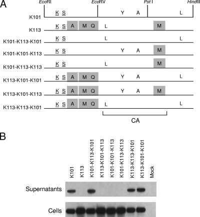
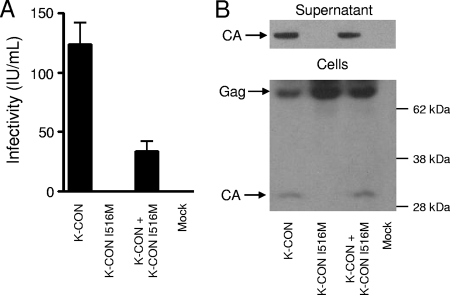
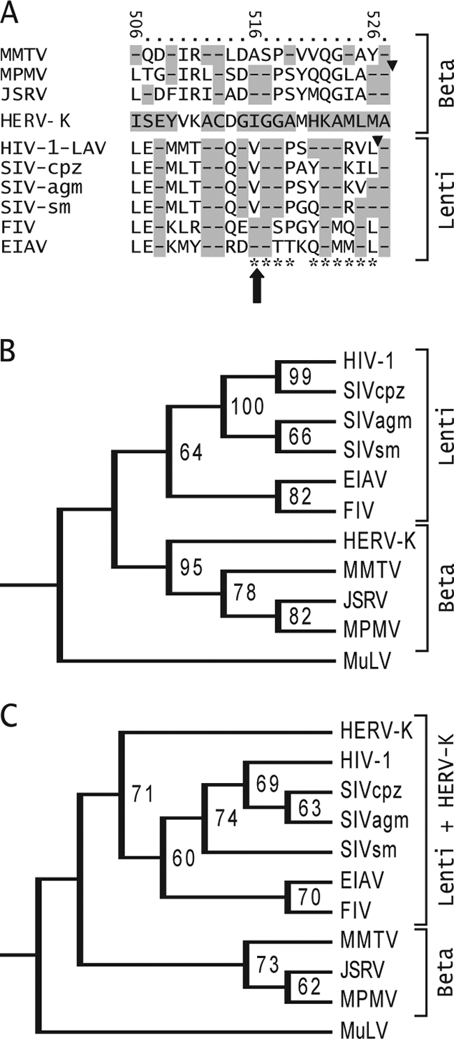
Human endogenous retrovirus K (HERV-K) is the most intact retrovirus in the human genome. However, no single HERV-K provirus in the human genome today appears to be infectious. Since the Gag protein is the central component for the production of retrovirus particles, we investigated the abilities of Gag from two HERV-K proviruses to support production of virus-like particles and viral infectivity. HERV-K113 has full-length open reading frames for all viral proteins, while HERV-K101 has a full-length gag open reading frame and is expressed in human male germ cell tumors. The Gag of HERV-K101 allowed production of viral particles and infectivity, although at lower levels than observed with a consensus sequence Gag. Thus, including HERV-K109, at least two HERV-K proviruses in human genome today have functional Gag proteins. In contrast, HERV-K113 Gag supported only very low levels of particle production, and no infectivity was detectable due to a single amino acid substitution (I516M) near the extreme C terminus of the CA protein within Gag. The sequence of this portion of HERV-K CA showed similarities to that of human immunodeficiency virus type 1 and other primate immunodeficiency viruses. The extreme C terminus of CA may be a general determinant of retrovirus particle production. In addition, precise mapping of the defects in HERV-K proviruses as was done here identifies the key polymorphisms that need to be analyzed to assess the possible existence of infectious HERV-K alleles within the human population.


Figures

FIG. 1.
FIG. 1.
Structures of the HERV-K Gag plasmids and positions of the individual HERV-K Gag mutations. (A) pCRU5-X-HA contained JSRV sequences, including the viral LTRs and env gene. The U3 region of the 5′ LTR was replaced with the CMV IE promoter to drive expression in mammalian cells. pCRU5-HERV-K gag was generated by cloning a HERV-K gag cDNA isolated from MGCT cells (13) into pCRU5-X-HA using the EcoRI (R) and HindIII (H) sites. The cloned HERV-K gag was sequenced and identified as K101 gag. K-CON, K108, and K113 gag genes were also cloned into this vector at the same positions. XbaI (X) and Nru I (N) sites were used to delete almost all of JSRV env from upstream of the start codon, up to and including the aspartate residue at position 577 (D577), to create pCRU5-K101 Δenv. pCRU5-K101-FS env was engineered by introducing a frameshift mutation (*) at the XmaI (M) site located at the alanine residue at position 207 (A207) in JSRV env. (B) Amino acid sequences of K101, K108, and K113 Gag proteins are aligned with the K-CON Gag sequence. The amino acid changes relative to the consensus Gag are indicated wherever one of the genomic proviral Gag proteins differs. The positions of the individual differences are also indicated. The corresponding amino acids for each position are also shown for K-CON. Amino acid variants that are shared among multiple HERV-K proviruses (shared polymorphisms) are underlined. The remaining amino acids are found only in the provirus indicated and not in any other HERV-K proviruses in the human genome. The bracket at the bottom shows the position of CA in Gag based on similarity to MPMV CA. (C) The infectious CHKCG plasmid (34) is composed of the HERV-K consensus sequence with the U3 region of the 5′ LTR replaced by the CMV IE promoter to drive expression in mammalian cells. The env gene was disrupted with a CMV IE-GFP cassette, resulting in a nonfunctional env. The SalI (S) and the PshAI (P) restriction sites used to clone K101 and K113 gag into CHKCG are indicated.
FIG. 2.
FIG. 2.
Expression of HERV-K Gag proteins and viral particle production. (A) Western blot analysis of 293T cells transfected with 2 μg of pCRU5 vector containing K-CON, K101, K108, or K113 was performed on both culture supernatants and whole-cell lysates (Cells) 36 h posttransfection. Proteins were separated by SDS-polyacrylamide gel electrophoresis, and the approximately 75-kDa unprocessed Gag product was visualized using a rabbit antiserum directed against HERV-K Gag, a secondary anti-rabbit-IgG-HRP antibody, and electrochemiluminescence. (B) Western blot analysis of 293T cells transfected with 10 μg of pCRU5 containing K101, K108, or K113 gag was performed on both cultured supernatants and whole-cell lysates (Cells) 48 h posttransfection. (C) Western blot analysis of 293T cells transfected with 2 μg of pCRU5 containing K-CON, K101, K108, or K113 gag, incubated at either 28°C or 37°C, was carried out 48 h posttransfection. (D) Western blot analysis of 293T cells transfected with 2 μg of pCRU5 containing K-CON, K101/Δenv, or K101/FS env was carried out 24 h and 48 h posttransfection.
FIG. 3.
FIG. 3.
K113 Gag proteins accumulate at the plasma membrane. (A) Representative example of staining pattern of cells expressing K-CON and K-113 Gag. Cells were fixed at 24 h posttransfection and processed as described in Materials and Methods. HERV-K Gag is in green, while nuclei (DAPI) are in blue. Gag is visible as dispersed dots in the cytoplasm (Cyt) or in the cytoplasm and the plasma membrane (Cyt+PM) of transfected cells. The majority of cells expressing K113 show accumulation of Gag staining at the plasma membrane. Panels a to f show representative examples of experiments performed in Cos cells, while panels g and h show 293T cells. Bars, 10 μm. (B) Quantitation of Gag staining pattern in confocal microscopy of HeLa, Cos and 293T cells expressing K-CON, K-101, and K-113 Gag at 24 h posttransfection. The graph represents the number of cells in which Gag proteins display a staining dispersed in the cytoplasm (Cyt) versus cells showing both cytoplasmic and intense plasma membrane staining (PM+Cyt), as shown in panel A. Approximately 100 cells were randomly chosen in each experiment. Data are averages from two independent experiments.
FIG. 4.
FIG. 4.
Infectivity and virus production by K101, K108, and K113 Gag proteins. (A) Infectious titers of HERV-K particles produced by transfection of 293T cells with Rec, VSV-G, and the indicated CHKCG-gag vectors. Producer cell supernatants were used to infect 293T target cells 48 h posttransfection, and GFP foci were counted 72 h postinfection and expressed as infectious units per milliliter (IU/ml). The data are means plus standard deviations (n = 3 [K101 and K113] or 2 [K-CON]). (B) Western blot analysis was performed on the producer cells used for panel A. Supernatants were collected, and whole-cell lysates were prepared from the producer cells at the time of target cell infection (48 h posttransfection). The approximately 75-kDa Gag precursor and 30-kDa processed Gag (CA) proteins are indicated. Only the processed form of Gag was detected in the culture supernatants.
FIG. 5.
FIG. 5.
Genetic mapping of the K113 defect in viral particle production. (A) K101 and K113 Gag proteins are depicted to indicate the amino acid residues at which they differ from the K-CON Gag sequence. Underlined amino acids represent shared polymorphisms among HERV-K Gag proteins. The unique mutations in K113 Gag are indicated by shaded boxes. The restriction sites for EcoRI (R), EcoRV (V), PstI (P), and HindIII (H) were used to generate the recombinants between K101 gag and K113 gag that are shown. The bracket at the bottom shows the position of CA in Gag based on similarity to MPMV CA. (B) Western blot analysis of 293T cells transfected with 2 μg of K101, K113, or the indicated recombinant gag pCRU5 vectors was performed on both cultured supernatants and whole-cell lysates (Cells) 24 h posttransfection. The approximately 75-kDa Gag unprocessed HERV-K Gag product was visualized using a rabbit antiserum directed against HERV-K Gag, a secondary anti-rabbit-IgG-HRP antibody, and enhanced chemiluminescence.
FIG. 6.
FIG. 6.
Infectivity and particle production of HERV-K viruses containing the K113 Gag I516M mutation. (A) Infectious titers of HERV-K particles produced by transfection of 293T cells with Rec, VSV-G, and the indicated CHKCG vectors. Producer cell supernatants were used to infect 293T target cells 48 h posttransfection, and GFP foci were counted 72 h postinfection and expressed as infectious units per milliliter (IU/ml). The data are means plus standard deviations (n = 4). (B) Western blot analysis of the producer cells used for panel A. Supernatants were collected, and whole-cell lysates were prepared at the time of target cell infection (48 h posttransfection). The approximately 75-kDa Gag precursor and 30-kDa processed CA proteins are indicated. Only the processed form was detected in the culture supernatants.
FIG. 7.
FIG. 7.
Similarity between the extreme C termini of HERV-K and other retroviruses. (A) Alignment of HERV-K Gag amino acids 506 to 527 with the corresponding Gag sequences of other retroviruses. The amino acids of HERV-K Gag are indicated, and the numbers at the top refer to position within HERV-K Gag. Other betaretroviruses are shown above HERV-K, and lentiviruses are shown below it. The large arrow shows the position of the I516M mutation unique to K113. Triangles show the positions of the protease cleavage sites that generate the C termini of MPMV and HIV-1 CA. Highlighted dashes indicate amino acid identity. The HIV-1 Gag amino acid positions identified by Melamed et al. to be highly detrimental to HIV-1 Gag particle production and infectivity are indicated by asterisks at the bottom (45). Retroviruses included in the alignment are as follows (GenBank accession numbers indicated): mouse mammary tumor virus (MMTV, AAF31467), MPMV (P07567), JSRV (CAA01899), HIV-1 LAV (AAB59747), simian immunodeficiency virus (SIV) from chimpanzee (SIV-cpz, AA013959), SIV from African green monkey (SIV-agm, BAF32563), SIV from sooty mangabey (SIV-sm, AAC68655), feline immunodeficiency virus (FIV, CAA48157), and equine anemia infectious virus (EIAV, AAA43011). (B) A phylogenetic tree of retrovirus CA amino acids sequences was generated using maximum parsimony in PAUP, version 4B10. Bootstrapping values were derived from 10,000 replications. The tree was rooted using the gammaretrovirus Moloney murine leukemia virus (AAB59942) as the outlier. (C) Tree generated using maximum parsimony on the amino acids at the extreme C terminus of CA. The sequences shown in panel A were used, along with the corresponding amino acids of Moloney murine leukemia virus (AAB59942) as the outlier.

Similar articles

Cited by

References

    1. Accola, M. A., S. Hoglund, and H. G. Gottlinger. 1998. A putative alpha-helical structure which overlaps the capsid-p2 boundary in the human immunodeficiency virus type 1 Gag precursor is crucial for viral particle assembly. J. Virol. 722072-2078. - PMC - PubMed
    1. Accola, M. A., B. Strack, and H. G. Gottlinger. 2000. Efficient particle production by minimal Gag constructs which retain the carboxy-terminal domain of human immunodeficiency virus type 1 capsid-p2 and a late assembly domain. J. Virol. 745395-5402. - PMC - PubMed
    1. Adamson, C. S., S. D. Ablan, I. Boeras, R. Goila-Gaur, F. Soheilian, K. Nagashima, F. Li, K. Salzwedel, M. Sakalian, C. T. Wild, and E. O. Freed. 2006. In vitro resistance to the human immunodeficiency virus type 1 maturation inhibitor PA-457 (Bevirimat). J. Virol. 8010957-10971. - PMC - PubMed
    1. Adamson, C. S., and E. O. Freed. 2007. Human immunodeficiency virus type 1 assembly, release, and maturation. Adv. Pharmacol. 55347-387. - PubMed
    1. Armbruester, V., M. Sauter, E. Krautkraemer, E. Meese, A. Kleiman, B. Best, K. Roemer, and N. Mueller-Lantzsch. 2002. A novel gene from the human endogenous retrovirus K expressed in transformed cells. Clin. Cancer Res. 81800-1807. - PubMed

Publication types

Substances

LinkOut - more resources