. 2009 Jan;101(1):8-19.
doi: 10.1152/jn.90934.2008. Epub 2008 Nov 12.

Maintenance of lateral stability during standing and walking in the cat

Affiliations

Maintenance of lateral stability during standing and walking in the cat

A Karayannidou et al. J Neurophysiol. 2009 Jan.

Abstract

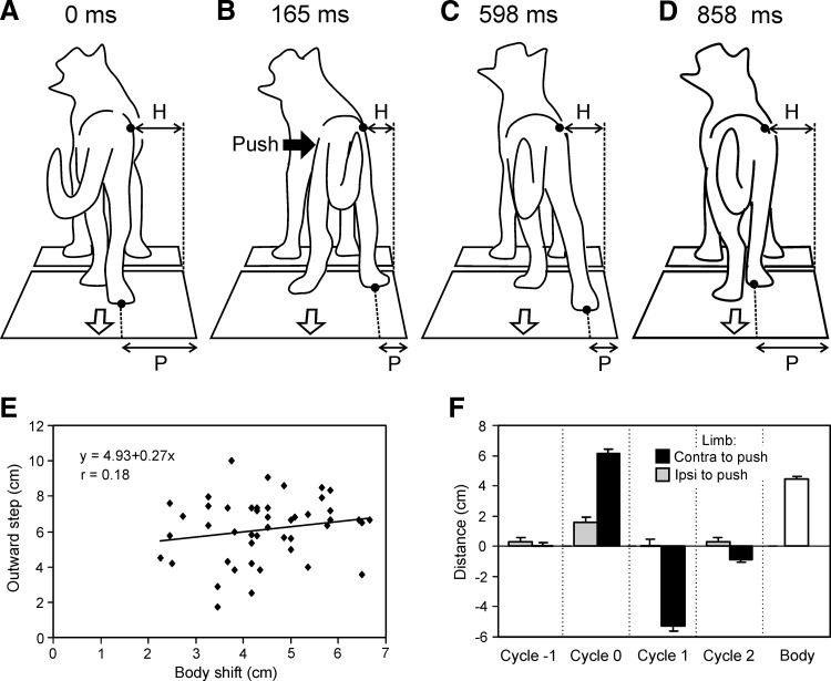
During free behaviors animals often experience lateral forces, such as collisions with obstacles or interactions with other animals. We studied postural reactions to lateral pulses of force (pushes) in the cat during standing and walking. During standing, a push applied to the hip region caused a lateral deviation of the caudal trunk, followed by a return to the initial position. The corrective hindlimb electromyographic (EMG) pattern included an initial wave of excitation in most extensors of the hindlimb contralateral to push and inhibition of those in the ipsilateral limb. In cats walking on a treadmill with only hindlimbs, application of force also caused lateral deviation of the caudal trunk, with subsequent return to the initial position. The type of corrective movement depended on the pulse timing relative to the step cycle. If the force was applied at the end of the stance phase of one of the limbs or during its swing phase, a lateral component appeared in the swing trajectory of this limb. The corrective step was directed either inward (when the corrective limb was ipsilateral to force application) or outward (when it was contralateral). The EMG pattern in the corrective limb was characterized by considerable modification of the hip abductor and adductor activity in the perturbed step. Thus the basic mechanisms for balance control in these two forms of behavior are different. They perform a redistribution of muscle activity between symmetrical limbs (in standing) and a reconfiguration of the base of support during a corrective lateral step (in walking).


Figures

FIG. 1.
FIG. 1.
Experimental design for testing lateral stability in different motor tasks. AC: postural tests during walking with hindlimbs. A and B: the side and back view of the walking cat, respectively, with mechanical sensors monitoring the anterior–posterior (AP) position of the left hindlimb, the medial–lateral (ML) position of the left hindlimb (LHL), and the ML position of the posterior part of the body (Bd). C: scheme of the step cycle (as derived from the AP position of limbs), with the phases of one-limb and two-limb support (nonshaded and shaded intervals, respectively). D: postural tests during walking with forelimbs. E and F: postural tests during standing. In different tests, lateral pushes were applied either in the hip area (A and E) or in the shoulder area (D). Correspondingly, video recording of the cat was done either from behind (A and E) or from the front (D).
FIG. 2.
FIG. 2.
Kinematic responses to push in the standing cat. AD: 4 sequential positions of the standing cat in response to the rightward push. The lateral deviation of the caudal trunk (H) is indicated. A: the position just before push. B: the maximal deviation of the caudal part of the trunk. C: under the effect of postural corrective mechanism, the trunk moved toward the initial position but passed it over. D: the trunk returned to the initial position. E: 3 examples of temporal patterns of postural responses (trunk deviation, H) with overshot. F: the maximal body deviation caused by push (Peak), the overshoot (Overshoot), and the final body position (Final) (mean ± SE), averaged over 21 responses recorded in 3 cats. Positive and negative values correspond to displacement in the direction of force and in the opposite direction, respectively.
FIG. 3.
FIG. 3.
Electromyographic (EMG) responses to push in the standing cat. A: a representative example of EMG responses in 6 selected hindlimb muscles during standing. B: average EMG responses to a leftward push in 8 hindlimb muscles (averaging over 12 trials in cat 3; mean ± SE).
FIG. 4.
FIG. 4.
Lateral oscillations of the trunk during stepping. A: averaged lateral oscillations of the pelvis in cat 2 (n = 51) and cat 4 (n = 49), in relation to a step cycle of the right hindlimb. B: positive correlation between the peak-to-peak (P-P) excursions of the posterior part of the trunk and the width of the step (the distance between the stance paws in the frontal plane), illustrated for cat 2 (n = 31).
FIG. 5.
FIG. 5.
Outward step. AD: 4 characteristic positions of the walking cat 1 resulting from the push applied in the hip region toward the right. A: configuration just before force application, at the moment when the right leg finished the stance phase. B: the push caused a rightward displacement of the trunk and landing of the foot at a more lateral position (P) than during unperturbed locomotion. C: extension of the leg in the subsequent stance phase caused a leftward displacement of the caudal trunk toward the initial position. D: during the subsequent swing phase, the right leg returned to the normal ML position. E and F: characteristics of outward steps of the hindlimbs. E: correlation between the postural perturbation (Body shift) and the response to this perturbation (Outward step). F: mean values (±SE) characterizing the lateral component of step in sequential cycles of the hindlimbs. Positive and negative values correspond to outward and inward displacement of the limb, respectively. Designation of cycles: (−1), the cycle before push; (0), the cycle including push; (1), the cycle next to the affected cycle; and (2), the cycle next to (1). The mean value of push-caused trunk displacement is also given (Body). For E and F, n = 3 and n = 46.
FIG. 6.
FIG. 6.
EMG pattern of the outward step. A: a representative example of EMG responses to a leftward push (Force) in 4 selected hindlimb muscles during walking (cat 3). Shown also are AP position of the left and right hindlimbs (L AP and R AP), ML position of the left hindlimb (L ML), and ML position of the trunk (Body). B and C: average EMG responses to a push in 8 hindlimb muscles. EMGs shown are for the limb performing the outward step (B) and for the opposite limb (C). Blue traces indicate EMGs recorded in the cycle preceding the outward step and red traces are those in the cycle with the outward step—averaging over 12 trials in cat 3. Stance and swing phases are shown by the filled (black) and the empty bar, respectively. The red bar indicates the push position in the cycle.
FIG. 7.
FIG. 7.
Inward step. AD: 4 characteristic positions of the walking cat resulting from the push applied in the hip region toward the right. A: configuration just before force application, at the end of the left leg's stance phase. B: the push caused a rightward displacement of the trunk and landing of the foot at a more medial position (P) than during unperturbed locomotion. C: leftward displacement of the caudal trunk toward the initial position in the subsequent stance phase. D: return to the normal ML position of the right leg in the subsequent swing phase. E and F: characteristics of inward steps of the hindlimbs. E: correlation between the postural perturbation (Body shift) and the response to this perturbation (Inward step). F: mean values characterizing the lateral component of step in sequential cycles of the hindlimbs. Designations as in Fig. 5. The mean value of push-caused trunk displacement is also given (Body). For E and F, n = 3 and n = 46.
FIG. 8.
FIG. 8.
EMG pattern of the inward step. A: a representative example of EMG responses to a rightward push in 4 selected hindlimb muscles during walking (cat 3) (designations as in Fig. 6A). B and C: average EMG responses to a push in 8 hindlimb muscles. EMGs shown are for the limb performing the inward step (A) and for the opposite limb (B). Blue traces indicate EMGs recorded in the cycle preceding the inward step and red traces are those in the cycle with the inward step—averaging over 12 trials in cat 3. Stance and swing phases are shown by the filled (black) and empty bar, respectively. The red bar indicates the push position in the cycle.
FIG. 9.
FIG. 9.
Duration of the cycle and its components in normal and perturbed steps. A: outward steps (n = 4, n = 36). B: inward steps (n = 4, n = 47). For the leg performing the lateral step, the mean value of cycle duration and the mean value of duration of swing and stance phases are shown. This was done for control (cycle before push) and for the affected cycle (which started with the lateral step). The values in affected cycles only slightly differed from control, but the differences were statistically significant (paired t-test, P < 0.05).
FIG. 10.
FIG. 10.
Characteristics of outward and inward steps of the forelimbs. A and C: correlation between the postural perturbation (Body shift) and the response to this perturbation (Lateral step) for outward steps (A) and inward steps (C). B and D: mean values characterizing the lateral component of step in sequential cycles of the forelimbs, for outward steps (B) and inward steps (D). Designation of cycles as in Fig. 5. The mean value of push-caused trunk displacement is also given (Body). In AD, n = 3. In A, n = 28; in B, n = 29; in C, n = 22; in D, n = 25.

Similar articles

Cited by

References

    1. Barbeau H, Rossignol S. Recovery of locomotion after chronic spinalization in the adult cat. Brain Res 412: 84–95, 1987. - PubMed
    1. Bard P Studies on the cerebral cortex. I. Localized control of placing and hopping reactions in the cat and their normal management by small cortical remnants. Arch Neurol Psychiatry 30: 40–74, 1933.
    1. Bard P, Macht MB. The behavior of chronically decerebrate cats. In: Neurological Basis of Behavior, edited by Wolstenholme GEW, O'Connor CM. London: Churchill, 1958, p. 55–71.
    1. Beloozerova IN, Sirota MG, Orlovsky GN, Deliagina TG. Activity of pyramidal tract neurons in the cat during postural corrections. J Neurophysiol 93: 1831–1844, 2005. - PubMed
    1. Beloozerova IN, Zelenin PV, Popova LB, Orlovsky GN, Grillner S, Deliagina TG. Postural control in the rabbit maintaining balance on the tilting platform. J Neurophysiol 90: 3783–3793, 2003. - PubMed

Publication types