Review
. 2008 Nov 12;28(46):11760-7.
doi: 10.1523/JNEUROSCI.3864-08.2008.

The immediate early gene arc/arg3.1: regulation, mechanisms, and function

Affiliations
Review

The immediate early gene arc/arg3.1: regulation, mechanisms, and function

Clive R Bramham et al. J Neurosci. .

Abstract

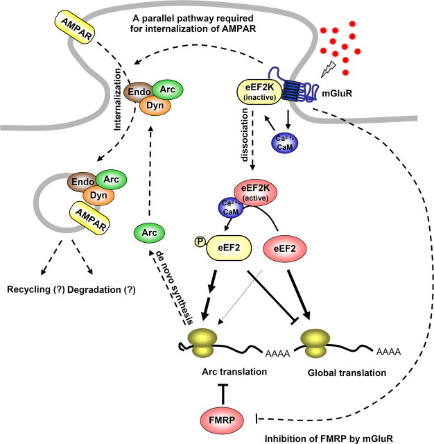
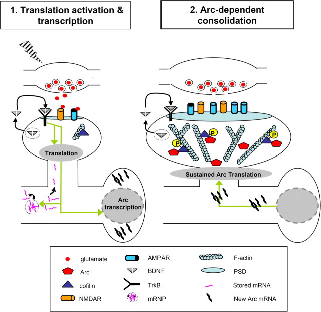
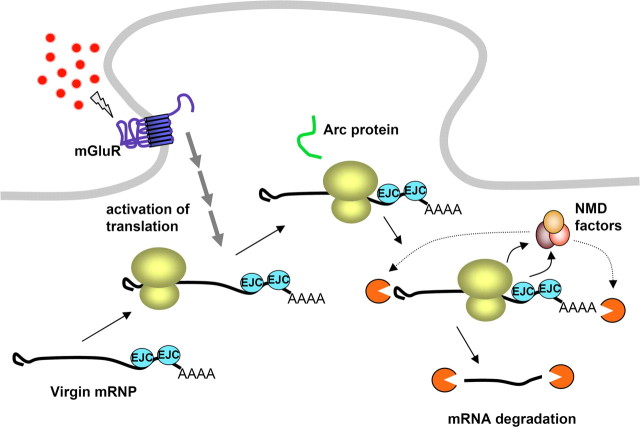
In a manner unique among activity-regulated immediate early genes (IEGs), mRNA encoded by Arc (also known as Arg3.1) undergoes rapid transport to dendrites and local synaptic translation. Despite this intrinsic appeal, relatively little is known about the neuronal and behavioral functions of Arc or its molecular mechanisms of action. Here, we attempt to distill recent advances on Arc spanning its transcriptional and translational regulation, the functions of the Arc protein in multiple forms of neuronal plasticity [long-term potentiation (LTP), long-term depression (LTD), and homeostatic plasticity], and its broader role in neural networks of behaving animals. Worley and colleagues have shown that Arc interacts with endophilin and dynamin, creating a postsynaptic trafficking endosome that selectively modifies the expression of AMPA-type glutamate receptors at the excitatory synapses. Both LTD and homeostatic plasticity in the hippocampus are critically dependent on Arc-mediated endocytosis of AMPA receptors. LTD evoked by activation of metabotropic glutamate receptors depends on rapid Arc translation controlled by elongation factor 2. Bramham and colleagues have shown that sustained translation of newly induced Arc mRNA is necessary for cofilin phosphorylation and stable expansion of the F-actin cytoskeleton underlying LTP consolidation in the dentate gyrus of live rats. In addition to regulating F-actin, Arc synthesis maintains the activity of key translation factors during LTP consolidation. This process of Arc-dependent consolidation is activated by the secretory neurotrophin, BDNF. Moore and colleagues have shown that Arc mRNA is a natural target for nonsense-mediated mRNA decay (NMD) by virtue of its two conserved 3'-UTR introns. NMD and other related translation-dependent mRNA decay mechanisms may serve as critical brakes on protein expression that contribute to the fine spatial-temporal control of Arc synthesis. In studies in behaving rats, Guzowski and colleagues have shown that location-specific firing of CA3 and CA1 hippocampal neurons in the presence of theta rhythm provides the necessary stimuli for activation of Arc transcription. The impact of Arc transcription in memory processes may depend on the specific context of coexpressed IEGs, in addition to posttranscriptional regulation of Arc by neuromodulatory inputs from the amygdala and other brain regions. In sum, Arc is emerging as a versatile, finely tuned system capable of coupling changes in neuronal activity patterns to diverse forms of synaptic plasticity, thereby optimizing information storage in active networks.


Figures

Figure 1.
Figure 1.
Model of Arc function in mGluR-LTD. Group I mGluRs activate eEF2K via calcium–calmodulin (CaM). eEF2K phosphorylates eEF2, which inhibits elongation generally but rapidly increases de novo Arc translation. Arc forms a complex with endophilin2/3 (Endo) and dynamin (Dyn) and induces the internalization of AMPAR. FMRP inhibits the translation of Arc at the basal state. Arc induction alone is not sufficient for mGluR-LTD, indicating that mGluR activates another pathway that is required to internalize AMPAR. In Fmr1 KO mice, the synthesis of Arc protein is constitutively de-repressed, and de novo synthesis of Arc is not required for mGluR-LTD. This figure was adapted with permission from Park et al. (2008), their Figure 8. FMRP, Fragile-X mental retardation protein.
Figure 2.
Figure 2.
Model of Arc function in LTP consolidation. In this two-stage model, translation activation is followed by Arc-dependent consolidation. In Translation activation: HFS (lightning bolt) causes activation of postsynaptic NMDAR receptors and TrkB receptors, leading to local translation activation as well as Arc transcription. Translation is modulated through regulation of translation factor activity, mobilization of mRNA (e.g., αCaMKII) from mRNPs and fine-localization of the translational machinery. Arc-dependent consolidation: Arc mRNA is transported to dendrites and translated in activated spines. Sustained translation of dendritically transported Arc is necessary for cofilin phosphorylation and local F-actin expansion. This figure was adapted with permission from Bramham and Wells (2007), their Figure 3. mRNP, Messenger ribonucleoprotein particle; TrkB, Tropomyosin-related kinase B; αCaMKII, α-subunit calcium/calmodulin-dependent protein kinase II.
Figure 3.
Figure 3.
Translation-dependent degradation of Arc mRNA via the NMD pathway. Virgin (not yet translated) Arc mRNA is stable and can accumulate in dendrites. However, juxtaposition of the ribosome with two EJCs in the 3′-UTR at the termination of translation leads to activation of NMD and rapid mRNA degradation. This mechanism could potentially limit each Arc mRNA to producing just a single copy of Arc protein.
Figure 4.
Figure 4.
Network regulation and function of Arc. a, Confocal projection image from CA3 of rat hippocampus showing the subcellular distribution of Arc RNA (red) using high-sensitivity FISH [Guzowski et al. (2005), their Fig. 1, reproduced with permission]. Note the presence of the two intense sites of Arc synthesis (transcription foci; white arrows) in the nucleus (DAPI; blue color) and the somatic/dendritic Arc mRNA (yellow arrows). The subcellular distribution of Arc RNA provides a “time stamp” of neural activity history for two behavioral epochs and provides the basis for the catFISH imaging approach. b, Complex network analysis (CNA) of gene expression changes associated with distinct stages of learning and memory [Miyashita et al. (2008), their Fig. 4, reproduced with permission]. Rats were trained in the spatial water maze task, and RNA from dissected dorsal hippocampi was used for microarray analysis. Details of the analysis are provided elsewhere (Miyashita et al., 2008b), but the data shown here represent a subset of differentially regulated genes (at p < 0.05 with fold changes of <1.5 or >1.5 relative to caged control values). The groups included the following: caged control (rats killed from the home cage); day 1, 30 min (rats killed 30 min after the first training session; early learning); day 1, 180 min (rats killed 180 min after an initial training session; early learning); day 5, 30 min (rats killed 30 min on the fifth day of training; stable reference memory retrieval); and day 5, reversal, 30 min (rats killed 30 min after spatial reversal learning on the fifth day of training; extinction of previous reference memory and new reversal learning). The CNA graph shows gene expression differentially regulated for all groups: day 1, 30 min (purple square); day 1, 180 min (purple octagon); day 5, 30 min (green square); and day 5, reversal, 30 min (green rounded-square). The lines (“edges”) connect behavioral groups with the genes (blue circle nodes) that are differentially regulated in that group. Red and green edges indicate upregulation and downregulation of gene expression, respectively, relative to caged control baseline levels. Note that the several genes in the center of the network are regulated across multiple stages of learning, as demonstrated by the high connectivity of these nodes. Of these “core” genes, several known IEGs are indicated as orange circle nodes (c-fos, Nr4a1, Homer 1a, junB, and zif268), and Arc is shown as a larger yellow circle node. In contrast, low-connectivity genes, represented by blue circles connected to only one group, are regulated only by a single behavior (i.e., in a distinct stage of learning and memory: “state specific”). The degree of similarity or difference of the gene expression networks between any two of the behavior groups (stages of learning and memory) can be culled from the number of shared and distinct regulated genes. Note that whereas Arc is upregulated 30 min after training on day 1, day 5, and day 5, reversal, the cohort of differentially regulated genes is distinct for each behavioral group. Thus, the “molecular context” of Arc RNA expression changes across stages of spatial water maze learning.

References

    1. Aicardi G, Argilli E, Cappello S, Santi S, Riccio M, Thoenen H, Canossa M. Induction of long-term potentiation and depression is reflected by corresponding changes in secretion of endogenous brain-derived neurotrophic factor. Proc Natl Acad Sci U S A. 2004;101:15788–15792. - PMC - PubMed
    1. Bingol B, Schuman EM. Synaptic protein degradation by the ubiquitin proteasome system. Curr Opin Neurobiol. 2005;15:536–541. - PubMed
    1. Bingol B, Schuman EM. Activity-dependent dynamics and sequestration of proteasomes in dendritic spines. Nature. 2006;441:1144–1148. - PubMed
    1. Bloomer WA, VanDongen HM, VanDongen AM. Activity-regulated cytoskeleton-associated protein Arc/Arg3.1 binds to spectrin and associates with nuclear promyelocytic leukemia (PML) bodies. Brain Res. 2007;1153:20–33. - PubMed
    1. Bourne JN, Harris KM. Balancing structure and function at hippocampal dendritic spines. Annu Rev Neurosci. 2008;31:47–67. - PMC - PubMed

Publication types

MeSH terms

Substances

LinkOut - more resources