

Review
doi: 10.1523/JNEUROSCI.3463-08.2008.
Habenula: crossroad between the basal ganglia and the limbic system
Affiliations
- PMID: 19005047
- PMCID: PMC2613689
- DOI: 10.1523/JNEUROSCI.3463-08.2008
Item in Clipboard
Review
Habenula: crossroad between the basal ganglia and the limbic system
J Neurosci.
.
Abstract
There is a growing awareness that emotion, motivation, and reward values are important determinants of our behavior. The habenula is uniquely positioned both anatomically and functionally to participate in the circuit mediating some forms of emotive decision making. In the last few years there has been a surge of interest in this structure, especially the lateral habenula (LHb). The new studies suggest that the LHb plays a pivotal role in controlling motor and cognitive behaviors by influencing the activity of dopamine and serotonin neurons. Further, dysfunctions of the LHb have also been implicated in psychiatric disorders, such as depression, schizophrenia, and drug-induced psychosis.
Figures
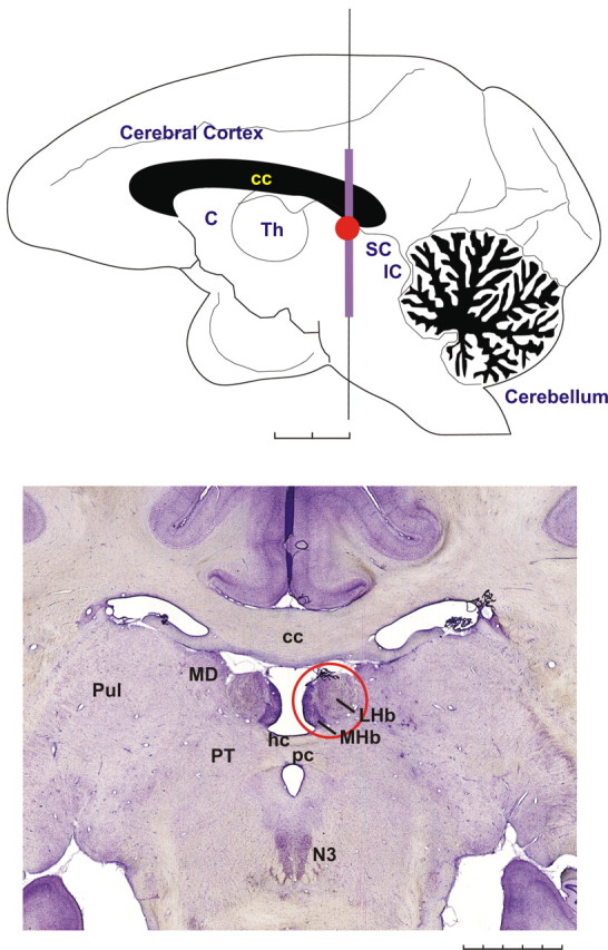
The habenula in the rhesus monkey. Top, The monkey's brain viewed from the mesial side. The location of the habenula is indicated by a red circle. C, Caudate nucleus; Th, thalamus; SC, superior colliculus; IC, inferior colliculus. Scale bar, 5 mm × 2. Bottom, A coronal histological section showing the habenula (red circle). The medially located dark region corresponds to the MHb, whereas the lateral part corresponds to the LHb. The vertical extent of this section corresponds to a violet line in the top figure. MD, Mediodorsal nucleus of the thalamus; Pul, pulvinar; PT, pretectum; N3, oculomotor nucleus; hc, habenular commissure; pc, posterior commissure. Scale bar, 1 mm × 5. This figure was reprinted with permission from Hikosaka (2007a), his Figure 1.
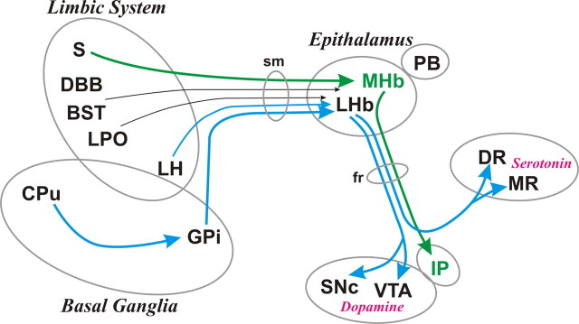
Afferent and efferent connections of the habenula. The MHb, LHb, and pineal body (PB) are collectively called the epithalamus. The MHb receives inputs mainly from the supracommissural septum (S) and sends outputs to the interpeduncular nucleus (IP). The LHb receives inputs mainly from the basal ganglia and limbic regions and sends outputs to the brain structures containing DA neurons and serotonin neurons. Afferent and efferent connections of the habenula are conveyed by the stria medullaris (sm) and fasciculus retroflexus (fr), respectively. Green and blue lines indicate the axonal connections associated with the MHb and LHb, respectively; black lines are associated with both. The thickness of the line implies the strength of the connection. Many other connections are not shown, including reverse connections (e.g., from DR/VTA to LHb). DR, Dorsal raphe; MR, medial raphe; S, septum; DBB, nucleus of diagonal band of Broca; BST, bed nucleus of stria terminalis; LPO, lateral preoptic area; LH, lateral hypothalamus; GPi, globus pallidus internal segment; CPu, caudate and putamen; sm, stria medullaris; fr, fasciculus retroflexus. This figure was modified with permission from Hikosaka (2007a), his Figure 2.
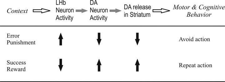
How the LHb control behaviors: a hypothesis. Events would occur from left to right. Upward arrows indicate increases; downward arrows indicate decreases. The sequence is by no means exclusive; for example, DA neuron activity can be influenced by many other inputs not shown here.
References
-
- Andres KH, von Düring M, Veh RW. Subnuclear organization of the rat habenular complexes. J Comp Neurol. 1999;407:130–150. - PubMed
-
- Araki M, McGeer PL, Kimura H. The efferent projections of the rat lateral habenular nucleus revealed by the PHA-L anterograde tracing method. Brain Res. 1988;441:319–330. - PubMed
-
- Bell RL, Omelchenko N, Sesack SR. Lateral habenula projections to the ventral tegmental area in the rat synapse onto dopamine and GABA neurons. Soc Neurosci Abstr. 2007;33:780–9.
Publication types
MeSH terms
Substances
Grants and funding
LinkOut - more resources
Full Text Sources