

Review
doi: 10.1523/JNEUROSCI.3879-08.2008.
Advanced neurotechnologies for chronic neural interfaces: new horizons and clinical opportunities
Affiliations
- PMID: 19005048
- PMCID: PMC3844837
- DOI: 10.1523/JNEUROSCI.3879-08.2008
Item in Clipboard
Review
Advanced neurotechnologies for chronic neural interfaces: new horizons and clinical opportunities
J Neurosci.
.
No abstract available
Figures
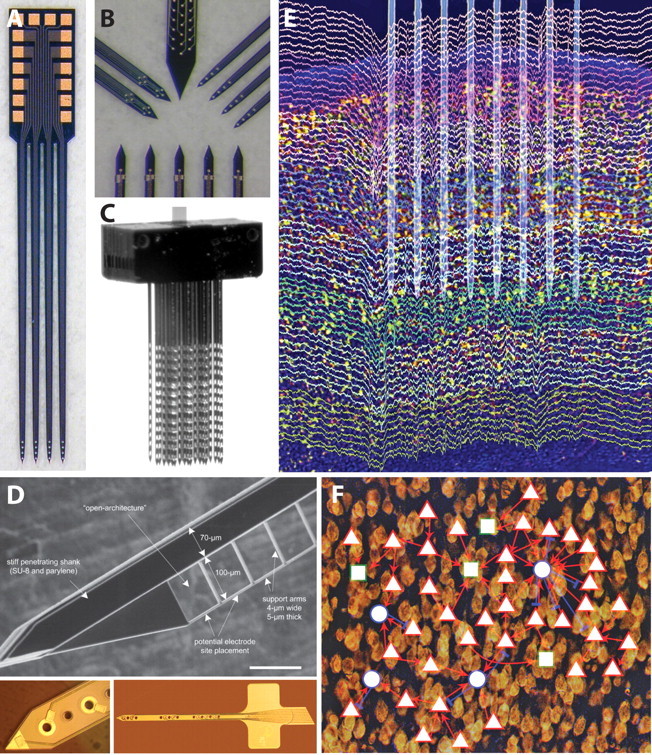
A, Photograph of a four-shank silicon neural probe having four electrode sites arranged near the tip, each terminated in a bond pad at the tab (NeuroNexus Technologies). B, High-magnification photographs illustrating four different types of sites layouts for specialized interfaces (NeuroNexus Technologies). C, Photograph of a modular 128-site, three-dimensional array made from several multishank planar arrays (NeuroNexus Technologies). D, Open architecture probe designs to improve tissue integration [Seymour and Kipke (2007), their Fig. 1A]. E, High-density recording of unit activity in rat neocortex. The placement of an eight-shank silicon device in layer V is overlaid on recordings color coded for different electrodes. Note the presence of spikes on several sites of the same shank and lack of the same spikes across the different shanks, indicating that electrodes placed ≥200 μm laterally record from different cell populations. Spikes and local field potentials are both visible in this wide-band (1 Hz to 10 kHz) recording (Fujisawa et al., 2008). Modified from Buzsáki (2004), his Figure 2A, reprinted with permission. F, Demonstration of functional connectivity in the cortex of behaving animals. A small network of pyramidal cells (red triangles) and putative inhibitory interneurons (blue circles) in layer V of the prefrontal cortex of the rat are mapped. Rectangles are unidentified units. Note a large “hub” formed by an interneuron and its multiple partners [Fujisawa et al. (2008), their supplemental Fig. 11].
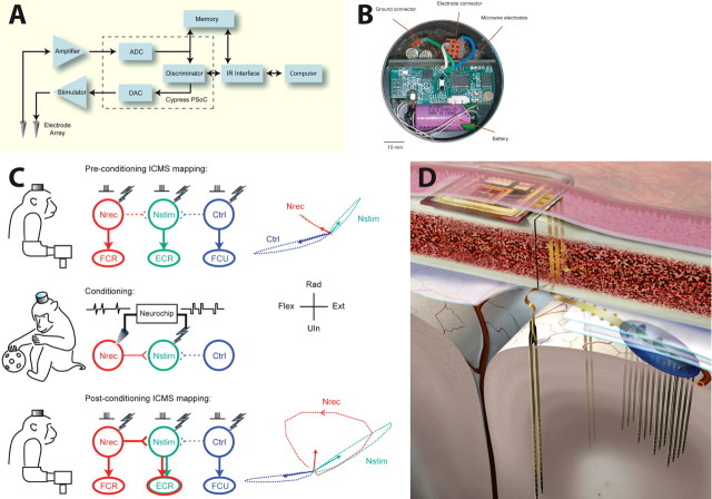
A, Schematic of signal flow in R-BCI (Mavoori et al., 2005). B, Neurochip circuit board, electrode connector, and battery in circular chamber implanted on monkey's head [Jackson et al., (2007), their Fig. 1A]. C, Continuous operation of a cortical recurrent BCI leads to long-lasting changes in physiological connections. Top, Intracranial microstimulation at three different motor cortex sites with the monkey at rest evoked three different muscle responses (center) and different isometric torques about the wrist (right). Arrows at right indicate means of 200 ms torque trajectories. Middle, Conditioning involved 2 d of triggering microstimuli at site Nstim for every spike recorded at Nrec during free behavior and sleep. Bottom, After conditioning, the output effects evoked from site Nrec had changed to include those from Nstim, an effect that lasted beyond a week [Jackson et al. (2006a), their Figs. 2, 5]. D, Illustration of a proposed fully implantable integrated microsystem (University of Michigan).
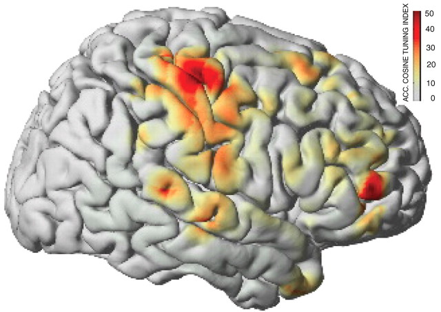
Brain activity captured by ECoG signals during a hand movement task in five human subjects. Color-coded shading (see color bar) illustrates as an average over all subjects how much information about movement direction is encoded by ECoG signals in different cortical areas. Most of that information is captured by hand representations of motor cortex. This figure was modified with permission from Schalk et al. (2007b) IOP Publishing, their Figure 5a. See Schalk et al. (2007b) for methodological details.
References
-
- Andersen RA, Burdick JW, Musallam S, Pesaran B, Cham JG. Cognitive neural prosthetics. Trends Cogn Sci. 2004;8:486–493. - PubMed
-
- Aoki F, Fetz EE, Shupe L, Lettich E, Ojemann GA. Increased gamma-range activity in human sensorimotor cortex during performance of visuomotor tasks. Clin Neurophysiol. 1999;110:524–537. - PubMed
-
- Barthó P, Hirase H, Monconduit L, Zugaro M, Harris KD, Buzsáki G. Characterization of neocortical principal cells and interneurons by network interactions and extracellular features. J Neurophysiol. 2004;92:600–608. - PubMed
Publication types
MeSH terms
Grants and funding
- R44 NS060269/NS/NINDS NIH HHS/United States
- P41 EB002030/EB/NIBIB NIH HHS/United States
- EB006356/EB/NIBIB NIH HHS/United States
- R43 NS060269/NS/NINDS NIH HHS/United States
- R01 NS044287/NS/NINDS NIH HHS/United States
- R37 NS012542/NS/NINDS NIH HHS/United States
- EB 002030/EB/NIBIB NIH HHS/United States
- R01 EB000856/EB/NIBIB NIH HHS/United States
- R43 NS054346/NS/NINDS NIH HHS/United States
- RR00166/RR/NCRR NIH HHS/United States
- NS054346/NS/NINDS NIH HHS/United States
- R44 NS054346/NS/NINDS NIH HHS/United States
- NS12542/NS/NINDS NIH HHS/United States
- R01 EB006356/EB/NIBIB NIH HHS/United States
- P51 RR000166/RR/NCRR NIH HHS/United States
- NS060269/NS/NINDS NIH HHS/United States
- R01 NS012542/NS/NINDS NIH HHS/United States
- EB000856/EB/NIBIB NIH HHS/United States
- NS044287/NS/NINDS NIH HHS/United States
LinkOut - more resources
Full Text Sources
Other Literature Sources