. 2008 Nov 12;28(46):11862-70.
doi: 10.1523/JNEUROSCI.3378-08.2008.

Light-induced rescue of breathing after spinal cord injury

Affiliations

Light-induced rescue of breathing after spinal cord injury

Warren J Alilain et al. J Neurosci. .

Abstract

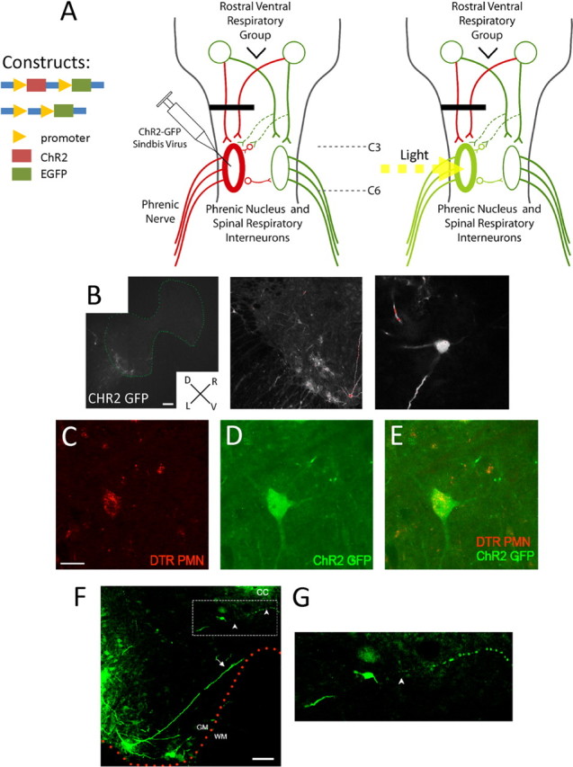
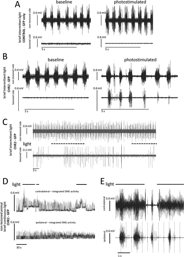
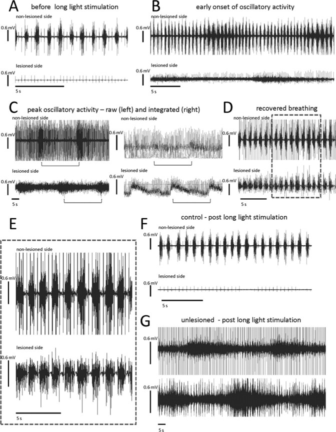
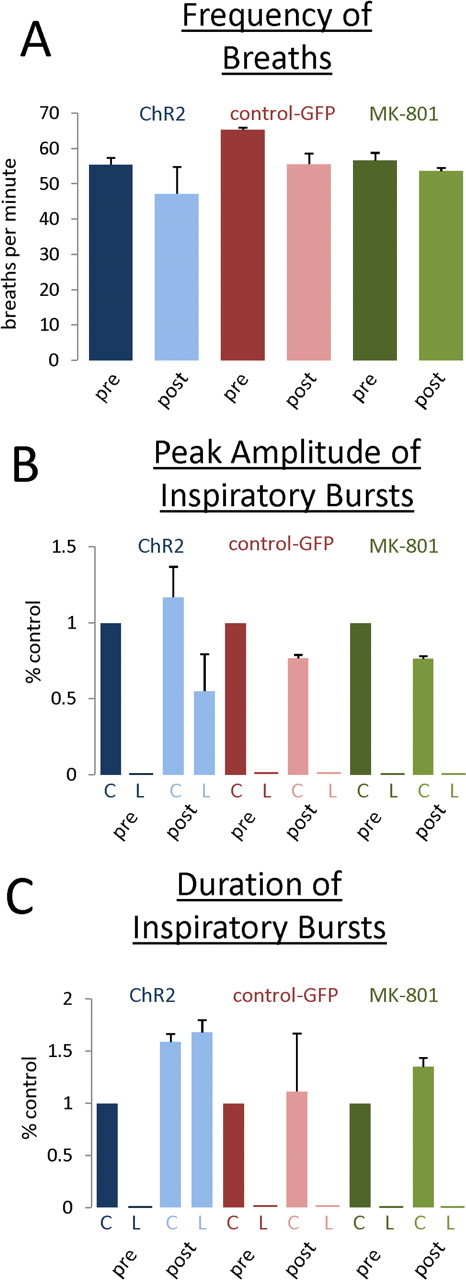
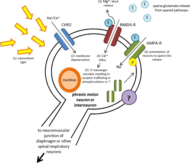
Paralysis is a major consequence of spinal cord injury (SCI). After cervical SCI, respiratory deficits can result through interruption of descending presynaptic inputs to respiratory motor neurons in the spinal cord. Expression of channelrhodopsin-2 (ChR2) and photostimulation in neurons affects neuronal excitability and produces action potentials without any kind of presynaptic inputs. We hypothesized that after transducing spinal neurons in and around the phrenic motor pool to express ChR2, photostimulation would restore respiratory motor function in cervical SCI adult animals. Here we show that light activation of ChR2-expressing animals was sufficient to bring about recovery of respiratory diaphragmatic motor activity. Furthermore, robust rhythmic activity persisted long after photostimulation had ceased. This recovery was accomplished through a form of respiratory plasticity and spinal adaptation which is NMDA receptor dependent. These data suggest a novel, minimally invasive therapeutic avenue to exercise denervated circuitry and/or restore motor function after SCI.


Figures

Figure 1.
Figure 1.
Expression of ChR2-GFP in cervical spinal cord neurons after injection of a Sindbis virus into C2 hemisected animals. A, Schematic of the C2 hemisection (black line), crossed phrenic pathway (dashed green lines), and ChR2-GFP photostimulation treatment protocol. After C2 hemisection, bulbospinal inputs to the ipsilateral phrenic nucleus are interrupted resulting in a quiescent phrenic nerve (red lines) and paralysis of the ipsilateral hemidiaphragm. At the same time of lesioning, ipsilateral C3–C6 spinal neurons, including contralateral projecting interneurons, are infected with a Sindbis virus to express ChR2 and GFP. After 4 d, the C3–C6 spinal cord is exposed to light to stimulate the phrenic nerve and reactivate the paralyzed ipsilateral hemidiaphragm. B, Treatment with Sindbis virus containing ChR2-GFP leads to GFP expression in ipsilateral C3–C6 spinal neurons. In addition, treatment with ChR2-GFP Sindbis virus leads to GFP expression in C3–C6 phrenic motor neurons retrogradely labeled with Dextran Texas Red. D, Dorsal; V, ventral; L, left; R, right. Scale bar, 200 μm. C, Dextran Texas Red-labeled phrenic motor neuron. Scale bar, 50 μm. D, GFP expression of Sindbis virus containing ChR2-GFP. E, Overlay of Dextran Texas Red-labeled phrenic motor neurons expressing GFP. F, Both interneurons and motor neurons infected with ChR2-GFP send neurites across or toward the midline and are in a position to potentially affect contralateral neurons and/or motor output. Arrows point to motor neuronal neurites projecting to the midline, and arrowheads point to interneuronal neurites. Scale bar, 100 μm. G, Enlarged image (dotted line rectangle) of interneurons with midline projecting neurites.
Figure 2.
Figure 2.
Photostimulation of ChR2-GFP-expressing spinal neurons leads to a return of hemidiaphragmatic EMG activity that can be reinitiated in C2-hemisected animals and can influence the contralateral hemidiaphragm, through midline projecting spinal neurons. A, In C2-hemisected animals treated with virus containing only the GFP vector, there is no respiratory activity ipsilateral to the lesion before and after photostimulation (only EKG activity is present). B, In C2-hemisected animals that were treated with virus containing the ChR2 and GFP vector, there is no activity before photostimulation. However, after intermittent photostimulation, there is a return of activity that is rhythmic and synchronous with the intact, contralateral side. EMG activity persisted for at least 1 min after the cessation of photostimulation. After photostimulation induced return of activity, there is a gradual cessation of EMG activity of the hemidiaphragm ipsilateral to the lesion. C, Photostimulation of spinal neurons infected to express ChR2 in C2-hemisected animals can return hemidiaphragmatic activity a number of times in the same animal, including after restored activity has ceased initially. Recovery was repeated up to five times in the same animal. D, E, In nonhemisected animals there is a significant increase of hemidiaphragmatic EMG activity contralateral to ChR2-GFP Sindbis virus injection with photostimulation (integrated EMG activity in D and raw EMG activity in E). There is a slight effect on EMG activity ipsilateral to the injection.
Figure 3.
Figure 3.
Intermittent photostimulation of ChR2-expressing spinal neurons leads to a pattern of EMG hemidiaphragmatic activity that is close to normal in C2-hemisected animals. A, Before photostimulation, there is no EMG activity ipsilateral to the lesion (bottom trace). Contralateral to the lesion, there is rhythmic EMG respiratory activity (top trace). B, In the same animal, during the photostimulation protocol of 5 min off, 5 min 0.5 Hz stimulation, a trace amount of EMG activity begins to develop ipsilateral to the lesion (lower trace). As the EMG activity begins to dwindle, the contralateral, intact side begins to display an increase of EMG activity (upper trace). C, This cycling of high intensity activity that wanes, while the contralateral side increases activity, continues with each period of high intensity activity being slightly more than the last (C compared with B), and this is after the last round of photostimulation. The left two traces are of the raw EMG signal, and the right is of the same time point but integrated and rectified. Brackets under traces indicate periods between onsets of increased diaphragmatic EMG activity. D, E, Eventually EMG activity becomes closer to normal patterned respiratory EMG activity. E, Inset of D. F, A trace of control-treated animal after photostimulation. G, A representative trace of the waxing and waning exhibited by non-C2-hemisected animals that expressed ChR2 and were photostimulated. Top trace is of the injected side.
Figure 4.
Figure 4.
Induction of respiratory plasticity and recovery of hemidiaphragmatic EMG activity results in increases of average peak amplitude and duration of inspiratory bursts after recovery of breathing which is NMDA receptor dependent. A, There was no change in the frequency of breaths before and after stimulation in ChR2-expressing animals, GFP-expressing animals, and MK-801-treated animals. B, After photostimulation, there was an increase of peak EMG amplitude during inspiratory bursts bilaterally in photostimulated ChR2 animals (blue bars). After blockade with MK-801, this increase was abolished (green bars) and brought back to control levels (red bars). C, After photostimulation, there was an increase in the duration of EMG inspiratory bursts bilaterally in photostimulated ChR2 animals (blue bars). After blockade with MK-801, the increase in duration was attenuated (green bars) and brought back to control levels (red bars). Measurements of post-photostimulated animals were made where normal patterned breathing had occurred, i.e., postoscillatory phasic activity. C, Control, nonlesioned side; L, lesioned side.
Figure 5.
Figure 5.
Our hypothetical model of light-induced activity-dependent plasticity. We hypothesize that (1) intermittent light stimulation and activation of the sodium channel ChR2 results in (2) membrane depolarization/activation followed by (3) the release of the Mg2+ block of the NMDA receptor, a ligand-gated Ca2+ channel. After release of the Mg+ block, (4) the resulting influx of Ca2+ will result in (5) induction of 2° messenger systems and cascade events, possibly insertion or phosphorylation of AMPA receptors, the primary mediator of the descending glutamatergic drive to the phrenic motor neurons, to the postsynaptic membrane or perhaps some new or unique form of activity-dependent synaptic plasticity. (6) Potentiation of the phrenic motor pool to subthreshold levels of glutamate from spared pathways/axons is achieved.

Similar articles

Cited by

References

    1. Alilain WJ, Goshgarian HG. Glutamate receptor plasticity and activity-regulated cytoskeletal associated protein regulation in the phrenic motor nucleus may mediate spontaneous recovery of the hemidiaphragm following chronic cervical spinal cord injury. Exp Neurol. 2008;212:348–357. - PMC - PubMed
    1. Arenkiel BR, Peca J, Davison IG, Feliciano C, Deisseroth K, Augustine GJ, Ehlers MD, Feng G. In vivo light-induced activation of neural circuitry in transgenic mice expressing channelrhodopsin-2. Neuron. 2007;54:205–218. - PMC - PubMed
    1. Bellingham MC. Synaptic inhibition of cat phrenic motoneurons by internal intercostal nerve stimulation. J Neurophysiol. 1999;82:1224–1232. - PubMed
    1. Bertram E. The relevance of kindling for human epilepsy. Epilepsia. 2007;48(Suppl 2):65–74. - PubMed
    1. Boyden ES, Zhang F, Bamberg E, Nagel G, Deisseroth K. Millisecond-timescale genetically targeted optical control of neural activity. Nat Neurosci. 2005;8:1263–1268. - PubMed

Publication types

MeSH terms