. 2008 Nov 12;28(46):11959-69.
doi: 10.1523/JNEUROSCI.3296-08.2008.

Maladaptive homeostatic plasticity in a rodent model of central pain syndrome: thalamic hyperexcitability after spinothalamic tract lesions

Affiliations

Maladaptive homeostatic plasticity in a rodent model of central pain syndrome: thalamic hyperexcitability after spinothalamic tract lesions

Gexin Wang et al. J Neurosci. .

Abstract

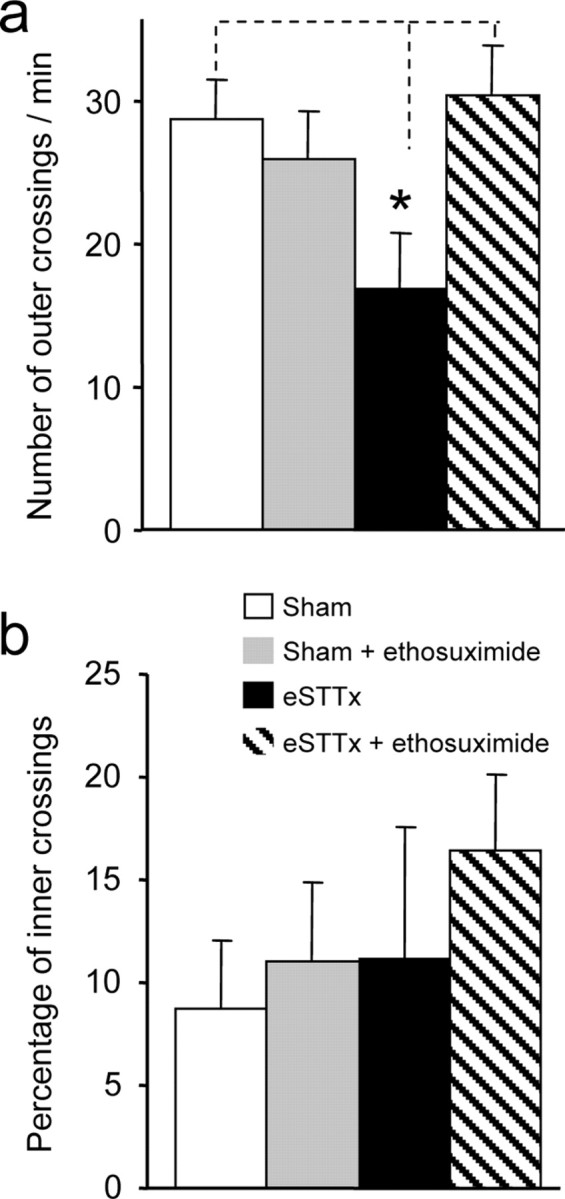
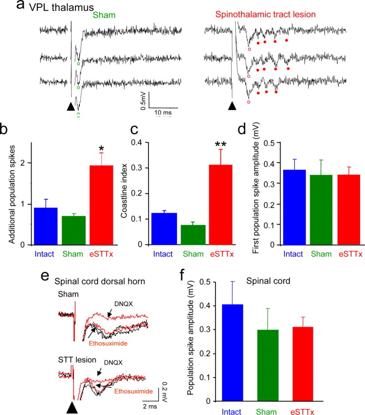
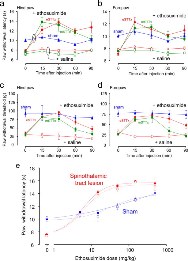
Central pain syndrome (CPS) is defined as pain associated with a lesion of the CNS and is a common consequence of spinal cord injuries. We generated a rodent model of CPS by making unilateral electrolytic or demyelinating lesions centered on the spinothalamic tract in rats. Thermal hyperalgesia and mechanical allodynia occurred in both hind paws and forepaws by 7 d postlesion and were maintained >31 d. Field potentials in the ventral posterior lateral nucleus (VPL) in thalamic brain slices from lesioned animals displayed an increased probability of burst responses. Ethosuximide, a T-type calcium channel blocker, eliminated busting in lesioned thalamic slices and attenuated lesion-induced hyperalgesia and allodynia. We conclude that CPS in this model results from an increase in the excitability of thalamic nuclei that have lost normal ascending inputs as the result of a spinal cord injury and suggest that ethosuximide will relieve human CPS by restoring normal thalamic excitability.


Figures

Figure 1.
Figure 1.
Electrolytic and demyelinating lesions of the ventrolateral spinal white matter. a, A representative Nissl-stained section illustrating the focal gliosis induced 3 d after an electrolytic lesion of the ventrolateral spinal white matter (eSTTx), the region of the spinothalamic tract, at the T8 or T9 vertebral level. Drawings of the injury sites in 15 rats used for pain behavior assessment, as assessed post hoc at 31 d postlesion. Animals in which the lesion site extended beyond the ventrolateral quadrant white matter were not analyzed. b, Visualization of LPC-induced demyelination in ventrolateral spinal cord with myelin staining 14 d after injection (left). Immunostaining with an antibody against neurofilament protein 200 in an adjacent section, showing that the LPC injection does not damage spinothalamic tract axons (right).
Figure 2.
Figure 2.
a–f, Hyperalgesia and allodynia to above- and below-level thermal and mechanical stimuli after electrolytic spinothalamic tract lesions. Paw withdrawal latencies are shown for ipsilateral (open symbols) and contralateral (closed symbols) thermal stimulation of hind paws (a) and forepaws (b) made at various times after spinothalamic tract lesions (eSTTx)(red) or sham surgeries (blue). After a transient increase in contralateral hind paw withdrawal latency at 3 d, lesioned rats respond to the thermal stimulus by withdrawing both ipsilateral and contralateral hind and forepaws more quickly at 7 d postlesion. This decrease in paw withdrawal latency to above- and below-level stimuli was maintained for at least 31 d postlesion (two-way RM ANOVA, Tukey's test, n = 15 lesioned, 16 sham, F = 79.3, p < 0.001 for lesioned vs sham contralateral hind paw; two-way RM ANOVA, Tukey's test, n = 5 lesioned, 8 sham, F = 110.58, p < 0.001 for lesioned vs sham ipsilateral forepaw). Stimulus-response curves were prepared by plotting the frequency of paw withdrawal elicited with repeated trials of mechanical stimuli of varying force delivered to the contralateral hind paw (c) and ipsilateral forepaw (d) at various times after spinothalamic tract lesions and in intact and sham-operated animals. A large leftward shift 7–31 d after spinothalamic lesions compared with either intact of sham animals was readily apparent. Data were fit with a sigmoid function and the force eliciting withdrawal in 50% of trials was taken as the paw withdrawal threshold (dashed lines). Plots of hind paw (e) and forepaw (f) withdraw thresholds for ipsilateral (open symbols) and contralateral (closed symbols) mechanical stimuli delivered at various times after electrolytic spinothalamic tract lesions (red) or sham surgery (blue). Paw withdrawal thresholds for both above- and below-level mechanical stimuli were decreased in lesioned animals beginning 3 d after lesion (two-way RM ANOVA, Tukey's test, n = 15 lesioned, 16 sham, F = 260.33 p < 0.001 for lesioned vs sham contralateral hind paw; two-way RM ANOVA, Tukey's test, n = 5 lesioned, 8 sham, F = 184.17, p < 0.01 for lesioned vs sham ipsilateral forepaw).
Figure 3.
Figure 3.
a–f, Hyperalgesia and allodynia to above- and below-level thermal and mechanical stimuli after demyelinating spinothalamic tract lesions. Paw withdrawal latencies are shown for ipsilateral (open symbols) and contralateral (closed symbols) thermal stimulation of hind paws (a) and forepaws (b) made at various times after demyelinating lesions (mSTTx)(red) or sham surgeries (blue). Lesioned rats respond to the thermal stimulus by withdrawing both ipsilateral and contralateral hind and forepaws more quickly at 14–31 d postlesion (two-way RM ANOVA, Tukey's test, n = 12 lesioned, 16 sham, F = 150.57, p < 0.001 for lesioned vs sham contralateral hind paw; two-way RM ANOVA, Tukey's test, n = 8 lesioned, 8 sham, F = 175.32, p < 0.01 for lesioned vs sham ipsilateral forepaw). Stimulus-response curves were prepared as in Figure 2 for mechanical stimuli delivered to the contralateral hind paw (c) and ipsilateral forepaw (d) at various times after demyelinating lesions and in intact and sham-operated animals. A large leftward shift 3–31 d after spinothalamic lesions compared with either intact of sham animals was readily apparent. Data were fit with a sigmoid function for calculation of the paw withdrawal threshold (dashed lines). Plots of hind paw (e) and forepaw (f) withdraw thresholds for ipsilateral (open symbols) and contralateral (closed symbols) mechanical stimuli delivered at various times after demyelinating lesions (red) or sham surgery (blue). Paw withdrawal thresholds for both above- and below-level mechanical stimuli were decreased in lesioned animals beginning 3 d after lesion (two-way RM ANOVA, Tukey's test, n = 12 lesioned, 16 sham, F = 278.48, p < 0.001 for lesioned vs sham contralateral hind paw; two-way RM ANOVA, Tukey's test, n = 8 lesioned, 8 sham, F = 142.30, p < 0.01 for lesioned vs sham ipsilateral forepaw).
Figure 4.
Figure 4.
Spinothalamic tract lesions induce subjective pain that is reversed by ethosuximide. a, Rats receiving an electrolytic spinothalamic tract lesion (eSTTx) 14 d earlier (n = 5) displayed significantly reduced locomotion, quantified as the number of crossings of the outer quadrants during a 5 min trial, comparing with sham animals (one-way ANOVA, n = 5 lesioned, 8 sham, F = 6.27, *p < 0.05 for lesioned vs sham). Intraperitoneal administration of ethosuximide (50 mg/kg) had no effect on locomotion in sham animals (n = 8), but prevented the decrease in locomotion in lesioned animals (n = 7). b, There were no significant differences in the percentage of crossings into the inner quadrants, an assay of anxiety, between lesioned and sham animals, either with or without ethosuximide (one-way ANOVA, n = 5 lesioned, 8 sham, F = 1.26).
Figure 5.
Figure 5.
Abnormal burst discharges in the ventral posterior lateral nucleus (VPL) after spinothalamic tract lesions. a, Field potential recordings made in three consecutive trials in the VPL of thalamic slices from a sham rat and a rat lesioned 14 d earlier. Local stimulation elicits only a single population spike in the sham slice but bursts of 4 or 5 spikes in the slice from the lesioned animal. Open circles indicate first population spikes, closed circles indicate later spikes. b, The average number of additional spikes occurring after the first spike per sweep was greater in slices from lesioned animals (eSTTx) than in slices from sham or intact animals (one-way ANOVA, n = 12 lesioned, 5 sham, 5 intact, *p < 0.01). c, A modified coastline index, a quantitative measure of bursting, was calculated for 100 ms period starting after the first population spike. The coastline index was significantly greater for responses in slices from lesioned animals than in slices from sham or intact animals (one-way ANOVA, n = 10 lesioned, 8 sham, 10 intact, F = 13.58, **p < 0.001, lesioned vs sham and intact). d, There were no significant differences in the amplitudes of the first population spike, indicating that comparable levels of stimulation were used in all three sets of slices. e, Field potentials, consisting of single population spikes, were recorded in response to stimulation at the dorsal root entry zone in lamina III-V of the dorsal horn in spinal cord slices from a sham rat and a rat in which the spinothalamic tract was lesioned 14 d earlier. Population spikes were eliminated by the glutamate receptor antagonist DNQX (40 μm), but were unaffected by ethosuximide in slices from lesioned animals, shams, or controls. f, There was no difference in population spike amplitude in slices from lesioned, sham, or intact animals (n = 7 lesioned, 7 sham, 5 intact).
Figure 6.
Figure 6.
Ethosuximide abolished thalamic burst discharges induced by spinothalamic tract lesion. a, Field potential recordings made in three consecutive trials in the VPL in a thalamic slices from a rat lesioned 14 d earlier before, during, and after application of ethosuximide (700 μm). The bursts of 4–6 spikes were reversibly blocked by ethosuximide, leaving the first population spike unaffected. b, Group data indicating that ethosuximide produces a 40% decrease in the coastline index of slices from lesioned animals (eSTTx)(paired t test, *p < 0.05, before vs after; n = 10 lesioned, 8 sham and 10 intact), but has no effect on the coastline index of responses in slices from sham or intact rats. c, Grouped data indicating that ethosuximide has no significant effect on the amplitude of the first population spike in any condition.
Figure 7.
Figure 7.
a–e, Ethosuximide eliminates hyperalgesia and allodynia induced by spinothalamic tract lesion. Contralateral hind (a) and forepaw (b) withdrawal latencies in response to thermal stimulation were measured at various times after administration of ethosuximide (15 mg/kg i.p., closed symbols) or saline (open symbols) in animals in which electrolytic (eSTTx, red, n = 6) or demyelinating (mSTTx, green, n = 4) spinothalamic lesions were made 14 d earlier, and sham-operated controls (blue, n = 5). Ethosuximide increased paw withdrawal latency significantly for 15–90 min for both hind and forepaws in eSTTx animals (two-way RM ANOVA, Tukey's test, F = 30.74; p < 0.001 in hind paw; F = 17, p < 0.01 in forepaw for ethosuximide vs saline), and also elevated the contralateral hind (c) and forepaw (d) paw withdrawal thresholds for mechanical stimuli in eSTTx animals (two-way RM ANOVA, Tukey's test, F = 30.74, in hind paw, F = 23.74 in forepaw, p < 0.001 for ethosuximide vs saline). Similarly, ethosuximide increased thermal paw withdrawal latency in both hind and forepaws in mSTTx animals (two-way RM ANOVA, Tukey's test, F = 30.03; p < 0.01 in hind paw; F = 35.29, p < 0.001 in forepaw for ethosuximide vs saline), but only significantly increased mechanical threshold at 30 min after injection (c, d) (two-way RM ANOVA, Tukey's test, intercomparison, q = 6.32, p < 0.001 in hind paw and q = 3.2, p < 0.05 in forepaw for ethosuximide vs saline). e, Dose–response curves were plotted for ipsilateral (open symbols) and contralateral (closed symbols) thermal paw withdrawal latency with various doses of ethosuximide. The ED50 of ethosuximide in increasing contralateral hind paw withdrawal latency was calculated at 5.2 mg/kg in lesioned animals (n = 4). Ethosuximide had some analgesic action in sham animals, but with a much higher ED50 of 67.5 mg/kg (n = 4).
Figure 8.
Figure 8.
Working model of central pain syndrome after spinal cord injury. Top, In intact animals, non-noxious peripheral stimulation signals are relayed via the spinothalamic tract to VPL of thalamus. The thalamocortical neurons in VPL faithfully convey this signal to cortex to form normal perception. Bottom, After an injury to the spinothalamic tract, denervation of thalamocortical neurons in the VPL causes homeostatic changes that produce hyperexcitability and strong activation of T-type Ca2+ channels. As a result of these changes, VPL neurons amplify non-noxious peripheral inputs thereby increasing output to the cortex, where it is perceived as painful.

Similar articles

Cited by

References

    1. Berić A, Dimitrijević MR, Lindblom U. Central dysesthesia syndrome in spinal cord injury patients. Pain. 1988;34:109–116. - PubMed
    1. Bonica JJ. History of pain concepts and pain therapy. Mt Sinai J Med. 1991;58:191–202. - PubMed
    1. Cannavero S, Bonicalzi V. Central pain syndrome: pathophysiology, diagnosis and management. Cambridge, UK: Cambridge UP; 2007.
    1. Coulter DA, Huguenard JR, Prince DA. Characterization of ethosuximide reduction of low-threshold calcium current in thalamic neurons. Ann Neurol. 1989;25:582–593. - PubMed
    1. Crunelli V, Leresche N. Childhood absence epilepsy: genes, channels, neurons and networks. Nat Rev Neurosci. 2002;3:371–382. - PubMed

Publication types

MeSH terms