. 2008 Nov 12;28(46):12097-106.
doi: 10.1523/JNEUROSCI.2635-08.2008.

Loss of gamma-secretase function impairs endocytosis of lipoprotein particles and membrane cholesterol homeostasis

Affiliations

Loss of gamma-secretase function impairs endocytosis of lipoprotein particles and membrane cholesterol homeostasis

Irfan Y Tamboli et al. J Neurosci. .

Abstract

Presenilins (PSs) are components of the gamma-secretase complex that mediates intramembranous cleavage of type I membrane proteins. We show that gamma-secretase is involved in the regulation of cellular lipoprotein uptake. Loss of gamma-secretase function decreased endocytosis of low-density lipoprotein (LDL) receptor. The decreased uptake of lipoproteins led to upregulation of cellular cholesterol biosynthesis by increased expression of CYP51 and enhanced metabolism of lanosterol. Genetic deletion of PS1 or transgenic expression of PS1 mutants that cause early-onset Alzheimer's disease led to accumulation of gamma-secretase substrates and mistargeting of adaptor proteins that regulate endocytosis of the LDL receptor. Consistent with decreased endocytosis of these receptors, PS1 mutant mice have elevated levels of apolipoprotein E in the brain. Thus, these data demonstrate a functional link between two major genetic factors that cause early-onset and late-onset Alzheimer's disease.


Figures

Figure 1.
Figure 1.
Increased cholesterol biosynthesis in PS-deficient cells via increased expression CYP51. A–C, MEFs were incubated in the presence or absence of 10 μm DAPT for 48 h where indicated. Cellular concentrations of cholesterol (A), desmosterol (B), and lanosterol (C) were determined by GC-FID (A) and GC-MS (B, C), respectively (dKO + hPS1; PS dKO MEFs stably expressing human PS1). Values represent means ± SD (n = 5). D, Transcriptional upregulation of CYP51 in PS dKO MEFs. mRNA levels of lanosterol 14α-demethylase (CYP51), lanosterol synthase (LS), and lanosterol Δ24-reductase (Sel-1) were analyzed by RT-PCR. mRNA of β-actin served as control. Quantitation of CYP51 mRNA expression revealed an increase to 402.3 ± 11.3% in PS dKO cells compared with PS WT cells (n = 3 for PS dKO and PS WT; p < 0.001). Significance values are indicated by asterisks as follows: *p < 0.05; **p < 0.01; ***p < 0.001.
Figure 2.
Figure 2.
PS deficiency results in decreased endocytosis of lipoproteins and increased expression of SREBP-2. A, Transcriptional regulation of CYP51 by LDL. MEF WT cells were incubated in presence or absence of 10 μg/ml human LDL for 16 h, and expression of CYP51 was analyzed by RT-PCR. The expression of CYP51 mRNA was significantly lower after incubation with exogenous LDL [38.8 ± 9.4% with LDL (n = 3) vs 100 ± 28.3% without LDL (n = 3), p < 0.05]. B, Cytochemical analysis of BODIPY-LDL endocytosis was performed in WT and PS dKO MEFs as described in the Materials and Methods. Scale bar, 50 μm. C, Western immunoblotting analysis of cellular apo B-100 levels. Purified human LDL (hLDL) was loaded as a positive control. Quantitation revealed significantly reduced levels of apo B-100 in PS dKO cells [68.6 ± 9.2% (n = 3) vs 100 ± 13.9% in PS WT cells (n = 3), p < 0.05]. D, Expression analysis of SREBP-2 full-length (SREBP-2 FL) and SREBP-2 N-terminal fragment (SREBP-2 NTF) in WT and PS dKO MEFs performed by Western immunoblotting. SREBP-2 FL was detected in crude membrane preparations, whereas SREBP-2 NTF was detected in cytosolic fractions. Quantitation showed significant increases in PS dKO cells for SREBP-2 FL (322.6 ± 49.3%, p < 0.01) and SREBP-2 NTF (241.1 ± 58.0%, p < 0.01) compared with PS WT cells. E, Detection of HMG-CoA reductase in WT and PS dKO MEFs by Western immunoblotting. Migration of HMG-CoA reductase is indicated by arrowheads. A lower molecular mass species was also detected in both cell types (asterisks). Levels of HMG-CoA reductase were significantly increased in PS dKO cells [240.9 ± 8.6% in PS dKO (n = 3) vs 100 ± 8.5% in PS WT (n = 3), p < 0.001]. F, The treatment of PS dKO MEFs with lanosterol (25 μm, 48 h) decreased the levels of HMG-CoA reductase [71.8 ± 6.9% in LDL-treated (n = 3) vs 100 ± 13.9% in untreated cells (n = 3); p < 0.05].
Figure 3.
Figure 3.
γ-Secretase-dependent distribution and endocytosis of the LDL receptor. A, Presenilin-dependent subcellular localization of LDLR was analyzed by immunocytochemistry. Prominent localization of LDLR at the cell surface in PS dKO cells is indicated by arrowheads. PS dKO cells have altered size and morphology compared with WT cells. These alterations were reverted by reexpression of hPS1. Scale bar, 25 μm. B, Cell surface proteins were labeled with sulfo-NHS-biotin and precipitated with streptavidin-conjugated agarose. Precipitated LDLR was detected by Western immunoblotting. β-Actin served as loading control. C, Decreased endocytosis of LDLR in PS-deficient cells. Surface proteins of MEF WT and PS dKO cells were labeled with sulfo-NHS-SS-biotin. Cells were incubated for the indicated time periods at 37°C to allow endocytosis, and residual biotin from cell surface was removed using reducing buffer. Internalized biotin-labeled proteins were precipitated and LDLR was detected by Western immunoblotting (see Materials and Methods section for details). D, LDLR endocytosis analysis in HEK293 cells expressing PS1 WT or a dominant-negative (DN) variant of PS1.
Figure 4.
Figure 4.
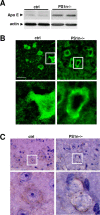
Regulation of apo E levels by PS in mouse brains. A, Western blot analysis of apo E in brains of WT and PS1 conditional knock-out mice (PS1n−/−) at age of 16 month. As determined by quantitative ECL imaging, apo E levels were 161.8 ± 55.4% in PS1n−/− (n = 4) versus 100 ± 20.5% in PS1 WT mice (n = 4); p < 0.05. The effect was sex independent. B, C, Neuronal localization of apo E in brain cortices of WT and PS1n−/− mice was analyzed by confocal microscopy (B) and immunohistochemistry (C, counterstain with hematoxylin) as described in Materials and Methods. Scale bars, 50 μm.
Figure 5.
Figure 5.
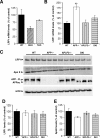
Deletion of AICD does not increase LRP1 expression in mouse brain. A, LRP1 mRNA expression analysis by qRT-PCR at embryonic day 15.5 in WT (n = 4), APP/APLP2 double knock-out (DKO; n = 4), and APP/APLP1/APLP3 triple knock-out (TKO; n = 4) mice brains. B, Expression analysis of LRP1 mRNA by qRT-PCR in brains of 22- to 24-week-old mice [WT (n = 7), APP-KO (n = 7), APLP2-KO (n = 7), DKI (n = 7)]. C, Expression of LRP1, apo E, APP, and β-actin was analyzed in brains of 22- to 26-week-old WT (n = 7), APP-KO (n = 3), APLP2-KO (n = 7), and DKI (n = 7) mice by Western immunoblotting. D, E, Quantitation of LRP1 (D) and apo E (E) expression by ECL imaging. Details are described in Materials and Methods. Values represent means ± SD. **p < 0.01.
Figure 6.
Figure 6.
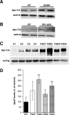
Increased levels of apo E in brains of PS1 FAD mutant mice. A, Western blot analysis of apo E levels in brains of WT and PS1 A246E mutant mice at age of 10 month. Quantitative ECL imaging revealed significantly increased levels in PS1 A246E mice [161.8 ± 55.4% (n = 5) vs 100 ± 20.5% (n = 5); p < 0.05]. B, Western blot analysis of apo E in brains of mice expressing PS1 WT or PS1-ΔE9 at age of 6 month. The effects on apo E were sex independent. C, D, Detection of apo E in human brains from control individuals and patients with familial AD harboring mutations in the PS1 gene by Western immunoblotting (FAD1-L174R, FAD2-T113-114 insertion mutation, FAD3-I437V, FAD4-T113-114 insertion mutation). Quantitation of apo E levels in PS1 FAD brain lysates compared with mean apo E levels from four control human brains was done by ECL imaging (D). Values represent means of three independent experiments ± SD. *p < 0.05; **p < 0.01.
Figure 7.
Figure 7.
Accumulation of APP CTFs causes mistargeting of adaptor proteins and decreased endocytosis of LDLR. A, HEK293 cells were transfected with cDNA encoding myc-tagged Fe65. After 48 h, cells were fixed, permeabilized, and stained for Fe65 with anti-myc antibody 9E10. Cells were counterstained with DAPI to localize nuclei. B, Cells were transfected with cDNAs encoding APP C99-EGFP and myc-tagged Fe65, and then incubated in the presence (bottom) or absence (top) of 10 μm DAPT. Expression of C99-EGFP caused relocalization of Fe65 to juxtanuclear compartments (top). In the presence of DAPT, prominent staining at the cell surface was also observed (bottom). C, D, APP CTF-dependent localization of ARH. HEK293 cells were transfected with cDNA encoding ARH alone (C) or together with APP C99-EGFP (D). Cells were incubated the presence (D, bottom) and absence (D, top) of 10 μm DAPT. Scale bars in A–D, 15 μm. E, LDLR endocytosis in HEK293 control cells and in cells expressing APP C99 was analyzed by biotinylation as described under Materials and Methods. Quantitative ECL imaging revealed that C99 expression resulted in significantly decreased endocytosis of LDLR (34.3 ± 18.0% vs 100 ± 11.6%; p < 0.05).

Similar articles

Cited by

References

    1. Annaert WG, Esselens C, Baert V, Boeve C, Snellings G, Cupers P, Craessaerts K, De Strooper B. Interaction with telencephalin and the amyloid precursor protein predicts a ring structure for presenilins. Neuron. 2001;32:579–589. - PubMed
    1. Bales KR, Dodart JC, DeMattos RB, Holtzman DM, Paul SM. Apolipoprotein E, amyloid, and Alzheimer disease. Mol Interv. 2002;2:363–375. - PubMed
    1. Bentahir M, Nyabi O, Verhamme J, Tolia A, Horré K, Wiltfang J, Esselmann H, De Strooper B. Presenilin clinical mutations can affect gamma-secretase activity by different mechanisms. J Neurochem. 2006;96:732–742. - PubMed
    1. Berger-Sweeney J, McPhie DL, Arters JA, Greenan J, Oster-Granite ML, Neve RL. Impairments in learning and memory accompanied by neurodegeneration in mice transgenic for the carboxyl-terminus of the amyloid precursor protein. Brain Res Mol Brain Res. 1999;66:150–162. - PubMed
    1. Biederer T, Cao X, Südhof TC, Liu X. Regulation of APP-dependent transcription complexes by Mint/X11s: differential functions of Mint isoforms. J Neurosci. 2002;22:7340–7351. - PMC - PubMed

Publication types