. 2008 Nov 12;28(46):12125-35.
doi: 10.1523/JNEUROSCI.3400-08.2008.

Crucial role of CB(2) cannabinoid receptor in the regulation of central immune responses during neuropathic pain

Affiliations

Crucial role of CB(2) cannabinoid receptor in the regulation of central immune responses during neuropathic pain

Ildiko Racz et al. J Neurosci. .

Abstract

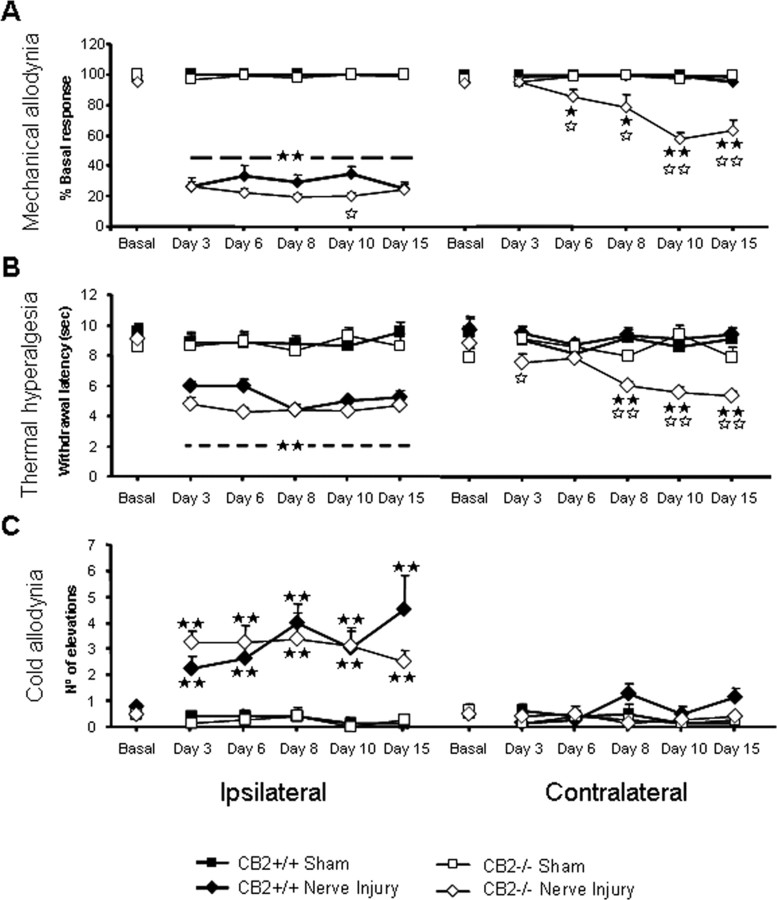
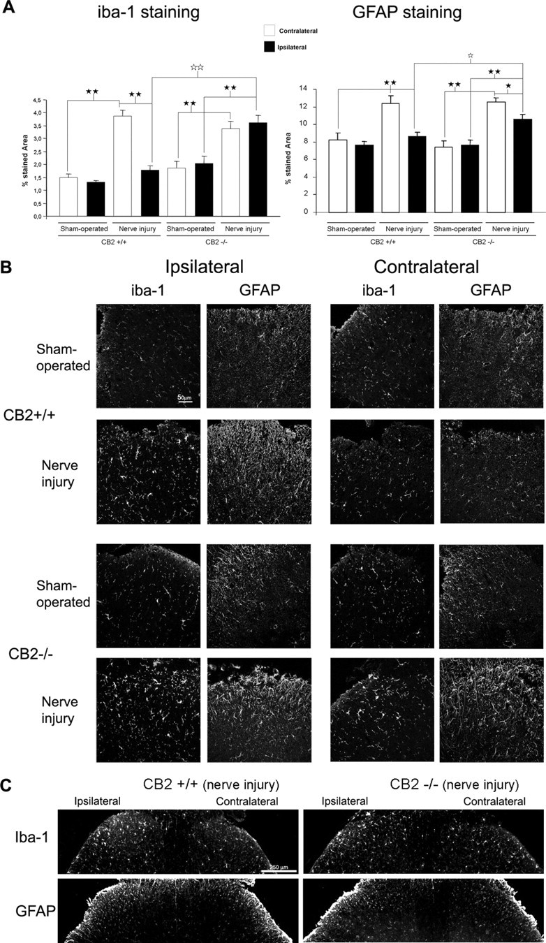
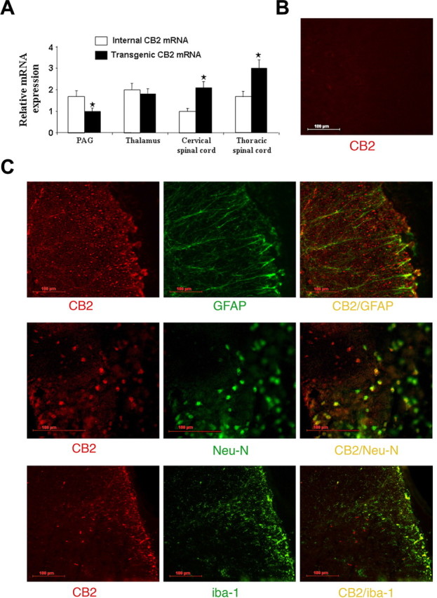
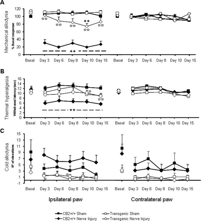
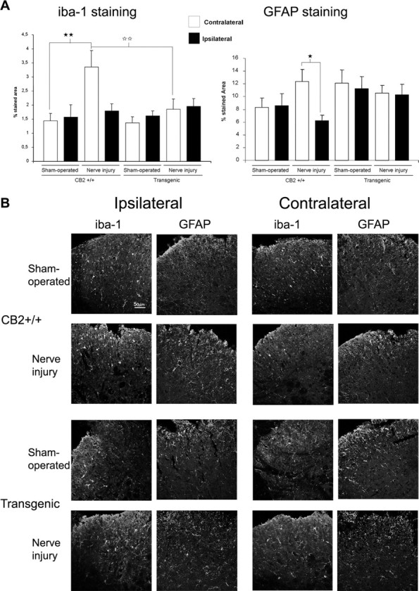
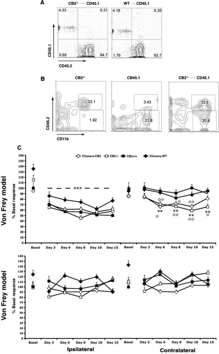
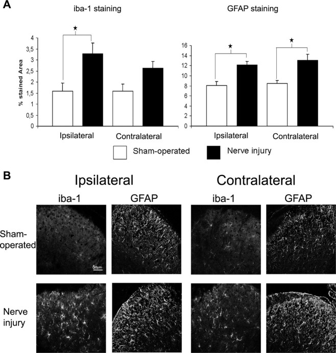
Neuropathic pain is a clinical manifestation of nerve injury difficult to treat even with potent analgesic compounds. Here, we used different lines of genetically modified mice to clarify the role played by CB(2) cannabinoid receptors in the regulation of the central immune responses leading to the development of neuropathic pain. CB(2) knock-out mice and wild-type littermates were exposed to sciatic nerve injury, and both genotypes developed a similar hyperalgesia and allodynia in the ipsilateral paw. Most strikingly, knock-outs also developed a contralateral mirror image pain, associated with an enhanced microglial and astrocytic expression in the contralateral spinal horn. In agreement, hyperalgesia, allodynia, and microglial and astrocytic activation induced by sciatic nerve injury were attenuated in transgenic mice overexpressing CB(2) receptors. These results demonstrate the crucial role of CB(2) cannabinoid receptor in modulating glial activation in response to nerve injury. The enhanced manifestations of neuropathic pain were replicated in irradiated wild-type mice reconstituted with bone marrow cells from CB(2) knock-outs, thus demonstrating the implication of the CB(2) receptor expressed in hematopoietic cells in the development of neuropathic pain at the spinal cord.


Figures

Figure 1.
Figure 1.
Development of neuropathic pain is enhanced in male CB2 −/− after sciatic nerve injury when compared with CB2 +/+. A single tight ligature around one-third or one-half of sciatic nerve was made to induce the neuropathic pain in CB2 −/− (n = 16) and CB2 +/+ mice (n = 16). Mice were tested in the ipsilateral and contralateral paw for evaluating mechanical allodynia (von Frey model; percentage of the basal CB2 +/+ sham-operated values) (A), thermal hyperalgesia (plantar test; withdrawal latency in seconds) (B), and cold allodynia (cold-plate test; number of paw elevations) (C) on days 3, 6, 8, 10, and 15 after surgery. The black stars represent comparison between sciatic nerve injury and sham-operated animals. The white stars represent comparison between genotypes. One star, p < 0.05; two stars, p < 0.01. Error bars indicate SEM.
Figure 2.
Figure 2.
Enhanced glial activation in spinal cord of CB2 −/− after sciatic nerve injury when compared with CB2 +/+ mice. A, Increase in iba-1 (microglia) (n = 5) and GFAP (astrocyte) (n = 5) staining in dorsal horn of lumbar spinal cord after sciatic nerve injury in CB2 −/− and in CB2 +/+ mice. White bars, Ipsilateral paw; black bars, contralateral paw. Iba-1 and GFAP visualized with CY3 anti-rabbit (red). Data are expressed as percentage of stained area. The black stars represent comparisons between sham-operated and nerve injury between ipsilateral and contralateral paw. The white stars represent comparisons between genotypes. One star, p < 0.05; two stars, p < 0.01. Error bars indicate SEM. B, Representative confocal images of the immunostaining of iba-1 and GFAP recorded with 40× objective in lumbar dorsal horn of CB2 +/+ and CB2 −/−. C, Representative low-magnification images of iba-1 and GFAP immunostaining recorded with 10× objective in the lumbar dorsal horn of sciatic nerve injury CB2 +/+ and CB2 −/− mice.
Figure 3.
Figure 3.
CB2 expression and colocalization in male transgenic mice overexpressing CB2 receptors. A, Relative abundance of endogenous and transgenic CB2 RNA in PAG, thalamus, cervical spinal cord, and thoracic spinal cord of transgenic mice was determined by real-time PCR quantification. After being analyzed using the ΔCt method, data were normalized to GAPDH expression. The group with the lower value was assigned a value of 1. The stars represent differences in relative transcript abundance between endogenous and transgenic CB2 receptor (one-way repeated-measures ANOVA). Error bars indicate SEM. B, Negative immunostaining for CB2 receptor in the lumbar spinal cord of CB2 −/− mice that serves as a control for CB2 staining in transgenic mice overexpressing CB2 receptors. C, Double-fluorescence immunostaining for CB2 receptor, GFAP (astrocyte), Neu-N (neuron), and iba-1 (microglia) in lumbar spinal cord sections from transgenic mice overexpressing CB2 receptors. First plot, Immunostaining for CB2 receptor (red; CY3 anti-rabbit) and for astrocyte marker GFAP (green; FITC anti-rabbit). Second plot, Same staining for CB2 receptor (red; CY3 anti-rabbit) and for neuron (fluorescein-conjugated Neu-N). Third plot, Same staining for CB2 receptor (red; CY3 anti-rabbit) and for microglial marker iba-1 (green; FITC anti-rabbit).
Figure 4.
Figure 4.
Behavioral manifestations of neuropathic pain in male transgenic mice overexpressing CB2 receptors. Development of neuropathic pain in male transgenic mice overexpressing CB2 receptors (n = 12) and wild-type mice (n = 12). A single tight ligature around one-third or one-half of sciatic nerve was made to induce the neuropathic pain. Mice were tested in the ipsilateral and contralateral paw for evaluating mechanical allodynia (von Frey model; percentage of the basal CB2 +/+ sham-operated values) (A), thermal hyperalgesia (plantar test; withdrawal latency in seconds) (B), and cold allodynia (cold-plate test; number of paw elevations) (C) on days 3, 6, 8, 10, and 15 after surgery. The black stars represent comparison between sciatic nerve injury and sham-operated animals. The white stars represent comparison between genotypes. One star, p < 0.05; two stars, p < 0.01. Error bars indicate SEM.
Figure 5.
Figure 5.
Expression of glial activation in spinal cord of CB2 +/+ and transgenic mice overexpressing CB2 receptors. A, The increase of microglia and astrocytic response in dorsal horn of lumbar spinal cord after sciatic nerve injury is only observed in CB2 +/+ mice. Iba-1 staining (microglia) (n = 4 per group) and GFAP staining (astrocytes) (n = 4 per group) were visualized with CY3 anti-rabbit (red). Ipsilateral dorsal horns were represented with white bars, and contralateral dorsal horns were represented with black bars. Data are expressed as percentage of stained area. Error bars indicate SEM. The black stars represent comparisons between sham-operated and nerve injury or between ipsilateral and contralateral paw. The white stars represent comparisons between genotypes. One star, p < 0.05; two stars, p < 0.01. B, Representative confocal images of the immunostaining of iba-1 (microglia) and GFAP (astrocyte) in lumbar dorsal horn of CB2 +/+ and transgenic mice overexpressing CB2 receptors recorded with the 40× objective.
Figure 6.
Figure 6.
Reconstitution efficiency of BM chimeric mice and behavioral manifestations of neuropathic pain after transplantation of BM cells lacking CB2 receptors in irradiated CB2 +/+ mice. A, Lethally irradiated recipient mice (CD45.1) were reconstituted with BM from CB2 −/− (left) or WT (right) animals expressing the congenic marker CD45.2. Flow cytometry of peripheral blood lymphocytes of BM chimeric mice was performed 8 weeks later by staining with anti-CD45.1 and -CD45.2 antibodies. Frequencies of CD45.2 donor-derived and CD45.1 recipient-derived cells are indicated in each quadrant. B, Presence of CB2-derived microglia in the spinal cord of BM-chimeric mice 14 d after surgery. Representative flow cytometry demonstrating CD45.2+CD11b+ cells in the spinal cord of CB2 −/− (CD45.2) control mice (left) and lethally irradiated CD45.1 congenic recipients after engraftment with CB2 −/− BM cells (chimera-CB2) (right). CD11b+ cells in the spinal cord of CD45.1 control mice (middle) are negative for the congenic marker CD45.2. Representative dot blots are shown. C, The following groups were studied: CB2 −/− and CB2 +/+ control animals, chimera-CB2 (CB2 +/+ mice receiving bone marrow from CB2 −/− animals), and chimera-WT (CB2 +/+ mice receiving bone marrow from CB2 +/+ mice). Development of neuropathic pain in CB2 +/+ (n = 10), CB2 −/− (n = 10), chimera-WT (n = 8), and chimera-CB2 (n = 12). Mechanical allodynia after partial sciatic nerve ligation was tested in the ipsilateral and contralateral paws on day 3, 6, 8, 10, and 15 after the surgery. The withdrawal thresholds are presented as percentage of the basal CB2 +/+ values. Error bars indicate SEM. The black stars represent comparison between sciatic nerve injury and sham-operated animals. The white stars represent comparison between genotypes. One star, p < 0.05; two stars, p < 0.01.
Figure 7.
Figure 7.
Histological changes in neuropathic pain after transplantation of BM cells lacking CB2 receptors into irradiated CB2 +/+ mice. A, Increase of microglia activation and astrocytic response in ipsilateral and contralateral dorsal horn of lumbar spinal cord after sciatic nerve injury in CB2 −/− BM-chimeric mice (black bars) (n = 6) compared with sham-operated BM-chimeric mice (white bars) (n = 3). Iba-1 was used to stain microglia cells and GFAP antibody for astrocyte staining. Iba-1 staining and GFAP staining visualized with CY3 anti-rabbit (red). Data are expressed as percentage of stained area. Error bars indicate SEM. Black stars represent comparisons between sham-operated and nerve injury. One star, p < 0.05; two stars, p < 0.01. B, Representative confocal images of the immunostaining of iba-1 (microglia) and GFAP (astrocyte) recorded with 40× objective in lumbar dorsal horn of sham-operated and nerve injured CB2 −/− BM-chimeric mice.

Similar articles

Cited by

References

    1. Agarwal N, Pacher P, Tegeder I, Amaya F, Constantin CE, Brenner GJ, Rubino T, Michalski CW, Marsicano G, Monory K, Mackie K, Marian C, Batkai S, Parolaro D, Fischer MJ, Reeh P, Kunos G, Kress M, Lutz B, Woolf CJ, et al. Cannabinoids mediate analgesia largely via peripheral type 1 cannabinoid receptors in nociceptors. Nat Neurosci. 2007;10:870–879. - PMC - PubMed
    1. Beltramo M, Bernardini N, Bertorelli R, Campanella M, Nicolussi E, Fredduzzi S, Reggiani A. CB2 receptor-mediated antihyperalgesia: possible direct involvement of neural mechanisms. Eur J Neurosci. 2006;23:1530–1538. - PubMed
    1. Bingham B, Jones PG, Uveges AJ, Kotnis S, Lu P, Smith VA, Sun SC, Resnick L, Chlenov M, He Y, Strassle BW, Cummons TA, Piesla MJ, Harrison JE, Whiteside GT, Kennedy JD. Species-specific in vitro pharmacological effects of the cannabinoid receptor 2 (CB2) selective ligand AM1241 and its resolved enantiomers. Br J Pharmacol. 2007;151:1061–1070. - PMC - PubMed
    1. Buckley NE, McCoy KL, Mezey E, Bonner T, Zimmer A, Felder CC, Glass M, Zimmer A. Immunomodulation by cannabinoids is absent in mice deficient for the cannabinoid CB(2) receptor. Eur J Pharmacol. 2000;396:141–149. - PubMed
    1. Carlisle SJ, Marciano-Cabral F, Staab A, Ludwick C, Cabral GA. Differential expression of the CB2 cannabinoid receptor by rodent macrophages and macrophage-like cells in relation to cell activation. Int Immunopharmacol. 2002;2:69–82. - PubMed

Publication types

MeSH terms

Substances