. 2009 Jan 15;113(3):744-54.
doi: 10.1182/blood-2008-03-145219. Epub 2008 Nov 12.

CYP1B1 expression promotes the proangiogenic phenotype of endothelium through decreased intracellular oxidative stress and thrombospondin-2 expression

Affiliations

CYP1B1 expression promotes the proangiogenic phenotype of endothelium through decreased intracellular oxidative stress and thrombospondin-2 expression

Yixin Tang et al. Blood. .

Abstract

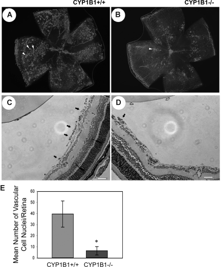
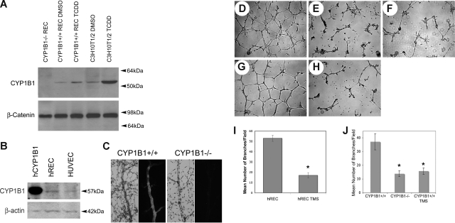
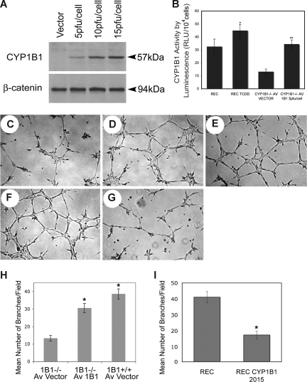
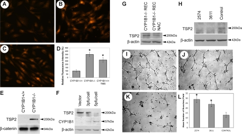
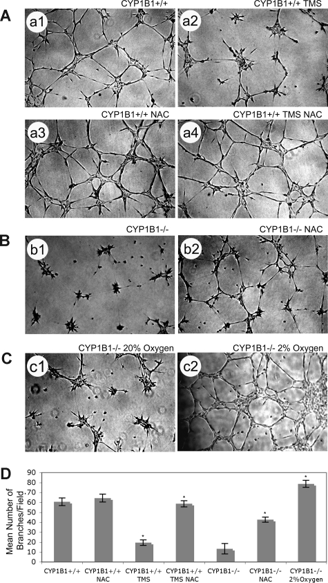
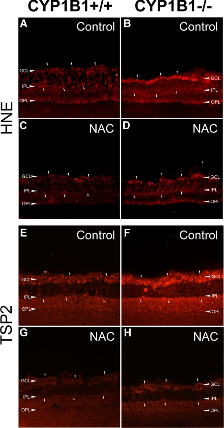
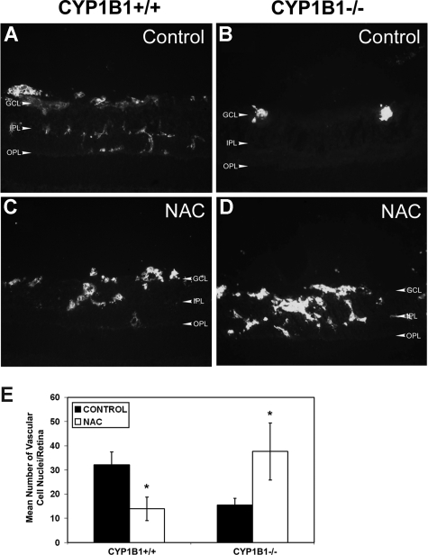
Reactive species derived from cell oxygenation processes play an important role in vascular homeostasis and the pathogenesis of many diseases including retinopathy of prematurity. We show that CYP1B1-deficient (CYP1B1(-/-)) mice fail to elicit a neovascular response during oxygen-induced ischemic retinopathy. In addition, the retinal endothelial cells (ECs) prepared from CYP1B1(-/-) mice are less adherent, less migratory, and fail to undergo capillary morphogenesis. These aberrant cellular responses were completely reversed when oxygen levels were lowered or an antioxidant added. CYP1B1(-/-) ECs exhibited increased oxidative stress and expressed increased amounts of the antiangiogenic factor thrombospondin-2 (TSP2). Increased lipid peroxidation and TSP2 were both observed in retinas from CYP1B1(-/-) mice and were reversed by administration of an antioxidant. Reexpression of CYP1B1 in CYP1B1(-/-) ECs resulted in down-regulation of TSP2 expression and restoration of capillary morphogenesis. A TSP2 knockdown in CYP1B1(-/-) ECs also restored capillary morphogenesis. Thus, CYP1B1 metabolizes cell products that modulate intracellular oxidative stress, which enhances production of TSP2, an inhibitor of EC migration and capillary morphogenesis. Evidence is presented that similar changes occur in retinal endothelium in vivo to limit neovascularization.


Figures

Figure 1
Figure 1
Attenuation of pathologic angiogenesis in CYP1B1−/− mice. CYP1B1+/+ and CYP1B1−/− mice were exposed to a cycle of hyperoxia and room air (OIR). The collagen IV–stained of whole mount retinas prepared from P17 CYP1B1+/+ and CYP1B1−/− mice are shown in panels A and B, respectively (×25). The hematoxylin/PAS–stained eye sections prepared from P17 CYP1B1+/+ and CYP1B1−/− mice are shown in panels C and D, respectively (×100). The arrows indicate growth of new vascular tufts, which is significantly diminished in CYP1B1−/− mice. The number of vascular cell nuclei present on the vitreous side of the retina penetrating the inner limiting membrane was determined as described in “Methods” at P17 and presented in panel E. Data in each bar are the mean number of vascular cell nuclei in 5 eyes of 5 mice (error bars indicate SD). Please note that there is a significant decrease in the degree of retinal neovascularization in CYP1B1−/− mice compared with CYP1B1+/+ mice (n = 20, *P < .05).
Figure 2
Figure 2
CYP1B1−/− EC fail to undergo capillary morphogenesis in Matrigel. CYP1B1 expression in mouse retinal ECs and C3H10T1/2 cells incubated with and without TCDD (A), and human retinal ECs and umbilical vein ECs (B), were evaluated by Western blot analysis of total cell lysates. The purified human recombinant CYP1B1 protein was used as positive control. β-catenin or β-actin was used as loading control. CYP1B1 or HE/PAS wholemount staining of retinal trypsin digests, prepared from CYP1B1+/+ (right panels) and CYP1B1−/− (left panels) are shown in panel C. Capillary morphogensis of CYP1B1+/+ (D) and CYP1B1−/− (E) retinal ECs were evaluated by plating the cells in Matrigel as described in “Methods.” After 17 hours of incubation, CYP1B1+/+ ECs formed well-organized capillary-like networks, while CYP1B1−/− ECs ability to organize was severely compromised (×40). A similar inhibition can be observed by incubating the CYP1B1+/+ ECs with TMS (F), a specific inhibitor of CYP1B1 activity. Human retinal ECs also form well-organized capillary networks in Matrigel (G), which was attenuated in the presence of TMS (H). The quantitative assessments of the data are shown in panels I and J. Data in each bar are the mean number of branches per 5 high-power fields (×100; error bars indicate standard deviation). Please note that the mean number of branch points formed by CYP1B1−/− ECs or CYP1B1+/+ ECs incubated with TMS were significantly lower than those formed by CYP1B1+/+ retinal ECs (n = 3, *P < .05). These experiments were repeated with 3 different preparations of ECs with similar results.
Figure 3
Figure 3
Expression of CYP1B1 restores the capillary morphogenesis defect observed in CYP1B1−/− EC. (A) Western blot analysis of whole cell lysates (20 μg) from CYP1B1−/− ECs infected with the adenoviruses expressing empty vector or CYP1B1 at different virus input. β-catenin was used for loading control. (B) CYP1B1 activity assay of CYP1B1+/+ (incubated with or without TCDD) and CYP1B1−/− ECs infected with the adenoviruses expressing empty vector or CYP1B1. Data in each bar are the mean relative luminescence (error bars indicate the standard deviation, n = 3, *P [CYP1B1+/+; control vs TCDD] and **P < .05 [CYP1B1−/−; vector vs CYP1B1]). The CYP1B1−/− ECs infected with adenovirus control (5 pfu/cell) (C), adenovirus expressing CYP1B1 (5 pfu/cell) (D), or CYP1B1+/+ ECs with vector control (5 pfu/cell) (E) were plated on Matrigel as described in “Methods” (×40). The capillary morphogenesis by CYP1B1+/+ cells infected with a retrovirus expressing control siRNA (F) or a mouse specific CYP1B1 siRNA 2015 (G) was similarly determined. The quantitative assessments of the data are shown in panels H and I. Data in each bar are the mean number of branches per 5 high-power fields (×100; error bars indicate SD). Note that the ability of CYP1B1−/− ECs to organize in Matrigel was significantly improved with reexpression of CYP1B1, while its siRNA knockdown resulted in attenuation of capillary morphogenesis in CYP1B1+/+ retinal ECs (n = 3, *P < .05). These experiments were repeated with 2 different preparations of ECs with similar results.
Figure 4
Figure 4
CYP1B1−/− ECs show higher oxidative stress with increased TSP2 expression. DHE staining of CYP1B1+/+ (A), CYP1B1−/− (B) ECs, and CYP1B1+/+ ECs incubated with TMS (C) (×400) are shown. The quantitative assessment of the is shown in panel D. The data in each bar are the mean fluorescence intensities determined as described in “Methods,” and error bars indicate standard deviation. A significant increased fluorescent intensity was observed in CYP1B1−/− cells (n = 20, *P < .05). CYP1B1+/+ cells incubated with TMS showed higher fluorescence compared with untreated CYP1B1+/+ cells, similar to CYP1B1−/− cells. The level of TSP2 was analyzed by Western blotting of serum-free conditioned medium prepared from CYP1B1+/+ and CYP1B1−/− ECs as described in “Methods” (E). A blot of cell lysates prepared from these cells was probed with β-catenin to control for loading. Western blot analysis of whole cell lysates of CYP1B1−/− ECs infected with the adenoviruses expressing empty vector or CYP1B1 is shown in F. Blots were probed with antibodies to TSP2, CYP1B1, and β-actin to control for loading. Please note expression of CYP1B1 in CYP1B1−/− ECs is inversely correlated with expression of TSP2. Western blot analysis of whole cell lysates from CYP1B1−/− ECs incubated with or without NAC for 2 days is shown in panel G. Blot was probed with TSP2, and β-actin was used as loading control. Western blot analysis of cell lysates prepared from CYP1B1−/− ECs expressing TSP2-specific siRNAs (2574 [I] or 3611 [J]) or control siRNA, probed with anti-TSP2 or β-actin (loading control) is shown in panel H. The capillary morphogenesis of these cells in Matrigel are shown in panels I-K. The quantitative assessment of the data are shown in panel L. Capillary morphogenesis was significantly restored in CYP1B1−/− cells expressing TSP2 siRNAs compared with control vector (n = 3, *P < .05). These experiments were repeated with 2 different preparations of ECs with similar results.
Figure 5
Figure 5
Antioxidant (NAC) or low oxygen overcome CYP1B1 deficiency and restore capillary morphogenesis of CYP1B1−/− ECs. NAC can overcome TMS inhibition of CYP1B1+/+ EC capillary morphogenesis (A). CYP1B1+/+ ECs, control (A1), with 5 μM TMS (A2), with 1 mM NAC (A3), or with both TMS and NAC (A4) were plated in Matrigel as described in “Methods” (×40). Please note a significant increase in the mean number of branches in cells incubated with both NAC and TMS, compared with cells incubated with TMS alone. CYP1B1−/− ECs, with (B2) or without (B1) NAC were plated in Matrigel as described in “Methods.” Please note a significant increase in the mean number of branches in CYP1B1−/− ECs incubated with NAC. CYP1B1−/− ECs were plated in Matrigel and cultured under low oxygen (2%, C2), compared with room air (20% oxygen, C1). Please note CYP1B1−/− ECs undergo extensive capillary morphogenesis when cultured in 2% oxygen. All cultures were photographed after 17 hours in digital format. Quantitative assessment of the data is shown in panel D. Data in each bar are the mean number of branches per 5 high-power fields (×100; error bars indicate the standard deviation; n = 3, *P < .05). These experiments were repeated with 2 different preparations of ECs with similar results.
Figure 6
Figure 6
Increased HNE and TSP2 staining in CYP1B1−/− mice retina. Frozen eye sections prepared from P17 CYP1B1+/+ (A,C,E,G) and CYP1B1−/− mice (B,D,F,H) exposed to OIR receiving solvent control (A,B,E,F) or NAC (C,D,G,H; 10 mg/kg in 0.1 mL IP) from P12 to P17, were stained with specific antibodies to HNE (A-D) and TSP2 (E-H) (×200). Please note the marked fluorescence staining for TSP2 and HNE (arrowheads) in CYP1B1−/− retina which are diminished upon administration of NAC compared with CYP1B1+/+ retinas. Sections were treated identically and images were obtained under identical conditions. These experiments were repeated twice with eyes from 4 different mice with similar results.
Figure 7
Figure 7
Restoration of retinal vascularization in CYP1B1−/− mice treated with NAC. Frozen eye sections from P17 CYP1B1+/+ (A,C) and CYP1B1−/− (B,D) mice exposed to OIR receiving solvent control (A,B) or NAC (10 mg/kg in 0.1 mL IP) from P12 to P17, were stained with anti-endoglin antibody (×400). Please note significant vascularization in CYP1B1−/− retina treated with NAC compared with solvent control. The degree of neovascularization was quantified as described in Figure 1 and shown in panel E (n = 15, *P < .05).

Similar articles

Cited by

References

    1. Campochiaro PA. Molecular targets for retinal vascular diseases. J Cell Physiol. 2007;210:575–581. - PubMed
    1. Smith LE, Wesolowski E, McLellan A, et al. Oxygen-induced retinopathy in the mouse. Invest Ophthalmol Vis Sci. 1994;35:101–111. - PubMed
    1. Kowluru RA, Chan PS. Oxidative stress and diabetic retinopathy. Exp Diabetes Res. 2007;2007:43603. - PMC - PubMed
    1. Al-Shabrawey M, Bartoli M, El-Remessy AB, et al. Inhibition of NAD(P)H oxidase activity blocks vascular endothelial growth factor overexpression and neovascularization during ischemic retinopathy. Am J Pathol. 2005;167:599–607. - PMC - PubMed
    1. Kowluru RA, Tang J, Kern TS. Abnormalities of retinal metabolism in diabetes and experimental galactosemia. VII. Effect of long-term administration of antioxidants on the development of retinopathy. Diabetes. 2001;50:1938–1942. - PubMed

Publication types

MeSH terms