. 2009 Jan;20(1):481-97.
doi: 10.1091/mbc.e08-08-0805. Epub 2008 Nov 12.

Polarized traffic of LRP1 involves AP1B and SNX17 operating on Y-dependent sorting motifs in different pathways

Affiliations

Polarized traffic of LRP1 involves AP1B and SNX17 operating on Y-dependent sorting motifs in different pathways

Maribel Donoso et al. Mol Biol Cell. 2009 Jan.

Abstract

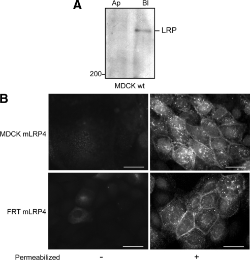
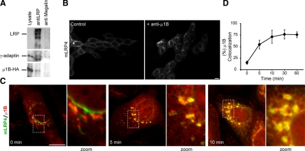
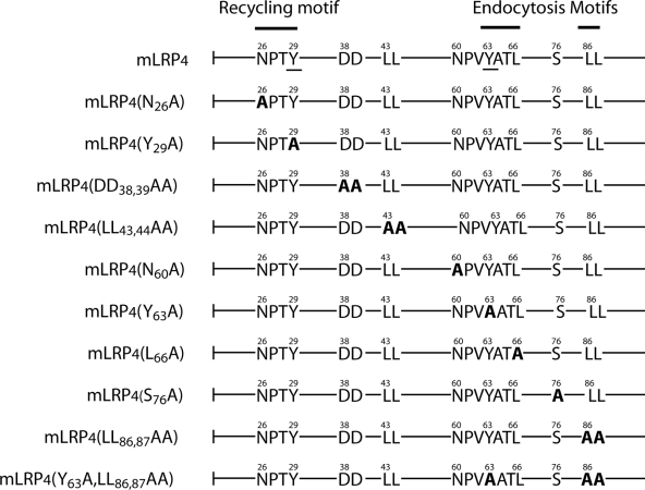
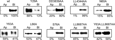
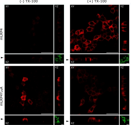
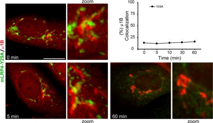
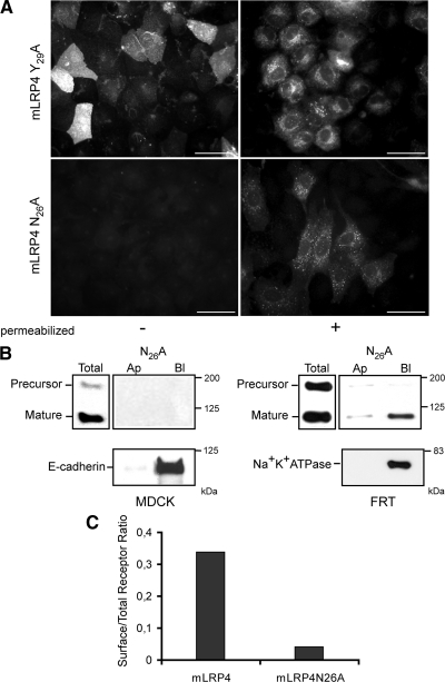
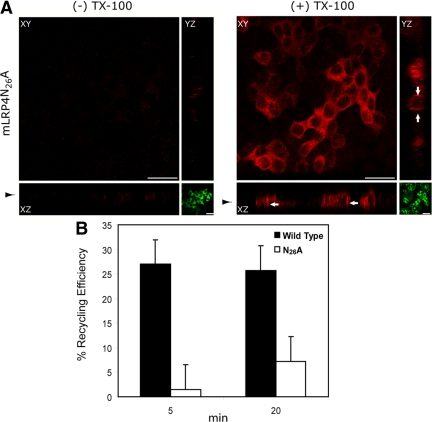
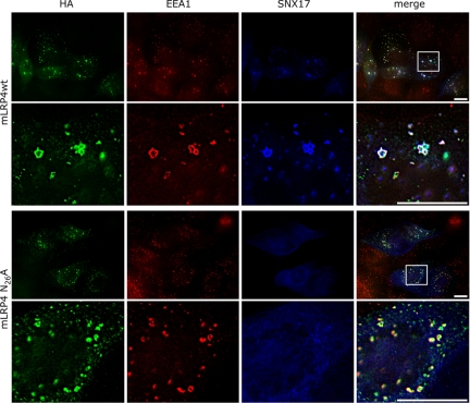
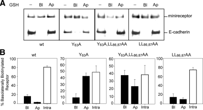
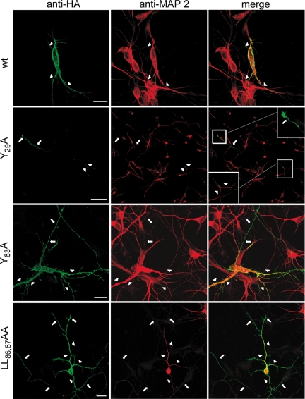
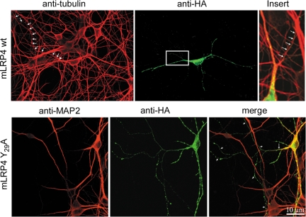
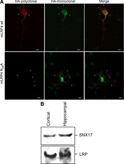
Low-density lipoprotein receptor-related protein 1 (LRP1) is an endocytic recycling receptor with two cytoplasmic tyrosine-based basolateral sorting signals. Here we show that during biosynthetic trafficking LRP1 uses AP1B adaptor complex to move from a post-TGN recycling endosome (RE) to the basolateral membrane. Then it recycles basolaterally from the basolateral sorting endosome (BSE) involving recognition by sorting nexin 17 (SNX17). In the biosynthetic pathway, Y(29) but not N(26) from a proximal NPXY directs LRP1 basolateral sorting from the TGN. A N(26)A mutant revealed that this NPXY motif recognized by SNX17 is required for the receptor's exit from BSE. An endocytic Y(63)ATL(66) motif also functions in basolateral recycling, in concert with an additional endocytic motif (LL(86,87)), by preventing LRP1 entry into the transcytotic apical pathway. All this sorting information operates similarly in hippocampal neurons to mediate LRP1 somatodendritic distribution regardless of the absence of AP1B in neurons. LRP1 basolateral distribution results then from spatially and temporally segregation steps mediated by recognition of distinct tyrosine-based motifs. We also demonstrate a novel function of SNX17 in basolateral/somatodendritic recycling from a different compartment than AP1B endosomes.


Figures

Figure 1.
Figure 1.
Basolateral expression of LRP1 and its minireceptors in polarized epithelial cells. (A) MDCK cells biotinylated either at the apical (Ap) or basolateral (Bl) domain at 4°C were lysed, and the surface biotinylated proteins were precipitated with streptavidin-agarose and resolved in 5% SDS-PAGE followed by immunoblot with anti-human LRP1. Endogenous LRP1 distributes at the basolateral domain. (B) MDCK and FRT cells stably expressing the mLRP4 minireceptor were grown until confluent on coverslips, and the distribution of the minireceptor was visualized by indirect immunofluorescence with monoclonal anti-HA. The minireceptor was detected only after cell permeabilization at the basolateral cell borders and in intracellular vesicles. Scale bars, 10 μm.
Figure 2.
Figure 2.
Physical and functional interaction between LRP1 and AP-1B. (A) MDCK cells were transiently transfected with a plasmid encoding μ1B-HA. Cells were lysed and the lysates were immunoprecipitated with anti-LRP1 or anti-megalin antibodies. The immunoprecipitates were resolved on 4–15% SDS-PAGE. The presence of LRP1 was detected by immunoblotting with anti-LRP antibody, and the complex AP-1B was evidenced by the presence of γ-adaptin and μ1-B. (B) FRT cells were microinjected with mLRP4 minireceptor plus RAP cDNAs with or without blocking-function anti-μ1B antibody. Cells were maintained for 1 h at 37°C, followed by 2 h at 20°C to accumulate the receptor at the TGN. After 1 h of exit at 37°C cells were fixed and processed for immunofluorescence with anti-HA to detect the minireceptor. After 60 min, release from TGN block mLRP4 was located on surface in control cells. In contrast, in μ1B-microinjected cells, mLRP4 was located mainly in a perinuclear localization, most likely RE, indicating that AP1B adaptor complex is involved in the intracellular trafficking of the receptor. (C and D) Kinetic analyses of mLRP4/μ1B colocalization of newly synthesized mLRP4 demonstrates that perinuclear accumulation was achieved during biosynthetic trafficking. Scale bar, 10 μm.
Figure 3.
Figure 3.
Schematic representation of mLRPs with the potential cytoplasmic basolateral sorting signals and their mutations. The sequence of the wild-type tail (mLRP4) is depicted with the already known recycling and endocytic-sorting motifs described in nonpolarized cells (Li et al., 2000; van Kerkhof et al., 2005) and the two tyrosines (Y29 and Y63, underlined) involved in basolateral distribution (Marzolo et al., 2003). All the mutants made are highlighted in bold where the critical residues were replaced by alanine.
Figure 4.
Figure 4.
Changes in the steady-state distribution of mLRP4 mutants uncover the critical basolateral sorting motifs within the LRP1 tail. MDCK cells stably expressing the different mLRP4 constructs were grown on filters to determine the membrane distribution of the receptors by domain-specific biotinylation. Biotinylated minireceptors were precipitated with streptavidin-agarose and resolved in 6% SDS-PAGE followed by immunoblot with monoclonal anti-HA. The receptor's relative expression level in each membrane domain was estimated by densitometry of the resulting bands. The critical basolateral determinants include the Y29 of the proximal NPxY, the Y63ATL66, and the distal LL86,87 motifs. Similar results were obtained from two to three clones of each construct.
Figure 5.
Figure 5.
The mLRP4Y29A mutant is directly addressed to the apical domain in MDCK cells. Three days after reaching confluency, MDCK cells plated on glass coverslips were microinjected in the nucleus with the expression plasmids mLRP4 or Y29A together with pcDNA-RAP and a plasmid encoding the dominant negative form of eps15 (D/N-eps15), GFP- Eps15 DD (EΔ95/295). After incubating the cells for 1 h at 37°C, the newly synthesized receptors were accumulated in the TGN at 20°C for 2 h in the presence of cycloheximide. TGN-to-cell-surface protein traffic was then allowed to proceed at 37°C for 60 min, and the cells were treated for indirect immunofluorescence. The apical or intracellular/basolateral surface distributions of the receptors were assessed in nonpermeabilized and permeabilized cells, respectively. Expression of the D/N-eps15 increased mLRP4wt detection at the cell surface. The cells were analyzed by confocal microscopy and representative x-y plane and x-z and y-z sections are shown (red). D/N-eps15 expression is shown in the x-y plane, medial to x-z sectioning (green). The mLRP4wt appears in intracellular vesicles and in the basolateral membrane, whereas the Y29A mutant was present only in intracellular vesicles and at the apical surface, indicating direct TGN-to-apical addressing. Scale bar: 20 μm. Arrowhead indicates selected x-y plane of x-z sectioning.
Figure 6.
Figure 6.
The mutant Y29A does not traffic through post-Golgi compartment to get the apical plasma membrane. FRT cells were microinjected with mLRP4-Y29A minireceptor plus the chaperone RAP cDNAs and the blocking-function anti-μ1B antibody. Cells were maintained for 1 h at 37°C, followed by 2 h at 20°C to accumulate the receptor at the TGN, and then the cells were fixed at the indicated time and processed for immunofluorescence with anti-HA to detect the minireceptor. Kinetic analyses of mLRP4-Y29A/μ1B colocalization of newly synthesized mLRP4-Y29A show that the μ1B compartment is not involved in biosynthetic trafficking of this mutant. Scale bar, 10 μm.
Figure 7.
Figure 7.
Mutations within the proximal NPxY motif of the LRP1 tail result in different receptor distributions. (A) FRT cells stably transfected with either mLRP4Y29A or mLRP4N26A were grown to confluence on coverslips and treated for indirect immunofluorescence with anti-HA under nonpermeabilized and permeabilized conditions, as indicated. Y29A distributed both at the apical cell surface (right panel) and intracellular vesicles (left panel), whereas N26A show only an intracellular localization, not being detectable at the cell surface. MDCK cells gave similar results (not shown). Scale bars, 10 μm. (B) Domain-specific cell surface biotinylation of MDCK and FRT cells expressing mLRP4N26A. In MDCK cells the protein was not detected at the cell surface. Controls confirmed the receptor expression (immunoblot of total lysate) and proper basolateral expression of E-cadherin. In FRT cells, however, a low amount of minireceptor was detected at the basolateral cell surface. Na+K+ ATPase was used as an endogenous basolateral marker. (C) MDCK cells expressing either mLRP4wt or the N26A mutant were consecutively incubated, either intact or after permeabilization with 0.1% saponin with an anti-HA and anti-mouse RPE-conjugated antibody, and analyzed by flow cytometry. The ratio of expression levels observed in intact versus permeabilized cells show 30% of the wild-type receptor versus no more than 4% of the N26A mutant at the cell surface.
Figure 8.
Figure 8.
The mutant N26A reaches the basolateral membrane, but its recycling efficiency is severely impaired. (A) MDCK cells were microinjected 3 d after reaching confluence with the plasmid encoding the D/N-eps15, GFP- EPS15 DD (EΔ95/295) as described and analyzed in Figure 5. Confocal microscopy and representative x-y plane and x-z and y-z sections are shown (red). D/N-eps15 expression is shown as x-y plane medial of x-z sectioning (green). The distribution of the N26A mutant was mainly intracellular, but was also detectable at the basolateral membrane (arrows). Scale bar, 20 μm. Arrowhead indicates selected x-y plane of x-z sectioning. (B) The recycling efficiency of the minireceptors was evaluated in MDCK cell lines by a fluorescence quenching assay. The mLRP4wt or N26A mutant were labeled with Alexa488-conjugated anti-HA antibody at 37°C for 20 min and then chased for the indicated time periods in the presence of quenching anti-Alexa488 IgG. Cells were processed for flow cytometry, and the recycling efficiency was calculated. The data were plotted and are shown in the graph, corresponding to two experiments measured in duplicate. Average values are shown; error bars, SEM.
Figure 9.
Figure 9.
Colocalization of minireceptors with endosomal markers EEA1 and SNX17. MDCK cells were microinjected with the plasmids encoding for RAP, SNX17-myc, and either the mLRP4wt or N26A minireceptors. Cells were processed for immunofluorescence to detect the HA-tagged minireceptor (green), the early endosome marker EEA1 (red), and the myc-tagged SNX17 (blue). In the amplified merged images, colocalization of the wild-type minireceptor with EEA1 in endosome-like structures was clearly visible; some of these structures also contain SNX17 (white dots). The N26A minireceptors practically did not colocalize with SNX17 and exhibited an increased colocalization with EEA1 (amplified merge image, yellow dots). Scale bars, 10 μm.
Figure 10.
Figure 10.
Basolateral to apical transcytosis of mLRPs in MDCK cells. MDCK cells stably expressing one of the following constructs: mLRP4wt, mLRP4LL86,87AA, mLRPY63A, or the double mutant minireceptor, were grown on filters to confluence. Cells were biotinylated with reducible biotin at 4°C, shifted to 37°C for 45 min, and reduced with glutathione either at the apical or basolateral surface. Cells were lysed, biotinylated proteins were precipitated with streptavidin beads, and the complexes resolved by reducing 6% SDS-PAGE and Western blot. (A) Minireceptor detection with anti-HA and E-cadherin blot. The absence of apical reduction of biotinylated E-cadherin indicates that the observed reduction of the minireceptor was due to its transcytosis from the basolateral to the apical surface. (B) Bands were quantified, and the total biotinylated minireceptor was taken as 100% (see Material and Methods). Minireceptor present in the basolateral surface, apical surface and within the cells was determined as a percentage of total. The determinations were performed in triplicate for each construct.
Figure 11.
Figure 11.
The somatodendritic distribution of mLRP in hippocampal neurons depends on the same motifs required for its basolateral distribution in epithelial cells. Primary cultured hippocampal neurons were transfected as described in Materials and Methods. The distribution of the transfected mLRP was determined by confocal microscopy, using anti-HA to detect the receptor at the cell surface (green) and, after cell permeabilization, using anti-MAP2 (red) to determine the somatodendritic domain. The restricted cell surface somatodendritic distribution of mLRP (arrowheads) was lost when the critical basolateral determinants, based on tyrosines and dileucines, were replaced by alanines. This was visualized by the axonal staining of the receptor in MAP2-negative structures (arrows). The most striking distribution was the mLRPY29A mutant, for which only the distal region of the axon was positively stained with anti-HA. Scale bars, 20 μm (wt, Y63A and LL86,87AA) and 100 μm (Y29A).
Figure 12.
Figure 12.
Intraneuronal distribution and unrestricted transport of the mLRP4Y29A mutant to the axons. Top, confocal micrographs showing the distribution of tubulin (red) and HA-tagged mLRP4wt (green) in 7 DIV (days in vitro) permeabilized cultured hippocampal neurons. HA-tagged mLRP4 localized to short, dendritic-like processes. Note that a thin long axon-like neurite (arrows) that emerges from a dendritic shaft (arrowhead) and that is positive for tubulin does not contain HA-tagged mLRP4. The inset on the right panel (merge color) shows a high-magnification view of the region where the axon-like neurite originates. Bottom, confocal micrographs showing the distribution of the somatodendritic marker MAP2 (red) and the mutant HA-tagged mLRP4Y29A (green) in 7 DIV cultured hippocampal neurons. Note that the ectopic mutant variant of mLRP4 not only localized to dendritic-like processes, but also to MAP2 (−), axon-like neurites (short arrows in the merge panel). The long arrow in the merge panel shows the dendritic site from which the axon merges. For these experiments cells were fixed and permeabilized 18 h after transfection. Scale bar, 10 μm.
Figure 13.
Figure 13.
Intraneuronal distribution and absence of cell surface expression of the mLRP4N26A mutant in hippocampal neurons. (A) Differential cell surface expression of mLRP4wt (top panel) and N26A (bottom panel) in hippocampal neurons in primary culture was evidenced by immunofluorescence detection of the surface minireceptor using a polyclonal anti-HA (red), followed by permeabilization and detection of the intracellular minireceptor using a monoclonal anti-HA (green). The wild-type mLRP4 was expressed in the somato-dendritic surface, but the N26A mutant was only intracellular and somatodendritic. Scale bars, 10 μm. (B) Hippocampal neurons cultured 7 d in vitro and cortical neurons were lysed, 100 μg of proteins were resolved in reducing SDS-PAGE, and the presence of endogenous SNX17 and LRP1 was determined by Western blot.
Figure 14.
Figure 14.
Working model for LRP1 intracellular trafficking in epithelial cells. The data obtained in this work and others previously published (Marzolo et al., 2003; Rodriguez-Boulan et al., 2005; van Kerkhof et al., 2005; Cancino et al., 2007) allow us to derive a working model for LRP1 trafficking. During the biosynthetic pathway, LRP1 is primarily segregated into basolateral-destined vesicles through the recognition of Y29, likely without an NPxY motif. This recognition step occurs at the TGN and likely requires a not yet known adaptor protein(s) that recognizes the critical tyrosine residue (1a). Most of LRP1 should be then segregated by AP1B in the RE (1b), probably recognized through the distal tyrosine-based signal YATL. When the sorting signal is mutated or the corresponding sorting machinery is functionally absent, the receptor fails to be correctly segregated and is directed to the apical membrane or to both domains (punctuate arrow). Once LRP1 arrives at the basolateral membrane, it is actively endocytosed and has to be postendocytically segregated from the basolateral sorting endosome (BSE) in a manner that depends on the proximal NPxY motif and SNX17 (2a; van Kerkhof et al., 2005). A failure in this step causes LRP1 to be trapped in this compartment. After BSE (2b), a secondary recycling step may occur at the common RE and depend on the Y63ATL66/LL86,87 motifs and probably on the AP1B adaptor complex as well (Marzolo et al., 2003) (3).

Similar articles

Cited by

References

    1. Ang A. L., Taguchi T., Francis S., Folsch H., Murrells L. J., Pypaert M., Warren G., Mellman I. Recycling endosomes can serve as intermediates during transport from the Golgi to the plasma membrane of MDCK cells. J. Cell Biol. 2004;167:531–543. - PMC - PubMed
    1. Aroeti B., Mostov K. E. Polarized sorting of the polymeric immunoglobulin receptor in the exocytotic and endocytotic pathways is controlled by the same amino acids. EMBO J. 1994;13:2297–2304. - PMC - PubMed
    1. Bacskai B. J., Xia M. Q., Strickland D. K., Rebeck G. W., Hyman B. T. The endocytic receptor protein LRP also mediates neuronal calcium signaling via N-methyl-D-aspartate receptors. Proc. Natl. Acad. Sci. USA. 2000;97:11551–11556. - PMC - PubMed
    1. Banker G. A., Cowan W. M. Rat hippocampal neurons in dispersed cell culture. Brain Res. 1977;126:397–425. - PubMed
    1. Bansal A., Gierasch L. M. The NPXY internalization signal of the LDL receptor adopts a reverse-turn conformation. Cell. 1981;67:1195–1201. - PubMed

Publication types

MeSH terms

Substances