. 2009 Jan;20(1):102-13.
doi: 10.1091/mbc.e08-07-0741. Epub 2008 Nov 12.

Deciliation is associated with dramatic remodeling of epithelial cell junctions and surface domains

Affiliations

Deciliation is associated with dramatic remodeling of epithelial cell junctions and surface domains

Christian E Overgaard et al. Mol Biol Cell. 2009 Jan.

Abstract

Stress-induced shedding of motile cilia (autotomy) has been documented in diverse organisms and likely represents a conserved cellular reaction. However, little is known about whether primary cilia are shed from mammalian epithelial cells and what impact deciliation has on polarized cellular organization. We show that several chemically distinct agents trigger autotomy in epithelial cells. Surprisingly, deciliation is associated with a significant, but reversible increase in transepithelial resistance. This reflects substantial reductions in tight junction proteins associated with "leaky" nephron segments (e.g., claudin-2). At the same time, apical trafficking of gp80/clusterin and gp114/CEACAM becomes randomized, basal-lateral delivery of Na,K-ATPase is reduced, and expression of the nonciliary apical protein gp135/podocalyxin is greatly decreased. However, ciliogenesis-impaired MDCK cells do not undergo continual junction remodeling, and mature cilia are not required for autotomy-associated remodeling events. Deciliation and epithelial remodeling may be mechanistically linked processes, because RNAi-mediated reduction of Exocyst subunit Sec6 inhibits ciliary shedding and specifically blocks deciliation-associated down-regulation of claudin-2 and gp135. We propose that ciliary autotomy represents a signaling pathway that impacts the organization and function of polarized epithelial cells.


Figures
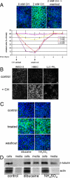
Figure 1.
Figure 1.
Deciliation of polarized epithelial cells. (A) MDCK II cells were cultured in the absence or presence of indicated concentrations of CH for the durations specified. To examine the reversibility of observed effects, CH was removed at the indicated time (“washout”), and cells were cultured for an additional 3 d. At various time points, cultures were fixed and labeled with anti-acetylated tubulin antibodies (to mark primary cilia) and DAPI (to label nuclei). Bar, 10 μM. The percentage of ciliated cells was quantified by dividing the total number of cilia by the number of nuclei counted in 10 randomly selected fields from duplicate filters. Error bars, SEM (B) Polarized cultures of the indicated cell lines were incubated in the absence (control) or presence (+ CH) of either 2 mM (IMCD-3 cells and HBECs) or 6 mM (LLC-PK1 cells) CH for 3 d and then fixed and labeled with anti-acetylated tubulin antibodies. Bars, 20 μM. (C) Polarized MDCK II cells were cultured in the absence (control) or presence (treated) of either 0.75 mM dibucaine for 30 min or 30 mM NH4SO4 for 3 h. For washout, cultures were incubated an additional 48 h in the absence of deciliating agents. (D) Polarized MDCK II cells were cultured as in C, but in serum-free culture medium. Cell extracts and culture media samples, normalized for total protein content, were resolved by SDS-PAGE, and Western blots were probed for β-tubulin and actin. Note that tubulin, but not actin, is present in the culture medium after treatment of cells with dibucaine or NH4SO4, indicating the presence of shed cilia in the media.
Figure 2.
Figure 2.
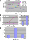
TJ remodeling accompanies deciliation. (A) Polarized cultures of MDCK II, LLC-PK1, and Caco-2 cells were incubated in the absence (control) or presence (+ CH) of 4 mM CH for indicated times, at which TER was measured. (B) Polarized cultures of HBECs were incubated in the absence or presence of 4 mM CH for indicated times, whereupon TER was measured. IMCD-3 cells were incubated without or with 2 mM CH for 3 d. (C) Polarized MDCK II cells were incubated in the absence or presence of 30 mM NH4SO4 for 24 h, followed by an additional 48 h in the absence of NH4SO4 for washout. Note that TER increase after NH4SO4-induced deciliation is of a similar magnitude as that after CH-induced deciliation (A). In all panels, n = 3 filters per condition; error bars, SEM.
Figure 3.
Figure 3.
TJ composition changes after epithelial deciliation. (A) Polarized MDCK II cells were treated without (control) or with (+ CH) 4 mM CH for 2 d and then fixed and labeled with antibodies to indicated claudin proteins. Confocal microscopic images were collected at identical settings for control and experimental samples. Note that intensity of staining of claudins-1 and -2 are decreased and that of claudin-4 is increased after deciliation. (B) Cultures, treated as in A were labeled with antibodies to indicated TJ proteins. Note that ZO-2 and -3 distributions appear to shift toward cytosolic distributions after deciliation. (C) Duplicate cultures, treated as in A or with indicated CH concentrations or treated and cultured an additional 2 d in the absence of CH (washout), were lysed and subjected to SDS-PAGE and immunoblotting with indicated anti-claudin antibodies. Note that expression of claudins-1 and -2 are significantly decreased in deciliated cultures, whereas that of other claudins is not substantially altered. (D) Polarized cultures of MDCK I cells were incubated in the presence of 4 mM CH for indicated times, at which TER was measured. n = 3 filters; error bars, SEM (E) Linearity of TJ segments from control or CH-treated cultures was measured using NIH ImageJ software. A straight line was defined as having a linearity = 1. n = 50 TJ segments per condition; error bars, SEM.
Figure 4.
Figure 4.
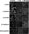
Monolayer dissolution assay. Confluent cultures of MDCK II cells were incubated in the absence (control) or presence of 2 mM CH for 3 d or of 30 mM NH4SO4 for 24 h. Media was aspirated, and cultures were incubated in calcium/magnesium-free Hanks' buffered saline solution at 37°C for 30 min before being fixed and labeled with antibodies to indicated proteins or FITC-phalloidin to label f-actin. Note the absence of intercellular contacts and accumulation of junction proteins in internal compartments in control cultures, but the retention of junctions in deciliated cultures. Bars, 5 μm.
Figure 5.
Figure 5.
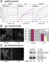
Alteration of apical protein expression and sorting accompanies deciliation. (A) Polarized cultures of MDCK II cells were treated as described in Figure 3, and secretion of gp80/clusterin was measured as described in Materials and Methods. Note that apical secretion of this protein (control) becomes randomized after deciliation (+ CH), but then returns to normal after regrowth of cilia (washout). n = 4 filters per condition; error bars, SEM. (B) Left, polarized MDCK II cells were fixed and labeled with antibodies to gp114/CEACAM. En face (top) and orthogonal (bottom) images are shown. Bar, 5 μm. Right, triplicate cultures of polarized MDCK II cells were biotinylated apically or basolaterally. Biotinylated proteins were precipitated on streptavidin agarose, resolved by SDS-PAGE, and immunoblotted for gp114/CEACAM. Levels of protein recovered were quantified by phosphorimaging, and results are presented as % of total surface-labeled gp114/CEACAM. Note that apical polarity of gp114/CEACAM is lost in deciliated cultures. n = 3; error bars, SEM. (C) Left, polarized MDCK II cells were fixed and labeled with antibodies to gp135/podocalyxin. En face (top) and orthogonal (bottom) images are shown. Bar, 10 μm. Right, surface biotinylation assay was performed as in B. Note that expression level of gp135/podocalyxin is greatly decreased in deciliated cultures.
Figure 6.
Figure 6.
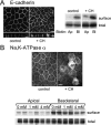
Deciliation is accompanied by selective effects on basal-lateral trafficking. (A) Left, polarized MDCK II cells were fixed and labeled with antibodies to E-cadherin. En face (top) and orthogonal (bottom) images are shown. Bar, 5 μm. Right, surface biotinylation assay. Note that expression level and polarized distribution of E-cadherin is not substantially altered after deciliation. (B) Top, polarized MDCK II cells were fixed and labeled with antibodies to N, K-ATPase α subunit. (Bottom) Quadruplicate cultures of polarized MDCK II cells were incubated in the presence of indicated concentrations of CH for 3 d, and then duplicate cultures were biotinylated either apically or basolaterally. Labeled proteins were recovered by precipitation with streptavidin agarose, resolved by SDS-PAGE, and immunoblotted with anti-N,K-ATPase α antibodies. Note that this protein is inefficiently retained at the basal-lateral membrane in deciliated cultures and instead accumulates within perinuclear endosomes.
Figure 7.
Figure 7.
Calcium signaling is involved in deciliation and TJ remodeling. (A) Ca2+ concentration was determined by digitized fluorescence microscopy of Fura-2–stained MDCK II cells incubated without or with 4 mM CH or NH4SO4 for indicated durations. n = 5 cells per condition; error bars, SEM. (B) Cells were preloaded with either 25 μM EGTA-AM or 25 μM BAPTA-AM before addition of 4 mM CH or 30 mM NH4SO4 or were left untreated (control). Twenty-four hours later, TER was measured and cultures were fixed and labeled with anti-acetylated tubulin antibodies. Note that preloading cells with calcium chelators largely prevented deciliation and blocked the associated rise in TER induced by CH, but had no effect on NH4SO4-induced deciliation and remodeling. Cilia were counted in five randomly selected fields per filter. n = 3 filters per condition; error bars, SEM. (C) Polarized MDCK II cells were cultured in HG-DMEM in the absence or presence of 1 μM thapsigargin for 24 h, at which time TER was measured and cultures were fixed and labeled with anti-acetylated tubulin antibodies. n = 3 filters per condition; error bars, SEM.
Figure 8.
Figure 8.
Mature primary cilia are not required for autotomy-associated epithelial remodeling. (A) Polarized MDCK II cells, or cultures with reduced expression of 14-3-3η or polaris/IFT88, were fixed and labeled with antibodies to acetylated tubulin (green) and claudin-2 (red). Note that mature primary cilia are absent in sh14-3-3η and shPolaris cells, but that these cells retain claudin-2 expression. Confocal images were collected at the focal plane of TJs to show claudin-2. Procilia do not label with anti-acetylated tubulin antibodies. Bar, 10 μm. (B) Polarized MDCK II, sh14-3-3η or shPolaris cells were cultured in the absence or presence of 4 mM CH, and TER was measured at indicated times. Note that sh14-3-3η or shPolaris cells tighten their junctions in response to deciliation-inducing signals, but that their response is somewhat slower than that of parental MDCK II cells. n = 3 filters per condition.
Figure 9.
Figure 9.
Inhibition of ciliary shedding prevents epithelial remodeling. (A) Blocking exocyst function prevents stress-induced deciliation. Polarized MDCK II cells or MDCK II cells with ∼90% reduced levels of Sec6 (MDCK II shSec6; top right) were incubated in the absence or presence of either 2 mM CH for 2 d or 30 mM NH4SO4 for 24 h. Cultures were fixed and labeled with anti-acetylated tubulin antibodies and DAPI (left), and the fraction of ciliated cells in the population was determined as described in Figure 1 (bottom right). “Control MDCK II” cells were transduced with lentiviral vectors encoding a nontargeting shRNA and were selected in puromycin, as described in Materials and Methods. (B) In cultures in which ciliary shedding is inhibited, TJs do not undergo remodeling and gp135/podocalyxin expression is maintained. Cells, cultured in the absence or presence of either 2 mM CH for 2 d, 30 mM NH4SO4 for 24 h, or 100 ng/ml EGF for 24 h, were monitored for TER at the time of harvest (left). In addition, cells were lysed and lysate was subjected to SDS-PAGE and immunoblotting with antibodies to claudin-2 and β-tubulin (top right) or gp135 (bottom right). n = 3 filters; error bars, SEM.

Similar articles

Cited by

References

    1. Balcarova-Stander J., Pfeiffer S. E., Fuller S. D., Simons K. Development of cell surface polarity in the epithelial Madin-Darby canine kidney (MDCK) cell line. EMBO J. 1984;3:2687–2694. - PMC - PubMed
    1. Balda M. S., Gonzalez-Mariscal L., Contreras R. G., Macias-Silva M., Torres-Marquez M. E., Garcia-Sainz J. A., Cereijido M. Assembly and sealing of tight junctions: possible participation of G-proteins, phospholipase C, protein kinase C and calmodulin. J. Membr. Biol. 1991;122:193–202. - PubMed
    1. Bergesse J. R., Domenech C. E., Balegno H. F. Chloral hydrate inhibition in vitro of ATPase in membrane of rat erythrocytes and in microsomes of dog kidney external medulla. Biochem. Pharmacol. 1983;32:3221–3225. - PubMed
    1. Blum J. J. Existence of a breaking point in cilia and flagella. J. Theor. Biol. 1971;33:257–263. - PubMed
    1. Burdett I.D. Internalisation of desmosomes and their entry into the endocytic pathway via late endosomes in MDCK cells. Possible mechanisms for the modulation of cell adhesion by desmosomes during development. J. Cell Sci. 1993;106(Pt 4):1115–1130. - PubMed

Publication types