. 2009 Jan;20(1):124-33.
doi: 10.1091/mbc.e08-06-0662. Epub 2008 Nov 12.

Fibroblast growth factor receptor-induced phosphorylation of ephrinB1 modulates its interaction with Dishevelled

Affiliations

Fibroblast growth factor receptor-induced phosphorylation of ephrinB1 modulates its interaction with Dishevelled

Hyun-Shik Lee et al. Mol Biol Cell. 2009 Jan.

Abstract

The Eph family of receptor tyrosine kinases and their membrane-bound ligands, the ephrins, have been implicated in regulating cell adhesion and migration during development by mediating cell-to-cell signaling events. The transmembrane ephrinB1 protein is a bidirectional signaling molecule that signals through its cytoplasmic domain to promote cellular movements into the eye field, whereas activation of the fibroblast growth factor receptor (FGFR) represses these movements and retinal fate. In Xenopus embryos, ephrinB1 plays a role in retinal progenitor cell movement into the eye field through an interaction with the scaffold protein Dishevelled (Dsh). However, the mechanism by which the FGFR may regulate this cell movement is unknown. Here, we present evidence that FGFR-induced repression of retinal fate is dependent upon phosphorylation within the intracellular domain of ephrinB1. We demonstrate that phosphorylation of tyrosines 324 and 325 disrupts the ephrinB1/Dsh interaction, thus modulating retinal progenitor movement that is dependent on the planar cell polarity pathway. These results provide mechanistic insight into how fibroblast growth factor signaling modulates ephrinB1 control of retinal progenitor movement within the eye field.


Figures

Figure 1.
Figure 1.
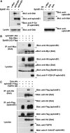
The C terminus of ephrinB1 mediates binding with Dsh. (A) Immunoprecipitates using anti-ephrinB1 (rabbit), anti-Dsh (rabbit), or anti-c-Myc (rabbit) antibodies in HT 29 human colon carcinoma cell lysates were immunoblotted with anti-Dsh (goat) and anti-ephrinB1 (goat) antibodies. Lysates were analyzed directly by SDS-polyacrylamide gel electrophoresis (PAGE) and immunoblotted with indicated antibodies to reveal endogenous expression levels of ephrinB1 and Dsh, respectively. (B) Oocytes were left uninjected (−) or injected (+) with FLAG-tagged ephrinB1WT or ephrinB1Δ6 (10 ng) and HA-tagged Dsh (10 ng) RNAs where indicated. Oocyte lysates were IPed with anti-HA antibody and then immunoblotted with anti-FLAG antibody to detect ephrinB1 proteins.
Figure 2.
Figure 2.
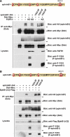
Cognate Eph receptor binding or FGF signaling blocks the interaction between ephrinB1 and Dsh through the tyrosine phosphorylation of the intracellular domain of ephrinB1. (A) EphB1-Fc was clustered using human Ig and added to the HT 29 cell culture medium. Immunoprecipitates using anti-ephrinB1 (rabbit), anti-Dsh (rabbit), or anti-c-Myc (rabbit) antibodies in HT 29 cell lysates were immunoblotted with anti-Dsh (goat), anti-phosphotyrosine-HRP-conjugated, and anti-ephrinB1 (goat) antibodies. Lysates were analyzed directly by SDS-PAGE and immunoblotted with indicated antibodies to reveal endogenous expression levels of ephrinB1 and dishevelled, respectively. (B) Oocytes were left uninjected (−) or injected (+) with ephrinB1-HA (10 ng), Dsh-Myc (10 ng), and EphB1(ΔC)-FLAG (10 ng) RNAs as indicated. Oocyte lysates were IPed with anti-Myc antibody and then immunoblotted with anti-HA antibody to detect ephrinB1 proteins. Oocyte lysates were analyzed directly by SDS-PAGE and immunoblotted with indicated antibodies. (C) Oocytes were left uninjected (−) or injected (+) with ephrinB1-FLAG (10 ng), Dsh-HA (10 ng) and FGFR1 KE (10 ng) RNAs as indicated. Oocyte lysates were IPed with anti-HA (top and second panels) or anti-FLAG (third and fourth panels) antibodies and then immunoblotted with either anti-FLAG or anti-HA antibodies to detect bound proteins. Oocyte lysates were analyzed directly by SDS-PAGE and immunoblotted with indicated antibodies.
Figure 3.
Figure 3.
FGFR1- or Eph-induced phosphorylation of tyrosines 324 and 325 in ephrinB1 disrupts the interaction with Dsh. (A) Oocytes were left uninjected (−) or injected (+) with ephrinB1WT-HA (10 ng) or ephrinB1Y324.5F-HA (10 ng), Dsh-Myc (10 ng) and FGFR1 KE (10 ng), or FGFR1 KD (10 ng) RNAs as indicated. Oocyte lysates were IPed with anti-Myc antibody and then immunoblotted with anti-HA antibody to detect ephrinB1 proteins. Oocyte lysates were analyzed directly by SDS-PAGE and immunoblotted with indicated antibodies. (B) Oocytes were left uninjected (−) or injected (+) with ephrinB1WT-HA (10 ng) or ephrinB1Y324.5F-HA (10 ng), Dsh-Myc (10 ng) and EphB1(ΔC)-FLAG (10 ng) RNAs as indicated. Oocyte lysates were IPed with anti-Myc antibody, then immunoblotted with anti-HA antibody to detect ephrinB1 proteins. Oocyte lysates were analyzed directly by SDS-PAGE and immunoblotted with indicated antibodies.
Figure 4.
Figure 4.
FGFR1-induced phosphorylation of tyrosines 324 and 325 in ephrinB1 restricts the movement of retinal progenitor cells. (A) The D1.1.1 blastomere was injected with FGFR1 KE (200 pg) and 200 pg of β-galactosidase with or without 50 pg of ephrinB1WT-HA or ephrinB1Y324.5F-HA RNA. Embryos were collected at stage 12.5 and stained for β-galactosidase (red). Histogram represents lateral scatter distances of RNA-injected D1.1.1 progeny as a percentage of controls. All control embryos showed D1.1.1 progeny broadly dispersed across the dorsal animal quadrant, whereas FGFR1 KE-injected embryos were tightly confined to the midline. EphrinB1WT RNA partially rescues cell dispersion that is restricted by FGFR1 KE. In contrast, ephrinB1Y324.5F more prominently rescues the FGFR1 KE-restriction. One hundred percent normal distribution represents the standard lateral distance observed for β-galactosidase expression in control D1.1.1 progeny. Data are shown as mean ± SD. (B) The D1.1.1 blastomere was injected with FGFR1 KE (200 pg) with 250 pg of GFP with or without 50 pg of ephrinB1WT-HA or ephrinB1Y324.5F-HA RNA. Immunofluorescence was performed on 37–38 stage embryos. The histogram denotes that >80% of control embryos showed D1.1.1 progeny in the retina, whereas injection of FGFR1 KE results in a significant reduction in the percentage of embryos displaying D1.1.1 progeny in the retina. EphrinB1WT RNA partially rescues the FGFR1 KE-induced block. In contrast, ephrinB1Y324.5F robustly rescues the FGFR1 KE-restricted phenotype. Embryos with D1.1.1 injections were considered positive when fluorescent progeny contributed >30% of the cells in the retina. White ovals denote retina. Data are shown as mean ± SD, and the number of embryos per sample (N) is denoted above the histogram.
Figure 5.
Figure 5.
Phosphorylation of both tyrosines is necessary for an FGFR1-induced alteration of cell fate. The D1.1.1 blastomere was injected with 200 pg of FGFR1 KE alone or with 50 pg of wild-type ephrinB1 or the Y324.5F mutant along with 250 pg of GFP RNA. Xenopus embryos were collected at stage 16 and subjected to whole mount in situ hybridization. Active FGFR1 reduces eye-specific transcription factors rx1 and pax6. In contrast, ventral neural fate marker pax2 is expanded on the injected side. A suboptimal amount of ephrinB1WT expression partially rescues FGFR1 KE-induced reduction of eye-specific markers and expansion of the ventral neural marker, but the ephrinB1Y324.5F mutant rescues these markers to a near normal pattern. The asterisk indicates injected side.
Figure 6.
Figure 6.
Phosphorylation on tyrosines 324 and 325 is necessary for blocking ephrinB1-driven movement of cells during gastrulation. The V1.1.1 blastomere was injected with 250 pg of ephrinB1WT-HA or ephrinB1Y324.5F-HA RNA with or without FGFR1 KE (400 pg) and 200 pg of β-galactosidase or 250 pg of GFP RNA (A and B, respectively). (A) Embryos were collected at stage 12.5 and stained for β-galactosidase (red). Histogram represents the percentage of embryos displaying ventral to dorsal (V-D) movements of RNA-injected V1.1.1 progeny. All control embryos display a tight restriction of V1.1.1 progeny to the ventral side, whereas ephrinB1WT or ephrinB1Y324.5F-injected embryos show V1.1.1 progeny dispersed to dorsal regions. FGFR1 KE RNA restricted ephrinB1WT-induced cell movement, whereas ephrinB1Y324.5F-expressing cells migrated dorsally. Embryos are considered positive for V-D movements when 30% of progeny cells cross the ventral-dorsal midline. Data are shown as mean ± SD, and the number of embryos per sample (N) is denoted above the histogram. (B) Embryos were collected at stage 37–38, sectioned, and immunofluorescence was performed. The histogram denotes that below 10% of control embryos showed V1.1.1 progeny in the retina. Injection of ephrinB1WT or ephrinB1Y324.5F mutant RNA results in a significant induction in the percentage of embryos displaying V1.1.1 progeny in the retina. FGFR1 KE RNA restricts ephrinB1WT-induced cell movement but it does not restrict ephrinB1Y324.5F-expressing cells. Embryos injected in V1.1.1 were considered positive when fluorescent progeny contributed >10% of the cells in the retina. White ovals denote retina. Data are shown as mean ± SD.
Figure 7.
Figure 7.
FGFR1-induced phosphorylation of tyrosines 324 and 325 in ephrinB1 is necessary to block PCP signaling. (A) GFP-Dsh RNA (250 pg) was injected singly or with RNAs encoding HA-tagged ephrinB1WT (400 pg) or ephrinB1Y324.5F (400 pg) in the presence of FGFR1 KE (500 pg) into each blastomere of two-cell stage embryos and collected at stage 10.5. Embryos were sectioned and immunostained for ephrinB1 (HA) or GFP-Dsh (GFP). Fluorescence microscopy shows cytoplasmic localization when GFP-Dsh is expressed alone. In contrast, coexpression of GFP-Dsh with ephrinB1WT or ephrinB1Y324.5F relocalizes Dsh to the membrane. Moreover, FGFR1 KE disrupts ephrinB1WT-induced membrane localization of Dsh but fails to disrupt ephrinB1Y324.5F-induced Dsh relocalization. (B) The D1.1.1 blastomere was injected with 200 pg of FGFR1 KE and 250 pg of Dsh or 50 pg of CA RhoA or 250 pg of DN RhoA RNA along with 250 pg of GFP RNA. At stage 37–38, the retina was examined, and a significant rescue by either Dsh or CA RhoA was observed as evidenced by the percentage of FGFR1 KE-injected embryos with fluorescent D1.1.1 retinal clones in the retina. White ovals denote retina. Data are shown as mean ± SD.

Similar articles

Cited by

References

    1. Amaya E., Musci T. J., Kirschner M. W. Expression of a dominant negative mutant of the FGF receptor disrupts mesoderm formation in Xenopus embryos. Cell. 1991;66:257–270. - PubMed
    1. Amaya E., Stein P. A., Musci T. J., Kirschner M. W. FGF signalling in the early specification of mesoderm in Xenopus. Development. 1993;118:477–487. - PubMed
    1. Arvanitis D., Davy A. Eph/ephrin signaling: networks. Genes Dev. 2008;22:416–429. - PMC - PubMed
    1. Bruckner K., Pasquale E. B., Klein R. Tyrosine phosphorylation of transmembrane ligands for Eph receptors. Science. 1997;275:1640–1643. - PubMed
    1. Cavodeassi F., Carreira-Barbosa F., Young R. M., Concha M. L., Allende M. L., Houart C., Tada M., Wilson S. W. Early stages of zebrafish eye formation require the coordinated activity of Wnt11, Fz5, and the Wnt/beta-catenin pathway. Neuron. 2005;47:43–56. - PMC - PubMed

Publication types

MeSH terms